The correct answer is e. Hemodialysis is not associated with high-flow priapism. The other answer choices are all known causes or associations of high-flow priapism.
Agenda
1. Definition
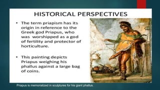
2. HistoricalBACKGROUND
3. PHISILIOGY OF ERECTION
4. Types of priapism and comparison between different types
5. Management of each type
6. Take home massage
7. Questions
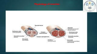
Physiology of erection
Cavernosal
arterysmooth
muscle
Relaxation
Dunder parasympathetic
nerve by release of NO
increase penile
blood flow asnd
corporal expantion
Increase intracaversosal pressure
Compression of subtunical veins
Full Rigidity and tumesent
5.
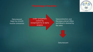
Physiology of erection
Detumescent
beginby smooth
muscle contraction
under sympathetic
control by
norepinephrine at alpha
receptor
Vasocontrection and
Decrease arterial inflow
and blood is drained by
the veins
Detumescent
6.
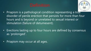
• Priapism isa pathological condition representing a true
disorder of penile erection that persists for more than four
hours and is beyond or unrelated to sexual interest or
stimulation (failure of detumesent).
• Erections lasting up to four hours are defined by consensus
as ‘prolonged’.
• Priapism may occur at all ages.
Definition
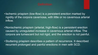
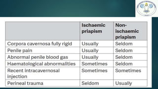
•Ischemic priapism (low-flow)is a persistent erection marked by
rigidity of the corpora cavernosa, with little or no cavernous arterial
inflow.
•Nonischemic priapism (arterial, high-flow) is a persistent erection
caused by unregulated increase in cavernous arterial inflow. The
corpora are tumescent but not rigid, and the erection is not painful.
•Stuttering priapism describes a pattern of recurrence. It described
recurrent prolonged and painful erections in men with SCD.
Definition
9.
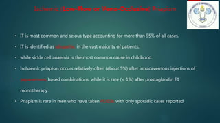
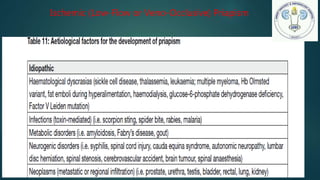
Ischemic (Low-Flow orVeno-Occlusive) Priapism
• IT is most common and seious type accounting for more than 95% of all cases.
• IT is identified as idiopathic in the vast majority of patients,
• while sickle cell anaemia is the most common cause in childhood.
• Ischaemic priapism occurs relatively often (about 5%) after intracavernous injections of
papaverinem based combinations, while it is rare (< 1%) after prostaglandin E1
monotherapy.
• Priapism is rare in men who have taken PDE5Is with only sporadic cases reported
10.
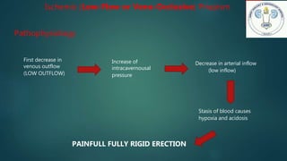
Ischemic (Low-Flow orVeno-Occlusive) Priapism
Pathophysiology
First decrease in
venous outflow
(LOW OUTFLOW)
Increase of
intracavernousal
pressure
Decrease in arterial inflow
(low inflow)
Stasis of blood causes
hypoxia and acidosis
PAINFULL FULLY RIGID ERECTION
Ischemic (Low-Flow orVeno-Occlusive) Priapism
prognosis
• Ischemic priapism is an emergency.
• The longer the duration of priapism the higher the rate of ED
• Interventions beyond 48 to 72 hours of onset may help relieve erection
and pain but have little benefit in preserving potency(100% ED).
• When left untreated, resolution may take days and erectile dysfunction
(ED) invariably results (dissolution of thrombus can be found in the
sinusoidal spaces)
15.
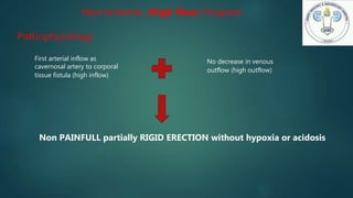
Non Ischemic (Highflow) Priapism
Pathophysiology
First arterial inflow as
cavernosal artery to corporal
tissue fistula (high inflow)
No decrease in venous
outflow (high outflow)
Non PAINFULL partially RIGID ERECTION without hypoxia or acidosis
16.
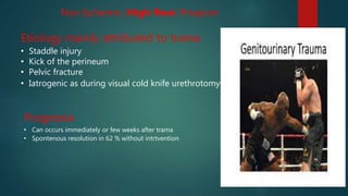
Non Ischemic (Highflow) Priapism
Etiology manily attributed to trama
• Staddle injury
• Kick of the perineum
• Pelvic fracture
• Iatrogenic as during visual cold knife urethrotomy
Prognosis
• Can occurs immediately or few weeks after trama
• Spontenous resolution in 62 % without intrtvention
17.
Stuttering (recurrent orintermittent) priapism
• It is recurrent ischemic priapism
• a distinct condition that is characterized by repetitive and painful episodes of
prolonged erections.
• Sickle cell disease is the most common cause
• Due to obstruction of venous outflow by sickled erythrocytes leads to
stagnation of blood within the sinusoids during erection
• Each episode should managed as usual
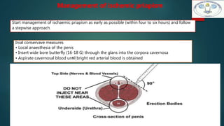
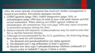
Start management ofischaemic priapism as early as possible (within four to six hours) and follow
a stepwise approach.
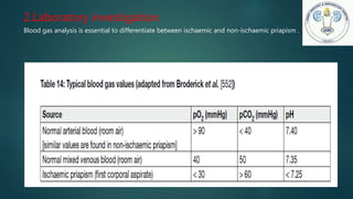
Inial conservave measures
• Local anaesthesia of the penis
• Insert wide bore butterfly (16-18 G) through the glans into the corpora cavernosa
• Aspirate cavernosal blood un•til bright red arterial blood is obtained
Management of ischemic priapism
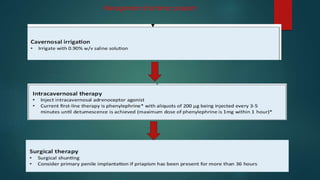
phenylephrinecan be concentratedas 200 ug/mL in saline and
administered intermittently as 0.5 mL to 1.0 mL, every 5 to 10 minutes to a
maximum dosage of 1 mg.
• This will permit up to 10 separate injections of 0.5 mL (100 ug each) or 5
separate injections of 1 mL (200 ug each).
• Aspirate the penis between successive injections until distal shaft is
empty.
• Blood pressure monitoring is recommended if repeated
sympathomimetic dosing is given. In patients with significant cardiovascular
risks, electrocardiogram monitoring is recommended.
28.
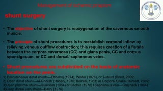
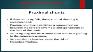
• The objectiveof shunt surgery is reoxygenation of the cavernous smooth
muscle.
• The principle of shunt procedures is to reestablish corporal inflow by
relieving venous outflow obstruction; this requires creation of a fistula
between the corpora cavernosa (CC) and glans penis, CC and corpus
sponsigosum, or CC and dorsal/ saphenous veins.
• Shunt procedures are subdivided on the basis of anatomic
location on the penis.
1) Percutaneous distal shunts—Ebbehoj (1974), Winter (1976), or T-shunt (Brant, 2009)
2) Open distal shunt—Al-Ghorab (Hanafy, 1976; Borrelli, 1983) or Corporal Snake (Burnett, 2009)
3)Open proximal shunt—Quackles (1964) or Sacher (1972) l Saphenous vein—Grayhack (1964)
4)Deep dorsal vein shunt—Barry (1976)
Management of ischemic priapism
shunt surgery
29.
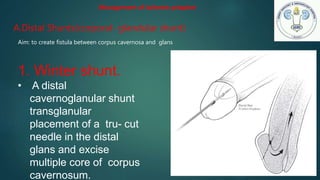
A.Distal Shunts(corporal- glandularshunt)
Aim: to create fistula between corpus cavernosa and glans
Management of ischemic priapism
1. Winter shunt.
• A distal
cavernoglanular shunt
transglanular
placement of a tru- cut
needle in the distal
glans and excise
multiple core of corpus
cavernosum.
30.
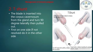
2. T shunt
•The blade is inserted into
the corpus cavernosum
from the gland and turn 90
degree laterally then pulled
out.
• First on one side If not
resolved do it in the other
side
Management of ischemic priapism
31.
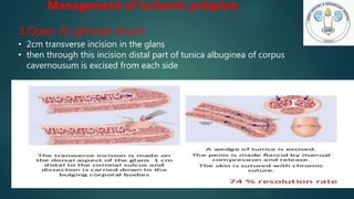
3.Open Al-ghorab shunt
•2cm transverse incision in the glans
• then through this incision distal part of tunica albuginea of corpus
cavernousum is excised from each side
Management of ischemic priapism
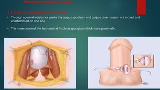
1.Corporo spongiosum shunt
•Through apernial incision or penile the corpus sponisum and corpus cavernousum are incised and
anastomosed on one side
• The more proximal the less urethral fistula as spongiusm thick more proximally.
Management of ischemic priapism
34.
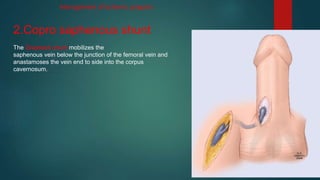
2.Copro saphenous shunt
TheGrayhack shunt mobilizes the
saphenous vein below the junction of the femoral vein and
anastamoses the vein end to side into the corpus
cavernosum.
Management of ischemic priapism
35.
• Complete flaccidity
•Visualization of bright red blood
• In cases where the examination may be equivocal (penile rigidity rather than
complete flaccidity) ,color Doppler ultrasonography or cavernous blood gas is
recommended to demonstrate patency of shunt and restoration of cavernous
inflows
Management of ischemic priapism
Assessing shunt patency
36.
Surgical Management ofIschemic Priapism with Immediate Penile
prosthesis
Consider penile prosthesis if:
1. The patient has failed aspiration and sympathomimetic intracavernous injection.
2. The patient has failed distal and proximal shunting.
3. Ischemia has been present for longer than 36 hours.
The advantages of early penile implantation
• preservation of penile length (before fibrosis)
• easier insertion.
Disadvantages
There are higher rates of revision surgery and complications
due to infection, urethral injury, device migration, and device erosion.
Management of ischemic priapism
37.
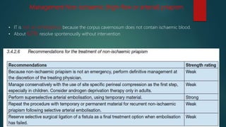
Management Non-ischaemic (high-flowor arterial) priapism
• IT is not an emergency because the corpus cavernosum does not contain ischaemic blood.
• About 62% resolve spontenouslly without intervention
38.
Surgical management ofnon ishemic priapism
• Surgery is technically challenging and may pose significant risks, mainly ED due to accidental
of the cavernous artery instead of the fistula.
• It is rarely performed and should only be considered when there are contraindications for selective
embolisation, no availability of the technique or embolisation failure
Management Non-ischaemic (high-flow or arterial) priapism
• Start managementof ischaemic priapism as early as possible (within four to six hours) and follow a stepwise
approach.
• Interventions 48 hours beyond the onset of ischemic priapism may relieve pain but will have little benefit in
preserving potency.
• Priapism following a PDE5 inhibitor usually occurs in men with other risk factors.
• Spontaneous resolution of high-flow arterial priapism generally occurs in two thirds of patients.
• Aspiration has no role in high-flow priapism other than for diagnosis; it plays no role in treatment.
Take home massage
43.
1. Ischemic priapismis a persistent erection
marked by each of the following clinical and
pathophysiologic characteristics EXCEPT:
a. rigidity of the corpora cavernosa.
b. bright red corporal blood.
c. hypoxic and acidotic corporal environment.
d. painful rigidity.
e. thrombus within the sinusoidal spaces.
Questions
44.
Questions
2. The associationsand pathophysiology of high-
flow priapism include each of
the following EXCEPT:
a. straddle injury.
b. coital trauma.
c. birth canal injury to the newborn male.
d. cold-knife urethrotomy.
e. hemodialysis.


![Erectile Dysfunction [Dr. Edmond Wong]](https://cdn.slidesharecdn.com/ss_thumbnails/ededmond-140716212750-phpapp02-thumbnail.jpg?width=640&height=640&fit=bounds)


