Зүрхний цахилгаан бичлэг
•Download as PPT, PDF•
2 likes•852 views

This document provides an overview of interpreting 12-lead electrocardiograms (ECGs) for myocardial infarction (MI). It reviews ECG waves, intervals, and segments. It defines ischemia, injury, and infarction and describes associated ECG changes. It identifies the five major infarct areas and corresponding lead changes. Color coding is used to indicate changes for anterior, inferior, lateral, posterior, and subendocardial MIs. Examples of ECG strips demonstrate single and combined infarct patterns. Cardiac enzymes that indicate infarction and their time courses are also reviewed.

Recommended
More Related Content
What's hot
What's hot (20)
Similar to Зүрхний цахилгаан бичлэг
Similar to Зүрхний цахилгаан бичлэг (20)
More from Анагаахын Шинжлэх Ухааны Үндэсний Их Сургууль
More from Анагаахын Шинжлэх Ухааны Үндэсний Их Сургууль (20)
Recently uploaded
Recently uploaded (20)
Зүрхний цахилгаан бичлэг
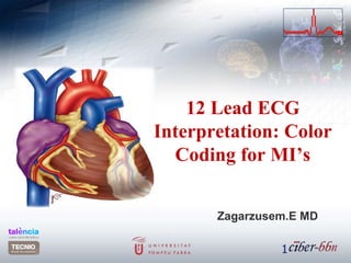
- 1. 12 Lead ECG Interpretation: Color Coding for MI’s Zagarzusem.E MD 1
- 2. Objectives review the ECG waveform and intervals Define myocardial ischemia, injury and infarction Identify the 5 major infarct areas on the 12 lead Name occluded arteries common to the area Differentiate ECG changes reflecting ischemia, injury and infarction Identify cardiac enzymes associated with ACS 2
- 3. 3
- 5. The 12-Lead view Each limb lead I, II, III, AVR, AVL, AVF records from a different angle All six limb leads intersect and visualize a frontal plane The six chest leads (precordial) V1, V2, V3, V4, V5, V6 view the body in the horizontal plane to the AV node The 12 lead ECG forms a camera view from 12 angles 5
- 6. Views from Augmented and Limb Leads- Frontal 6
- 7. Precordial lead snapshots Think of each precordial lead as a horizontal view of the heart at the AV node With the limb leads and the precordial leads you have a snapshot of heart portions 7
- 8. Unipolar and Bipolar Limb leads I, II, III are bipolar and have a negative and positive pole Electrical potential differences are measured between the poles AVR, AVL and AVF are unipolar No negative lead The heart is the negative pole Electrical potential difference is measured betweeen the lead and the heart Chest leads are unipolar The heart also is the negative pole 2004 Anna Story8
- 9. Lead Placement is Important Each positive electrode acts as a camera looking at the heart Ten leads attached for twelve lead diagnostics. The monitor combines 2 leads. Mnemonic for limb leads White on right Smoke(black) over fire(red) Snow(white) on grass(green) 2004 Anna Story 9
- 11. Arrangement of Leads on the EKG
- 18. The ECG Tracing: Waves P- wave Marks the beginning of the cardiac cycle and measures the electrical impulse that causes atrial depolarization and mechanical contraction QRS- Complex Measures the impulse that causes ventricular depolarization Q-wave- may or may not be evident on the ECG R-wave- first upward deflection following P wave S-wave- the first downward deflection following the R- wave T- wave Marks ventricular repolarization that ends the cardiac cycle 18
- 19. Intervals and Segments P-R interval- Time interval for impulse to go from the SA to the AV node normal 0.12-0.20 secs QRS Interval Time interval for impulse to go from AV node to stimulate Purkinjie fibers Less than 0.12 secs QT Interval Time interval from beginning of depolarization to the end of repolarization Should not exceed ½ the length of the R-R ST segment end of the S to the beginning of the T 19
- 21. MI Definition A result of occlusion of arterial flow to the myocardium. Ischemia, injury and necrosis is result Occlusion occurs via spasm, blood clot or stenosis 21
- 22. ECG Changes : Ischemia T-wave inversion ( flipped T) ST segment depression T wave flattening Biphasic T-waves 22 Baseline Inversion-tongoroh Flattening-havtgairah Depressed-dooshloh
- 23. ECG Changes: Injury ST segment elevation of greater than 1mm in at least 2 contiguous leads Heightenedөндөрсөх or peaked T waves Directly related to portions of myocardium rendered electrically inactive 23 Baseline
- 24. ECG Changes: Infarct Significant Q-wave where none previously existed Why? Impulse traveling away from the positive lead Necrotic tissue is electrically dead No Q-wave in Subendocardial infarcts Why? Not full thickness dead tissue But will see a ST depression Often a precursor to full thickness MI Criteria Depth of Q wave should be 25% the height of the R wave Width of Q wave is 0.04 secs Diminished height of the R wave 24
- 25. Evolving MI and Hallmarks of AMI 25 1 year •Q wave •ST Elevation •T wave inversion
- 26. Dissecting the 12 Lead ECG Horizontal marks time Vertical marks amplitude 6 limb leads 6 precordial leads Positioning measures 12 perspectives or views of the heart The 12 perspectives are arranged in vertical columns Limb leads are I, II, III, AVR, AVL, AVF Precordial leads are V1, V2, V3, V4, V5, V6 26
- 27. A Normal 12 Lead ECG 27
- 28. A Normal 12 Lead ECG 28
- 29. Color Coding ECG’s Anterior Yellow indicates V1, V2, V3, V4 Anterior infarct with ST elevation Left Anterior Descending Artery (LAD) V1 and V2 may also indicate septal involvement which extends from front to the back of the heart along the septum Left bundle branch block Right bundle branch block 2nd Degree Type2 Complete Heart Block 29
- 30. Anterior MI 30
- 31. Color Coding ECG- Inferior Blue indicates leads II, III, AVF Inferior Infarct with ST elevations Right Coronary Artery (RCA) 1st degree Heart Block 2nd degree Type 1, 2 3rd degree Block N/V common, Brady 31
- 32. Inferior MI 32
- 33. As an aside…. Right sided EKG Ever heard of it? Ever done one? Think about it….. For your cases that are clearly inferior MI’s Obtain a dextrocardiogram whenever ST segment elevation is noted in Inferior leads 33
- 34. Right Sided EKG???? RVI occurs around 40% in inferior MI’s Significance Larger area of infarct Both ventricles Different treatment Right leads “look” directly at Right Ventricle and can show ST elevations in leads II. III. AVF, V4R , V5R and V6R Occlusion in RCA and proximal enough to involve the RV 34 The single most accurate tool used in measuring RVI. 90% sensitive and specific
- 35. Clinical Triad of RVI Hypotension Jugular vein distention Dry lung sounds 35
- 36. Color Coding ECG- Lateral Red indicates leads I, AVL, V5, V6 Lateral Infarct with ST elevations Left Circumflex Artery Rarely by itself Usually in combo 36
- 37. Lateral MI 37
- 38. Color Coding ECG- Posterior Green indicates leads V1, V2 Posterior Infarct with ST Depressions and/ tall R wave RCA and/or LCX Artery Understand Reciprocal changes The posterior aspect of the heart is viewed as a mirror image and therefore depressions versus elevations indicate MI Rarely by itself usually in combo 38
- 39. Posterior MI 39
- 40. Color Coding ECG- SubEndo No color for SubEndocardial infarcts since they are not transmural Look for diffuse or localized changes and non – Q wave abnormalities T-wave inversions ST segment depression 40
- 41. SubEndo MI 41
- 42. More than one color shows abnormality A combination of infarcts such as: Anterolateral yellow and red Inferoposterior blue and green Anteroseptal yellow and green 42
- 43. Putting it ALL together 43
- 44. 44
- 45. Practice 1 45 Anterior MI with lateral involvement ST elevations V2, V3, V4 ST elevations II, AVL, V5 Click for answer
- 46. Practice 2 46 Anteroseptal MI ST elevations V1, V2, V3, V4 Click for answer
- 47. Practice 3 47 Click for answer Inferior MI ST elevation 2,3 AVF
- 48. Practice 4 48 Click for answer Inferior lateral MI ST elevations 2, 3, AVF ST elevations V5
- 49. Practice 5 49 •Acute inferior MI •Lateral ischemia Click for answer
- 66. Cardiac Enzymes Indicating Infarct Normals CPK- 10-155u/liter begin rise 3-6 hours and peaks 12-24 with return to norm 3-5 days CPK-MB < than 5% IU/liter LDH 85-200 IU/liter Begin rise 12 hours, peaks 36-72 and normal around 10 days LDH 1- 18.1% - 29% of total LDH 2- 27.4% to 37.5% of total 66
- 67. Cardiac Enzymes Indicating Infarct Troponins- Now the Gold Standard! Rises after 3-6 hours Negative Troponin within 6 hours of onset of S&S rules out the MI Peaks at about 20 hours May be raised for 14 days 67
- 68. Cardiac Enzymes Indicating Infarct Troponin T 84% sensitivity for MI 8 hours after onset of symptoms 22% for unstable angina Advantages Highly sensitive for detecting myocardial ischemia Levels may help to stratify risks Disadvantages Less specific than Troponin I Increased in angina Increased in chronic renal failure 68
- 69. Cardiac Enzymes Indicating Infarct Troponin I 90% sensitivity for MI 8 hours after onset of S&S and 95% specificity Level greater than 1.2 suggest MI Negative rules out MI Obtain two negative troponin values 4 hours apart Normally exceedingly low Even a small elevation indicates myocardial damage 69
- 71. References Хүний физиологи-Г.Дашзэвэг Дотор өвчний оношзүй-Б.Гомбосүрэн www.slideshare.net www.wikipedia.org 71
Editor's Notes
- 1
- 2