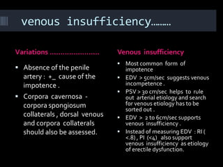
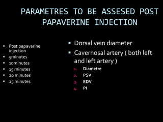
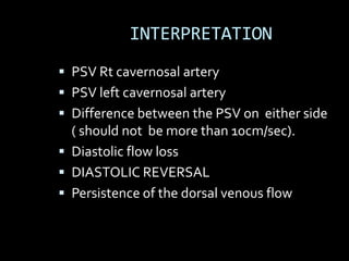
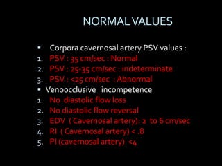
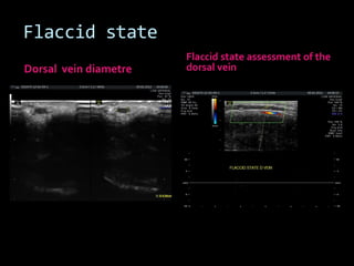
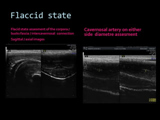
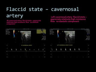
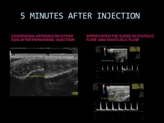
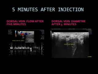
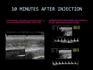
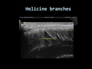
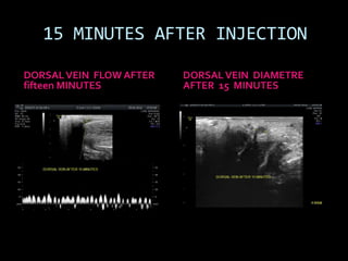
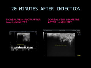
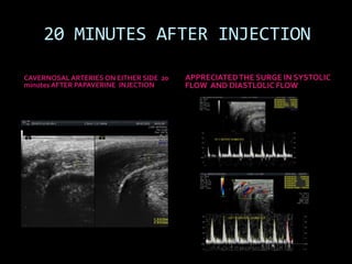
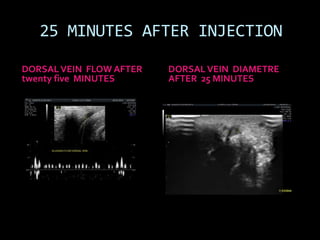
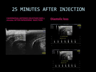
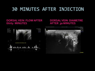
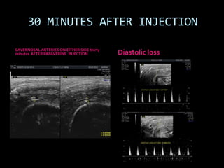
SlideShare a Scribd company logo
Penile doppler – A practical approach


35 yr male patient with h/o
depression
And Erectile dysfunction.
Grey scale / color doppler
assessment was done
to assess vasogenic / other
etiology of the erectile
dysfunction.


                               Dr Ritesh Mahajan
                                Free lance radiology
                              Approach towards basic imaging
PENILE ANATOMY………………………..
Penile vascular anatomy……….
 Internal pedundle artery
  through bulbar artery supplies  Venous drainage is through
  base of the penis . Penile         efferent venules – emisssary
  artery divides into two            veins - dorsal veins . Base of
  cavernosal arteries and            the penis through crural
  continues as dorsal artery .       veins drains into the
  There are helicine arteries that   periprostatic venous plexus
  run through the substance of       in to the internal iliac veins .
  the corpora .
                                    The glans region has it’s
 Cavernosal arteries are            drainage into the external
  paramedian in location.            iliac venous system.
 Cavernosal and dorsal
                                    Penile venous system is
  arteries show more
  variability than venous            more constant than the
  drainage of the penis .            arterial anatomy.
Basis of normal erection……….
                                   Flaccid state : Intracavernosal arterial
 After neural impulse              resistance is high . Cavernosal arterial flow
                                    has low systolic, dampened diastolic flow .
 Vasodilatation                   After giving vasoactive agent : Increased
 Increased blood supply            dilatation of the cavernosal arterial tree is
                                    there with increased systolic and diastolic
 Increased intracavernosal         component of the flow and velocities.
  pressure                         There is sinusoidal expansion of the arterial
 Efferent venous channel are       flow with obstructed venous egress
  obstructed by taut tunica        Further rise in cavernosal pressure leads to
                                    systolic dampening and loss of diastolic
  albuginea.                        component .
 On doppler study                 With rigid erection – there is near total loss
  predictable spectral              of diastolic flow and at times reversal .
  waveform corrborates with        As far as venous flow is concerned : flaccid
  changes in the intra              state has sluggish flow. With vasoactive
  cavernosal pressure .             agent there is increase in the dorsal venous
                                    flow and with rigid erection the venous
                                    flow can stop . Retrograde venous flow is
                                    also appreciated in normal individuals.
Basis of normal erection……
Phases of erection ………….    After neural impulse there is
                             rise in the intracavernosal
                             presssure –There is cavernosal
 Flaccid                    arterial dilatation and rise in
                             the systolic and diastolic flow
 Latent                     . The dorsal venous flow also
                             rises initially . With rise in the
 Tumescent                  cavernosal pressure –
                             distended sinusoids abut the
 Rigid                      tunica albuginea and this
 Detumescence               leads to cessation of the
                             venous egress and leads to
                             rigid erection. With rigid
                             erection ,this diastolic
                             component of the cavernosal
                             arterial flow is lost and at
                             times reverses also .
Penile imaging ………………………..
                 ERECTILE DYSFUNCTION
                       ETIOLOGY

 Psychogenic
 Endocrine
 Pharmacological
 Neurological
 Vascular
Organic etiology – Vasogenic
  etiology is important and
  penile Doppler assessment
  can be of use to ascertain
  the same .
Penile imaging …………………………….

Diagnostic work up for erectile
dysfunction                       Penile anatomy
 Medical / drug history .         Three distensible corpora
                                     chambers -
 Routine / endocrine blood       1. Corpora spongiosum enveloping
  analysis.                           the urethera. This does not play
                                      significant role in erection.
 Non invasive testing            2. Corpora cavernosa – dorsal in
                                      position –paired .
 Brachial – penile indices        Mid line septum separates the
 Nocturnal penile                    two corpora cavernosa . Thick
                                      fascia (tunica albugenia) encircles
  tumescence.                         the corpora cavernosa and bucks
                                      fascia covers corpora cavernosa
                                      and spongiosa .
Basic methodology of penile doppler


 Linear transducer parallel     Grey scale assessment
  to skin surface is used .       involves assessment of
  Both ventral and dorsal         echogenic tunica
  transducer position             albuginea. Midlevel echoes
  approaches can be used.         of the corpora cavernosa .
 Slow flow detection             Assess mid line septum .
  settings are to be used.       Cavernosal arteries are
 Longitudinal and                assessed by echogenic
  parasagitttal image             walls and with paramedian
  acquisition is to be done .     location.
Brief about doppler examination…….

 Complete discussion of         Velocity measurements are
    the examination with the      done along the base of the penis
                                  . Angle of assessment <60
    patient is to be done .       degree.
   Assessment of the privacy    PSV, EDV, RI , PI is done for
                                  cavernosal arteries on either side
    is to be done .               .
   Quiet examination setting    Look for cavernosal artery
                                  stenosis , occlusion, retrograde
    is necessary .                arterial flow , dampened spectral
                                  flow.
   Pharmacological agents :
                                 Cavernosal artery dilatation
    papaverine, phentolamine,     <75% of the base arterial
     prostaglandin E              diametre is indirect e/o
                                  vasogenic etiology of erectile
   Eye technique : visual        dysfunction.
    inspection is important .
Grey scale            sonography…..
Grey scale sonography
Good for assessment of

 Peyronie’s disease.
 Penile trauma
 Penile neoplasm
venous insufficiency………

Variations …………………..            Venous insufficiency
                                 Most common form of
 Absence of the penile             impotence
  artery : +_ cause of the         EDV > 5cm/sec suggests venous
  impotence .                       incompetence .
                                   PSV > 30 cm/sec helps to rule
 Corpora cavernosa -               out arterial etiology and search
  corpora spongiosum                for venous etiology has to be
                                    sorted out .
  collaterals , dorsal venous      EDV > 2 to 6cm/sec supports
  and corpora collaterals           venous insufficiency .
  should also be assessed.         Instead of measuring EDV : RI (
                                    <.8) , PI (<4) also support
                                    venous insufficiency as etiology
                                    of erectile dysfunction.
Grey scale and basic
        doppler assessment
                            BASIC COLOR DOPPLER ASSESMENT –
GREY SCALE                  DONE AT BASE OF THE PENIS

 PLAQUE / CALCIFICATION.    Imaging especially for
                                 doppler is done along the
 MID LEVEL ECHOES OF
                                 base of the penis .
  CORPORA CAVERNOSA
                                The sequence of the
 TUNICA ALBUGENIA               imaging is as following :
  /BUCKS FASCIA             1.    Flaccid state
                            2.    Papaverine injection
                            3.    Post injection imaging is
                                  done at 5 , 10,15,20,25
                                  minutes .
PARAMETRES TO BE ASSESED IN
     THE FLACCID STATE
         Dorsal vein diameter
         Cavernosal artery ( both left
          and left artery )
          1.   Diametre
          2.   PSV
          3.   EDV
          4.   PI
          5.   Dorsal cavernosal collaterals
          6.   Cavernosal spongiosal collaterals
PARAMETRES TO BE ASSESED POST
              PAPAVERINE INJECTION

 Post papaverine
                     Dorsal vein diameter
    injection
                     Cavernosal artery ( both left
   5minutes
   10minutes         and left artery )
   15 minutes        1.   Diametre
   20 minutes        2.   PSV
   25 minutes        3.   EDV
                      4.   PI
INTERPRETATION
 PSV Rt cavernosal artery
 PSV left cavernosal artery
 Difference between the PSV on either side
  ( should not be more than 10cm/sec).
 Diastolic flow loss
 DIASTOLIC REVERSAL
 Persistence of the dorsal venous flow
NORMAL VALUES

 Corpora cavernosal artery PSV values :
1. PSV : 35 cm/sec : Normal
2. PSV : 25-35 cm/sec : indeterminate
3. PSV : <25 cm/sec : Abnormal
 Venoocclusive incompetence
1. No diastolic flow loss
2. No diastolic flow reversal
3. EDV ( Cavernosal artery): 2 to 6 cm/sec
4. RI ( Cavernosal artery) < .8
5. PI (cavernosal artery) <4
PRECAUTIONS

 Inject papaverine only once
 Keep region of injection pressed
 Use insulin syringe
 Alcohol swab to clean
 Keep watch for priapism ( urologist
  /anesthetist support ) .
Flaccid state
                       Flaccid state assessment of the
Dorsal vein diametre   dorsal vein
Flaccid state
Flacid state assesment of the corpora /     Cavernosal artery on either
bucks fascia / intercavernosal connection   side diametre assesment
Sagittal / axial images
Flaccid state – cavernosal
artery          Left cavernosal artery flaccid state –
                appreciate relatively high resistance
 Rt cavernosal artery flaccid state – appreciate
relatively high resistance flow no diastolic       flow no diastolic component
component
Ancilliary findings

   No e/o dorsal cavernosal collaterals . No
   e/o cavernosal spongiosal collaterals
Injection of the papaverine injection in the
             left corpora cavernosa
 INSULIN SYRINGE
USED
INJECTION DONE IN
LEFT CORPORA
CAVERNOSA
GUIDED INJECTION
DONE AVOIDING THE
LEFT SIDE
CAVERNOSAL ARTERY
ANESTHETIST WAS
INVOLVED IN THE
INTERVENTION .
 ALCOHOL SWAB WAS
USED .
PRECAUTIONS WERE
TAKEN TO AVOID SPILL.
5 MINUTES AFTER INJECTION

CAVERNOSAL ARTERIES ON EITHER     APPRECIATED THE SURGE IN SYSTOLIC
SIDE AFTER PAPAVERINE INJECTION   FLOW AND DIASTLOLIC FLOW
5 MINUTES AFTER INJECTION

DORSAL VEIN FLOW AFTER   DORSAL VEIN DIAMETRE
FIVE MINUTES             AFTER 5 MINUTES
10 MINUTES AFTER INJECTION

CAVERNOSAL ARTERIES ON EITHER SIDE 10   APPRECIATED THE SURGE IN SYSTOLIC
minutes AFTER PAPAVERINE INJECTION      FLOW AND DIASTLOLIC FLOW
10 MINUTES AFTER INJECTION

DORSAL VEIN FLOW AFTER   DORSAL VEIN DIAMETRE
TEN MINUTES              AFTER 10 MINUTES
Helicine branches
15 MINUTES AFTER INJECTION

DORSAL VEIN FLOW AFTER   DORSAL VEIN DIAMETRE
fifteen MINUTES          AFTER 15 MINUTES
15 MINUTES AFTER INJECTION
CAVERNOSAL ARTERIES ON EITHER SIDE AFTER 15
                                              APPRECIATE THE SURGE IN SYSTOLIC
minutes of PAPAVERINE INJECTION               FLOW AND DIASTLOLIC FLOW
20 MINUTES AFTER INJECTION

DORSAL VEIN FLOW AFTER   DORSAL VEIN DIAMETRE
twenty MINUTES           AFTER 20 MINUTES
20 MINUTES AFTER INJECTION

CAVERNOSAL ARTERIES ON EITHER SIDE 20   APPRECIATED THE SURGE IN SYSTOLIC
minutes AFTER PAPAVERINE INJECTION      FLOW AND DIASTLOLIC FLOW
25 MINUTES AFTER INJECTION

DORSAL VEIN FLOW AFTER   DORSAL VEIN DIAMETRE
twenty five MINUTES      AFTER 25 MINUTES
25 MINUTES AFTER INJECTION
CAVERNOSAL ARTERIES ON EITHER SIDE 5
minutes AFTER PAPAVERINE INJECTION     Diastolic loss
30 MINUTES AFTER INJECTION

DORSAL VEIN FLOW AFTER   DORSAL VEIN DIAMETRE
thirty MINUTES           AFTER 30 MINUTES
30 MINUTES AFTER INJECTION

CAVERNOSAL ARTERIES ON EITHER SIDE thirty
minutes AFTER PAPAVERINE INJECTION          Diastolic loss

More Related Content

What's hot

Doppler ultrasound of carotid arteries
Doppler ultrasound of carotid arteriesDoppler ultrasound of carotid arteries
Doppler ultrasound of carotid arteriesSamir Haffar
 
Doppler ultrasound of lower limb arteries
Doppler ultrasound of lower limb arteriesDoppler ultrasound of lower limb arteries
Doppler ultrasound of lower limb arteriesSamir Haffar
 
Imaging of urethral pathologies
Imaging of urethral pathologiesImaging of urethral pathologies
Imaging of urethral pathologies
Sunil Kumar
 
Doppler ultrasound of acute scrotum
Doppler ultrasound of acute scrotumDoppler ultrasound of acute scrotum
Doppler ultrasound of acute scrotum
Samir Haffar
 
Unilateral small kidney
Unilateral small kidneyUnilateral small kidney
Unilateral small kidney
maimusirdan
 
Presentation1, mri imaging of the prostate
Presentation1, mri imaging of the prostatePresentation1, mri imaging of the prostate
Presentation1, mri imaging of the prostate
Abdellah Nazeer
 
Presentation1.pptx, radiological imaging of prostatic diseases
Presentation1.pptx, radiological imaging of prostatic diseasesPresentation1.pptx, radiological imaging of prostatic diseases
Presentation1.pptx, radiological imaging of prostatic diseasesAbdellah Nazeer
 
Doppler of the portal system
Doppler of the portal systemDoppler of the portal system
Doppler of the portal system
Dr. Muhammad Bin Zulfiqar
 
Role of medical imaging in management of arteriovenous fistula Dr. Muhammad B...
Role of medical imaging in management of arteriovenous fistula Dr. Muhammad B...Role of medical imaging in management of arteriovenous fistula Dr. Muhammad B...
Role of medical imaging in management of arteriovenous fistula Dr. Muhammad B...
Dr. Muhammad Bin Zulfiqar
 
Imaging of kidny i htn by dr.abd alla shady md
Imaging of kidny i htn by dr.abd alla shady mdImaging of kidny i htn by dr.abd alla shady md
Imaging of kidny i htn by dr.abd alla shady md
FarragBahbah
 
ULTRASOUND SCROTUM
ULTRASOUND SCROTUMULTRASOUND SCROTUM
ULTRASOUND SCROTUM
Lohith Varma
 
Renal masses imaging
Renal masses imagingRenal masses imaging
Renal masses imaging
Satish Naga
 
Doppler ultrasound of the portal system - Normal findings
Doppler ultrasound of the portal system - Normal findingsDoppler ultrasound of the portal system - Normal findings
Doppler ultrasound of the portal system - Normal findingsSamir Haffar
 
Venous Doppler upper limb
Venous Doppler upper limb Venous Doppler upper limb
Venous Doppler upper limb
Milan Silwal
 
Presentation1.pptx, radiological imaging of peri natal acute ischemia and hyp...
Presentation1.pptx, radiological imaging of peri natal acute ischemia and hyp...Presentation1.pptx, radiological imaging of peri natal acute ischemia and hyp...
Presentation1.pptx, radiological imaging of peri natal acute ischemia and hyp...Abdellah Nazeer
 
Renal doppler usg
Renal doppler usgRenal doppler usg
Renal doppler usg
Dr. Mohit Goel
 
Radiology Spotters
Radiology Spotters Radiology Spotters
Radiology Spotters
Anish Choudhary
 
Doppler ultrasound of the kidneys
Doppler ultrasound of the kidneysDoppler ultrasound of the kidneys
Doppler ultrasound of the kidneys
Samir Haffar
 
Doppler ultrasound in peripheral arterial disease
Doppler ultrasound in peripheral arterial diseaseDoppler ultrasound in peripheral arterial disease
Doppler ultrasound in peripheral arterial disease
Dr. Naveed Quetta
 
Ultrasound of the abdominal wall hernias
Ultrasound of the abdominal wall herniasUltrasound of the abdominal wall hernias
Ultrasound of the abdominal wall herniasSamir Haffar
 

What's hot (20)

Doppler ultrasound of carotid arteries
Doppler ultrasound of carotid arteriesDoppler ultrasound of carotid arteries
Doppler ultrasound of carotid arteries
 
Doppler ultrasound of lower limb arteries
Doppler ultrasound of lower limb arteriesDoppler ultrasound of lower limb arteries
Doppler ultrasound of lower limb arteries
 
Imaging of urethral pathologies
Imaging of urethral pathologiesImaging of urethral pathologies
Imaging of urethral pathologies
 
Doppler ultrasound of acute scrotum
Doppler ultrasound of acute scrotumDoppler ultrasound of acute scrotum
Doppler ultrasound of acute scrotum
 
Unilateral small kidney
Unilateral small kidneyUnilateral small kidney
Unilateral small kidney
 
Presentation1, mri imaging of the prostate
Presentation1, mri imaging of the prostatePresentation1, mri imaging of the prostate
Presentation1, mri imaging of the prostate
 
Presentation1.pptx, radiological imaging of prostatic diseases
Presentation1.pptx, radiological imaging of prostatic diseasesPresentation1.pptx, radiological imaging of prostatic diseases
Presentation1.pptx, radiological imaging of prostatic diseases
 
Doppler of the portal system
Doppler of the portal systemDoppler of the portal system
Doppler of the portal system
 
Role of medical imaging in management of arteriovenous fistula Dr. Muhammad B...
Role of medical imaging in management of arteriovenous fistula Dr. Muhammad B...Role of medical imaging in management of arteriovenous fistula Dr. Muhammad B...
Role of medical imaging in management of arteriovenous fistula Dr. Muhammad B...
 
Imaging of kidny i htn by dr.abd alla shady md
Imaging of kidny i htn by dr.abd alla shady mdImaging of kidny i htn by dr.abd alla shady md
Imaging of kidny i htn by dr.abd alla shady md
 
ULTRASOUND SCROTUM
ULTRASOUND SCROTUMULTRASOUND SCROTUM
ULTRASOUND SCROTUM
 
Renal masses imaging
Renal masses imagingRenal masses imaging
Renal masses imaging
 
Doppler ultrasound of the portal system - Normal findings
Doppler ultrasound of the portal system - Normal findingsDoppler ultrasound of the portal system - Normal findings
Doppler ultrasound of the portal system - Normal findings
 
Venous Doppler upper limb
Venous Doppler upper limb Venous Doppler upper limb
Venous Doppler upper limb
 
Presentation1.pptx, radiological imaging of peri natal acute ischemia and hyp...
Presentation1.pptx, radiological imaging of peri natal acute ischemia and hyp...Presentation1.pptx, radiological imaging of peri natal acute ischemia and hyp...
Presentation1.pptx, radiological imaging of peri natal acute ischemia and hyp...
 
Renal doppler usg
Renal doppler usgRenal doppler usg
Renal doppler usg
 
Radiology Spotters
Radiology Spotters Radiology Spotters
Radiology Spotters
 
Doppler ultrasound of the kidneys
Doppler ultrasound of the kidneysDoppler ultrasound of the kidneys
Doppler ultrasound of the kidneys
 
Doppler ultrasound in peripheral arterial disease
Doppler ultrasound in peripheral arterial diseaseDoppler ultrasound in peripheral arterial disease
Doppler ultrasound in peripheral arterial disease
 
Ultrasound of the abdominal wall hernias
Ultrasound of the abdominal wall herniasUltrasound of the abdominal wall hernias
Ultrasound of the abdominal wall hernias
 

Viewers also liked

Erectile Dysfunction
Erectile DysfunctionErectile Dysfunction
Erectile Dysfunction
fhammoud
 
Male genital trauma
Male genital traumaMale genital trauma
Male genital trauma
SCGH ED CME
 
TRUS in Evaluation of Male Infertility
TRUS in Evaluation of Male InfertilityTRUS in Evaluation of Male Infertility
TRUS in Evaluation of Male Infertility
Siewhong Ho
 
Presentation1.pptx, radiological imaging of male infertility.
Presentation1.pptx, radiological imaging of male infertility.Presentation1.pptx, radiological imaging of male infertility.
Presentation1.pptx, radiological imaging of male infertility.Abdellah Nazeer
 
Erectile dysfunction and Premature Ejaculation
Erectile dysfunction and Premature Ejaculation Erectile dysfunction and Premature Ejaculation
Erectile dysfunction and Premature Ejaculation
Dr. Amit Chougule
 
Erectile Dysfunction Symptoms And Treatment
Erectile Dysfunction Symptoms And TreatmentErectile Dysfunction Symptoms And Treatment
Erectile Dysfunction Symptoms And Treatment
Manas Das
 
Erectile Dysfunction Treatment Information by Premier Men's Medical Center
Erectile Dysfunction Treatment Information by Premier Men's Medical CenterErectile Dysfunction Treatment Information by Premier Men's Medical Center
Erectile Dysfunction Treatment Information by Premier Men's Medical Center
Premier Men's Medical Center
 
Priapism 2015
Priapism 2015Priapism 2015
Priapism 2015
Harish Patel
 
Emergencies (non traumatic)
Emergencies (non traumatic)Emergencies (non traumatic)
Emergencies (non traumatic)
Mohammed Abd El Wadood
 
Lectura de caso : Rotura peneana
Lectura de caso : Rotura peneanaLectura de caso : Rotura peneana
Lectura de caso : Rotura peneana
Heidy Saenz
 
Durex condoms
Durex condomsDurex condoms
Durex condomspeepee99
 
Us disfunción erectil
Us disfunción erectilUs disfunción erectil
Us disfunción erectil
patovaro
 
Vascular Ultrasound
Vascular UltrasoundVascular Ultrasound
Vascular Ultrasound
Steve Henao MD FACS FACC
 
Peyronies disease
Peyronies diseasePeyronies disease
Peyronies disease
Ramayya Pramila
 
Erectile Dysfunction [Dr. Edmond Wong]
Erectile Dysfunction [Dr. Edmond Wong]Erectile Dysfunction [Dr. Edmond Wong]
Erectile Dysfunction [Dr. Edmond Wong]
Edmond Wong
 
Ecografia de pene
Ecografia de peneEcografia de pene
Ecografia de pene
Rodrigo Sazo
 
Ultrasound in infertility
Ultrasound in infertilityUltrasound in infertility
Ultrasound in infertilityRupal Shah
 
Exámenes urológicos de laboratorio
Exámenes urológicos de laboratorioExámenes urológicos de laboratorio
Exámenes urológicos de laboratorio
Javier Jaramillo
 

Viewers also liked (20)

Erectile Dysfunction
Erectile DysfunctionErectile Dysfunction
Erectile Dysfunction
 
Male genital trauma
Male genital traumaMale genital trauma
Male genital trauma
 
Priapism
PriapismPriapism
Priapism
 
TRUS in Evaluation of Male Infertility
TRUS in Evaluation of Male InfertilityTRUS in Evaluation of Male Infertility
TRUS in Evaluation of Male Infertility
 
Presentation1.pptx, radiological imaging of male infertility.
Presentation1.pptx, radiological imaging of male infertility.Presentation1.pptx, radiological imaging of male infertility.
Presentation1.pptx, radiological imaging of male infertility.
 
Erectile dysfunction and Premature Ejaculation
Erectile dysfunction and Premature Ejaculation Erectile dysfunction and Premature Ejaculation
Erectile dysfunction and Premature Ejaculation
 
Erectile Dysfunction Symptoms And Treatment
Erectile Dysfunction Symptoms And TreatmentErectile Dysfunction Symptoms And Treatment
Erectile Dysfunction Symptoms And Treatment
 
Erectile Dysfunction Treatment Information by Premier Men's Medical Center
Erectile Dysfunction Treatment Information by Premier Men's Medical CenterErectile Dysfunction Treatment Information by Premier Men's Medical Center
Erectile Dysfunction Treatment Information by Premier Men's Medical Center
 
Priapism 2015
Priapism 2015Priapism 2015
Priapism 2015
 
Emergencies (non traumatic)
Emergencies (non traumatic)Emergencies (non traumatic)
Emergencies (non traumatic)
 
Lectura de caso : Rotura peneana
Lectura de caso : Rotura peneanaLectura de caso : Rotura peneana
Lectura de caso : Rotura peneana
 
Durex condoms
Durex condomsDurex condoms
Durex condoms
 
Us disfunción erectil
Us disfunción erectilUs disfunción erectil
Us disfunción erectil
 
Priapism
PriapismPriapism
Priapism
 
Vascular Ultrasound
Vascular UltrasoundVascular Ultrasound
Vascular Ultrasound
 
Peyronies disease
Peyronies diseasePeyronies disease
Peyronies disease
 
Erectile Dysfunction [Dr. Edmond Wong]
Erectile Dysfunction [Dr. Edmond Wong]Erectile Dysfunction [Dr. Edmond Wong]
Erectile Dysfunction [Dr. Edmond Wong]
 
Ecografia de pene
Ecografia de peneEcografia de pene
Ecografia de pene
 
Ultrasound in infertility
Ultrasound in infertilityUltrasound in infertility
Ultrasound in infertility
 
Exámenes urológicos de laboratorio
Exámenes urológicos de laboratorioExámenes urológicos de laboratorio
Exámenes urológicos de laboratorio
 

Similar to Penile doppler a review

Systemic venous anomalies
Systemic venous anomaliesSystemic venous anomalies
Systemic venous anomaliesdrranjithmp
 
Color doppler in fetal hypoxia
Color doppler in fetal hypoxiaColor doppler in fetal hypoxia
Color doppler in fetal hypoxia
NARENDRA MALHOTRA
 
Lacture varicose
Lacture  varicose Lacture  varicose
Lacture varicose
Narendra Singh Patel
 
Presentation1.pptx, radiological imaging of hydrocephalus.
Presentation1.pptx, radiological imaging of hydrocephalus.Presentation1.pptx, radiological imaging of hydrocephalus.
Presentation1.pptx, radiological imaging of hydrocephalus.Abdellah Nazeer
 
Venography/ Phlebography- Avinesh Shrestha
Venography/ Phlebography- Avinesh ShresthaVenography/ Phlebography- Avinesh Shrestha
Venography/ Phlebography- Avinesh Shrestha
Avinesh Shrestha
 
Detecting Deep Venous Disease with Duplex Ultrasound
Detecting Deep Venous Disease with Duplex UltrasoundDetecting Deep Venous Disease with Duplex Ultrasound
Detecting Deep Venous Disease with Duplex Ultrasound
Vein Global
 
The peripheral veins
The peripheral veinsThe peripheral veins
The peripheral veins
Vrishit Saraswat
 
Anatomy of Blood vessels of abdomen pelvic cavities. Portacaval & Cavacaval A...
Anatomy of Blood vessels of abdomen pelvic cavities. Portacaval & Cavacaval A...Anatomy of Blood vessels of abdomen pelvic cavities. Portacaval & Cavacaval A...
Anatomy of Blood vessels of abdomen pelvic cavities. Portacaval & Cavacaval A...
Eneutron
 
C O L O R D O P P L E R I N F E T A L H Y P O X I A
C O L O R  D O P P L E R  I N  F E T A L  H Y P O X I AC O L O R  D O P P L E R  I N  F E T A L  H Y P O X I A
C O L O R D O P P L E R I N F E T A L H Y P O X I A
NARENDRA MALHOTRA
 
Duplex ultrasound of Vericose vein
 Duplex ultrasound of  Vericose vein Duplex ultrasound of  Vericose vein
Duplex ultrasound of Vericose vein
Ravi patel
 
Priapism.ppt sumit
Priapism.ppt sumitPriapism.ppt sumit
Priapism.ppt sumit
Sumit Hadgaonkar
 
Renal doppler
Renal doppler Renal doppler
Renal doppler
ParameshwarKeerthi2
 
C O L O R D O P P L E R I N F E T A L H Y P O X I A
C O L O R  D O P P L E R  I N  F E T A L  H Y P O X I AC O L O R  D O P P L E R  I N  F E T A L  H Y P O X I A
C O L O R D O P P L E R I N F E T A L H Y P O X I ANARENDRA MALHOTRA
 
Ultrasound Assessment Of Chronic Venous Disease
Ultrasound Assessment Of Chronic Venous DiseaseUltrasound Assessment Of Chronic Venous Disease
Ultrasound Assessment Of Chronic Venous Diseasejavier.fabra
 
Renal doppler HOD.pptx
Renal doppler HOD.pptxRenal doppler HOD.pptx
Renal doppler HOD.pptx
dypradio
 
CEREBROSPINAL FLUID (CSF)
CEREBROSPINAL FLUID (CSF)CEREBROSPINAL FLUID (CSF)
CEREBROSPINAL FLUID (CSF)
Dr. Aniket Shilwant
 
Venous Disorders- gunabhi.ppt
Venous Disorders- gunabhi.pptVenous Disorders- gunabhi.ppt
Venous Disorders- gunabhi.ppt
GUNABHIDAS1
 
PHYSIOLOGY OF CSF PRODUCTION AND CIRCULATION, ALTERATIONS IN VARIOUS PATHOLOGY
PHYSIOLOGY OF CSF PRODUCTION AND CIRCULATION, ALTERATIONS IN VARIOUS PATHOLOGYPHYSIOLOGY OF CSF PRODUCTION AND CIRCULATION, ALTERATIONS IN VARIOUS PATHOLOGY
PHYSIOLOGY OF CSF PRODUCTION AND CIRCULATION, ALTERATIONS IN VARIOUS PATHOLOGY
Unnikrishnan Prathapadas
 

Similar to Penile doppler a review (20)

Systemic venous anomalies
Systemic venous anomaliesSystemic venous anomalies
Systemic venous anomalies
 
Color doppler in fetal hypoxia
Color doppler in fetal hypoxiaColor doppler in fetal hypoxia
Color doppler in fetal hypoxia
 
Lacture varicose
Lacture  varicose Lacture  varicose
Lacture varicose
 
Presentation1.pptx, radiological imaging of hydrocephalus.
Presentation1.pptx, radiological imaging of hydrocephalus.Presentation1.pptx, radiological imaging of hydrocephalus.
Presentation1.pptx, radiological imaging of hydrocephalus.
 
Venography/ Phlebography- Avinesh Shrestha
Venography/ Phlebography- Avinesh ShresthaVenography/ Phlebography- Avinesh Shrestha
Venography/ Phlebography- Avinesh Shrestha
 
Detecting Deep Venous Disease with Duplex Ultrasound
Detecting Deep Venous Disease with Duplex UltrasoundDetecting Deep Venous Disease with Duplex Ultrasound
Detecting Deep Venous Disease with Duplex Ultrasound
 
The peripheral veins
The peripheral veinsThe peripheral veins
The peripheral veins
 
Anatomy of Blood vessels of abdomen pelvic cavities. Portacaval & Cavacaval A...
Anatomy of Blood vessels of abdomen pelvic cavities. Portacaval & Cavacaval A...Anatomy of Blood vessels of abdomen pelvic cavities. Portacaval & Cavacaval A...
Anatomy of Blood vessels of abdomen pelvic cavities. Portacaval & Cavacaval A...
 
C O L O R D O P P L E R I N F E T A L H Y P O X I A
C O L O R  D O P P L E R  I N  F E T A L  H Y P O X I AC O L O R  D O P P L E R  I N  F E T A L  H Y P O X I A
C O L O R D O P P L E R I N F E T A L H Y P O X I A
 
Duplex ultrasound of Vericose vein
 Duplex ultrasound of  Vericose vein Duplex ultrasound of  Vericose vein
Duplex ultrasound of Vericose vein
 
Priapism.ppt sumit
Priapism.ppt sumitPriapism.ppt sumit
Priapism.ppt sumit
 
Renal doppler
Renal doppler Renal doppler
Renal doppler
 
C O L O R D O P P L E R I N F E T A L H Y P O X I A
C O L O R  D O P P L E R  I N  F E T A L  H Y P O X I AC O L O R  D O P P L E R  I N  F E T A L  H Y P O X I A
C O L O R D O P P L E R I N F E T A L H Y P O X I A
 
Ultrasound Assessment Of Chronic Venous Disease
Ultrasound Assessment Of Chronic Venous DiseaseUltrasound Assessment Of Chronic Venous Disease
Ultrasound Assessment Of Chronic Venous Disease
 
Renal doppler HOD.pptx
Renal doppler HOD.pptxRenal doppler HOD.pptx
Renal doppler HOD.pptx
 
Renal physiology
Renal physiology Renal physiology
Renal physiology
 
CEREBROSPINAL FLUID (CSF)
CEREBROSPINAL FLUID (CSF)CEREBROSPINAL FLUID (CSF)
CEREBROSPINAL FLUID (CSF)
 
Advanced usg lounge
Advanced usg loungeAdvanced usg lounge
Advanced usg lounge
 
Venous Disorders- gunabhi.ppt
Venous Disorders- gunabhi.pptVenous Disorders- gunabhi.ppt
Venous Disorders- gunabhi.ppt
 
PHYSIOLOGY OF CSF PRODUCTION AND CIRCULATION, ALTERATIONS IN VARIOUS PATHOLOGY
PHYSIOLOGY OF CSF PRODUCTION AND CIRCULATION, ALTERATIONS IN VARIOUS PATHOLOGYPHYSIOLOGY OF CSF PRODUCTION AND CIRCULATION, ALTERATIONS IN VARIOUS PATHOLOGY
PHYSIOLOGY OF CSF PRODUCTION AND CIRCULATION, ALTERATIONS IN VARIOUS PATHOLOGY
 

More from Ritesh Mahajan

Ultrasound november 17
Ultrasound november 17Ultrasound november 17
Ultrasound november 17
Ritesh Mahajan
 
Usg image gallery nov 16
Usg image gallery nov 16Usg image gallery nov 16
Usg image gallery nov 16
Ritesh Mahajan
 
Usg image gallery advanced usg lounge
Usg image gallery  advanced usg loungeUsg image gallery  advanced usg lounge
Usg image gallery advanced usg lounge
Ritesh Mahajan
 
Ultrasound image gallery 2016 (2)
Ultrasound image gallery 2016 (2)Ultrasound image gallery 2016 (2)
Ultrasound image gallery 2016 (2)
Ritesh Mahajan
 
Ultrasound image gallery 2016
Ultrasound image gallery 2016Ultrasound image gallery 2016
Ultrasound image gallery 2016
Ritesh Mahajan
 
Ultrasound image gallery
Ultrasound image galleryUltrasound image gallery
Ultrasound image gallery
Ritesh Mahajan
 
3 d power doppler angiogram
3 d power doppler  angiogram3 d power doppler  angiogram
3 d power doppler angiogram
Ritesh Mahajan
 
Anterior ectopic anus advanced usg lounge
Anterior  ectopic anus  advanced usg loungeAnterior  ectopic anus  advanced usg lounge
Anterior ectopic anus advanced usg lounge
Ritesh Mahajan
 
Fetal anomaly archive advanced usg lounge
Fetal anomaly archive advanced usg loungeFetal anomaly archive advanced usg lounge
Fetal anomaly archive advanced usg lounge
Ritesh Mahajan
 
Transcranial doppler advanced usg lounge
Transcranial doppler advanced usg loungeTranscranial doppler advanced usg lounge
Transcranial doppler advanced usg lounge
Ritesh Mahajan
 
Advanced usg lounge fetal pulmonary sequestration
Advanced usg lounge fetal pulmonary sequestrationAdvanced usg lounge fetal pulmonary sequestration
Advanced usg lounge fetal pulmonary sequestration
Ritesh Mahajan
 
Advanced usg lounge 3 d image gallery
Advanced usg lounge  3 d image galleryAdvanced usg lounge  3 d image gallery
Advanced usg lounge 3 d image gallery
Ritesh Mahajan
 
New microsoft office power point presentation
New microsoft office power point presentationNew microsoft office power point presentation
New microsoft office power point presentation
Ritesh Mahajan
 
Fetal anomaly series prune belly syndrome.
Fetal anomaly series prune  belly syndrome.Fetal anomaly series prune  belly syndrome.
Fetal anomaly series prune belly syndrome.
Ritesh Mahajan
 
ADVANCED USG LOUNGE FETAL FEET
ADVANCED USG LOUNGE  FETAL FEET ADVANCED USG LOUNGE  FETAL FEET
ADVANCED USG LOUNGE FETAL FEET
Ritesh Mahajan
 
Advanced usg lounge gynaecology imaging series
Advanced usg lounge gynaecology imaging seriesAdvanced usg lounge gynaecology imaging series
Advanced usg lounge gynaecology imaging series
Ritesh Mahajan
 
Torsion testis final
Torsion testis finalTorsion testis final
Torsion testis final
Ritesh Mahajan
 
Acute tenosynovitis
Acute tenosynovitisAcute tenosynovitis
Acute tenosynovitis
Ritesh Mahajan
 
Anencephaly
AnencephalyAnencephaly
Anencephaly
Ritesh Mahajan
 
Pedicle artery sign
Pedicle  artery signPedicle  artery sign
Pedicle artery sign
Ritesh Mahajan
 

More from Ritesh Mahajan (20)

Ultrasound november 17
Ultrasound november 17Ultrasound november 17
Ultrasound november 17
 
Usg image gallery nov 16
Usg image gallery nov 16Usg image gallery nov 16
Usg image gallery nov 16
 
Usg image gallery advanced usg lounge
Usg image gallery  advanced usg loungeUsg image gallery  advanced usg lounge
Usg image gallery advanced usg lounge
 
Ultrasound image gallery 2016 (2)
Ultrasound image gallery 2016 (2)Ultrasound image gallery 2016 (2)
Ultrasound image gallery 2016 (2)
 
Ultrasound image gallery 2016
Ultrasound image gallery 2016Ultrasound image gallery 2016
Ultrasound image gallery 2016
 
Ultrasound image gallery
Ultrasound image galleryUltrasound image gallery
Ultrasound image gallery
 
3 d power doppler angiogram
3 d power doppler  angiogram3 d power doppler  angiogram
3 d power doppler angiogram
 
Anterior ectopic anus advanced usg lounge
Anterior  ectopic anus  advanced usg loungeAnterior  ectopic anus  advanced usg lounge
Anterior ectopic anus advanced usg lounge
 
Fetal anomaly archive advanced usg lounge
Fetal anomaly archive advanced usg loungeFetal anomaly archive advanced usg lounge
Fetal anomaly archive advanced usg lounge
 
Transcranial doppler advanced usg lounge
Transcranial doppler advanced usg loungeTranscranial doppler advanced usg lounge
Transcranial doppler advanced usg lounge
 
Advanced usg lounge fetal pulmonary sequestration
Advanced usg lounge fetal pulmonary sequestrationAdvanced usg lounge fetal pulmonary sequestration
Advanced usg lounge fetal pulmonary sequestration
 
Advanced usg lounge 3 d image gallery
Advanced usg lounge  3 d image galleryAdvanced usg lounge  3 d image gallery
Advanced usg lounge 3 d image gallery
 
New microsoft office power point presentation
New microsoft office power point presentationNew microsoft office power point presentation
New microsoft office power point presentation
 
Fetal anomaly series prune belly syndrome.
Fetal anomaly series prune  belly syndrome.Fetal anomaly series prune  belly syndrome.
Fetal anomaly series prune belly syndrome.
 
ADVANCED USG LOUNGE FETAL FEET
ADVANCED USG LOUNGE  FETAL FEET ADVANCED USG LOUNGE  FETAL FEET
ADVANCED USG LOUNGE FETAL FEET
 
Advanced usg lounge gynaecology imaging series
Advanced usg lounge gynaecology imaging seriesAdvanced usg lounge gynaecology imaging series
Advanced usg lounge gynaecology imaging series
 
Torsion testis final
Torsion testis finalTorsion testis final
Torsion testis final
 
Acute tenosynovitis
Acute tenosynovitisAcute tenosynovitis
Acute tenosynovitis
 
Anencephaly
AnencephalyAnencephaly
Anencephaly
 
Pedicle artery sign
Pedicle  artery signPedicle  artery sign
Pedicle artery sign
 

Recently uploaded

New Directions in Targeted Therapeutic Approaches for Older Adults With Mantl...
New Directions in Targeted Therapeutic Approaches for Older Adults With Mantl...New Directions in Targeted Therapeutic Approaches for Older Adults With Mantl...
New Directions in Targeted Therapeutic Approaches for Older Adults With Mantl...
i3 Health
 
Ozempic: Preoperative Management of Patients on GLP-1 Receptor Agonists
Ozempic: Preoperative Management of Patients on GLP-1 Receptor Agonists  Ozempic: Preoperative Management of Patients on GLP-1 Receptor Agonists
Ozempic: Preoperative Management of Patients on GLP-1 Receptor Agonists
Saeid Safari
 
Report Back from SGO 2024: What’s the Latest in Cervical Cancer?
Report Back from SGO 2024: What’s the Latest in Cervical Cancer?Report Back from SGO 2024: What’s the Latest in Cervical Cancer?
Report Back from SGO 2024: What’s the Latest in Cervical Cancer?
bkling
 
basicmodesofventilation2022-220313203758.pdf
basicmodesofventilation2022-220313203758.pdfbasicmodesofventilation2022-220313203758.pdf
basicmodesofventilation2022-220313203758.pdf
aljamhori teaching hospital
 
micro teaching on communication m.sc nursing.pdf
micro teaching on communication m.sc nursing.pdfmicro teaching on communication m.sc nursing.pdf
micro teaching on communication m.sc nursing.pdf
Anurag Sharma
 
Non-respiratory Functions of the Lungs.pdf
Non-respiratory Functions of the Lungs.pdfNon-respiratory Functions of the Lungs.pdf
Non-respiratory Functions of the Lungs.pdf
MedicoseAcademics
 
Surat @ℂall @Girls ꧁❤8527049040❤꧂@ℂall @Girls Service Vip Top Model Safe
Surat @ℂall @Girls ꧁❤8527049040❤꧂@ℂall @Girls Service Vip Top Model SafeSurat @ℂall @Girls ꧁❤8527049040❤꧂@ℂall @Girls Service Vip Top Model Safe
Surat @ℂall @Girls ꧁❤8527049040❤꧂@ℂall @Girls Service Vip Top Model Safe
Savita Shen $i11
 
Prix Galien International 2024 Forum Program
Prix Galien International 2024 Forum ProgramPrix Galien International 2024 Forum Program
Prix Galien International 2024 Forum Program
Levi Shapiro
 
263778731218 Abortion Clinic /Pills In Harare ,
263778731218 Abortion Clinic /Pills In Harare ,263778731218 Abortion Clinic /Pills In Harare ,
263778731218 Abortion Clinic /Pills In Harare ,
sisternakatoto
 
Physiology of Special Chemical Sensation of Taste
Physiology of Special Chemical Sensation of TastePhysiology of Special Chemical Sensation of Taste
Physiology of Special Chemical Sensation of Taste
MedicoseAcademics
 
HOT NEW PRODUCT! BIG SALES FAST SHIPPING NOW FROM CHINA!! EU KU DB BK substit...
HOT NEW PRODUCT! BIG SALES FAST SHIPPING NOW FROM CHINA!! EU KU DB BK substit...HOT NEW PRODUCT! BIG SALES FAST SHIPPING NOW FROM CHINA!! EU KU DB BK substit...
HOT NEW PRODUCT! BIG SALES FAST SHIPPING NOW FROM CHINA!! EU KU DB BK substit...
GL Anaacs
 
Alcohol_Dr. Jeenal Mistry MD Pharmacology.pdf
Alcohol_Dr. Jeenal Mistry MD Pharmacology.pdfAlcohol_Dr. Jeenal Mistry MD Pharmacology.pdf
Alcohol_Dr. Jeenal Mistry MD Pharmacology.pdf
Dr Jeenal Mistry
 
Couples presenting to the infertility clinic- Do they really have infertility...
Couples presenting to the infertility clinic- Do they really have infertility...Couples presenting to the infertility clinic- Do they really have infertility...
Couples presenting to the infertility clinic- Do they really have infertility...
Sujoy Dasgupta
 
Ophthalmology Clinical Tests for OSCE exam
Ophthalmology Clinical Tests for OSCE examOphthalmology Clinical Tests for OSCE exam
Ophthalmology Clinical Tests for OSCE exam
KafrELShiekh University
 
ARTIFICIAL INTELLIGENCE IN HEALTHCARE.pdf
ARTIFICIAL INTELLIGENCE IN  HEALTHCARE.pdfARTIFICIAL INTELLIGENCE IN  HEALTHCARE.pdf
ARTIFICIAL INTELLIGENCE IN HEALTHCARE.pdf
Anujkumaranit
 
The Normal Electrocardiogram - Part I of II
The Normal Electrocardiogram - Part I of IIThe Normal Electrocardiogram - Part I of II
The Normal Electrocardiogram - Part I of II
MedicoseAcademics
 
Hemodialysis: Chapter 3, Dialysis Water Unit - Dr.Gawad
Hemodialysis: Chapter 3, Dialysis Water Unit - Dr.GawadHemodialysis: Chapter 3, Dialysis Water Unit - Dr.Gawad
Hemodialysis: Chapter 3, Dialysis Water Unit - Dr.Gawad
NephroTube - Dr.Gawad
 
ANATOMY AND PHYSIOLOGY OF URINARY SYSTEM.pptx
ANATOMY AND PHYSIOLOGY OF URINARY SYSTEM.pptxANATOMY AND PHYSIOLOGY OF URINARY SYSTEM.pptx
ANATOMY AND PHYSIOLOGY OF URINARY SYSTEM.pptx
Swetaba Besh
 
Pharynx and Clinical Correlations BY Dr.Rabia Inam Gandapore.pptx
Pharynx and Clinical Correlations BY Dr.Rabia Inam Gandapore.pptxPharynx and Clinical Correlations BY Dr.Rabia Inam Gandapore.pptx
Pharynx and Clinical Correlations BY Dr.Rabia Inam Gandapore.pptx
Dr. Rabia Inam Gandapore
 
POST OPERATIVE OLIGURIA and its management
POST OPERATIVE OLIGURIA and its managementPOST OPERATIVE OLIGURIA and its management
POST OPERATIVE OLIGURIA and its management
touseefaziz1
 

Recently uploaded (20)

New Directions in Targeted Therapeutic Approaches for Older Adults With Mantl...
New Directions in Targeted Therapeutic Approaches for Older Adults With Mantl...New Directions in Targeted Therapeutic Approaches for Older Adults With Mantl...
New Directions in Targeted Therapeutic Approaches for Older Adults With Mantl...
 
Ozempic: Preoperative Management of Patients on GLP-1 Receptor Agonists
Ozempic: Preoperative Management of Patients on GLP-1 Receptor Agonists  Ozempic: Preoperative Management of Patients on GLP-1 Receptor Agonists
Ozempic: Preoperative Management of Patients on GLP-1 Receptor Agonists
 
Report Back from SGO 2024: What’s the Latest in Cervical Cancer?
Report Back from SGO 2024: What’s the Latest in Cervical Cancer?Report Back from SGO 2024: What’s the Latest in Cervical Cancer?
Report Back from SGO 2024: What’s the Latest in Cervical Cancer?
 
basicmodesofventilation2022-220313203758.pdf
basicmodesofventilation2022-220313203758.pdfbasicmodesofventilation2022-220313203758.pdf
basicmodesofventilation2022-220313203758.pdf
 
micro teaching on communication m.sc nursing.pdf
micro teaching on communication m.sc nursing.pdfmicro teaching on communication m.sc nursing.pdf
micro teaching on communication m.sc nursing.pdf
 
Non-respiratory Functions of the Lungs.pdf
Non-respiratory Functions of the Lungs.pdfNon-respiratory Functions of the Lungs.pdf
Non-respiratory Functions of the Lungs.pdf
 
Surat @ℂall @Girls ꧁❤8527049040❤꧂@ℂall @Girls Service Vip Top Model Safe
Surat @ℂall @Girls ꧁❤8527049040❤꧂@ℂall @Girls Service Vip Top Model SafeSurat @ℂall @Girls ꧁❤8527049040❤꧂@ℂall @Girls Service Vip Top Model Safe
Surat @ℂall @Girls ꧁❤8527049040❤꧂@ℂall @Girls Service Vip Top Model Safe
 
Prix Galien International 2024 Forum Program
Prix Galien International 2024 Forum ProgramPrix Galien International 2024 Forum Program
Prix Galien International 2024 Forum Program
 
263778731218 Abortion Clinic /Pills In Harare ,
263778731218 Abortion Clinic /Pills In Harare ,263778731218 Abortion Clinic /Pills In Harare ,
263778731218 Abortion Clinic /Pills In Harare ,
 
Physiology of Special Chemical Sensation of Taste
Physiology of Special Chemical Sensation of TastePhysiology of Special Chemical Sensation of Taste
Physiology of Special Chemical Sensation of Taste
 
HOT NEW PRODUCT! BIG SALES FAST SHIPPING NOW FROM CHINA!! EU KU DB BK substit...
HOT NEW PRODUCT! BIG SALES FAST SHIPPING NOW FROM CHINA!! EU KU DB BK substit...HOT NEW PRODUCT! BIG SALES FAST SHIPPING NOW FROM CHINA!! EU KU DB BK substit...
HOT NEW PRODUCT! BIG SALES FAST SHIPPING NOW FROM CHINA!! EU KU DB BK substit...
 
Alcohol_Dr. Jeenal Mistry MD Pharmacology.pdf
Alcohol_Dr. Jeenal Mistry MD Pharmacology.pdfAlcohol_Dr. Jeenal Mistry MD Pharmacology.pdf
Alcohol_Dr. Jeenal Mistry MD Pharmacology.pdf
 
Couples presenting to the infertility clinic- Do they really have infertility...
Couples presenting to the infertility clinic- Do they really have infertility...Couples presenting to the infertility clinic- Do they really have infertility...
Couples presenting to the infertility clinic- Do they really have infertility...
 
Ophthalmology Clinical Tests for OSCE exam
Ophthalmology Clinical Tests for OSCE examOphthalmology Clinical Tests for OSCE exam
Ophthalmology Clinical Tests for OSCE exam
 
ARTIFICIAL INTELLIGENCE IN HEALTHCARE.pdf
ARTIFICIAL INTELLIGENCE IN  HEALTHCARE.pdfARTIFICIAL INTELLIGENCE IN  HEALTHCARE.pdf
ARTIFICIAL INTELLIGENCE IN HEALTHCARE.pdf
 
The Normal Electrocardiogram - Part I of II
The Normal Electrocardiogram - Part I of IIThe Normal Electrocardiogram - Part I of II
The Normal Electrocardiogram - Part I of II
 
Hemodialysis: Chapter 3, Dialysis Water Unit - Dr.Gawad
Hemodialysis: Chapter 3, Dialysis Water Unit - Dr.GawadHemodialysis: Chapter 3, Dialysis Water Unit - Dr.Gawad
Hemodialysis: Chapter 3, Dialysis Water Unit - Dr.Gawad
 
ANATOMY AND PHYSIOLOGY OF URINARY SYSTEM.pptx
ANATOMY AND PHYSIOLOGY OF URINARY SYSTEM.pptxANATOMY AND PHYSIOLOGY OF URINARY SYSTEM.pptx
ANATOMY AND PHYSIOLOGY OF URINARY SYSTEM.pptx
 
Pharynx and Clinical Correlations BY Dr.Rabia Inam Gandapore.pptx
Pharynx and Clinical Correlations BY Dr.Rabia Inam Gandapore.pptxPharynx and Clinical Correlations BY Dr.Rabia Inam Gandapore.pptx
Pharynx and Clinical Correlations BY Dr.Rabia Inam Gandapore.pptx
 
POST OPERATIVE OLIGURIA and its management
POST OPERATIVE OLIGURIA and its managementPOST OPERATIVE OLIGURIA and its management
POST OPERATIVE OLIGURIA and its management
 

Penile doppler a review

  • 1. Penile doppler – A practical approach 35 yr male patient with h/o depression And Erectile dysfunction. Grey scale / color doppler assessment was done to assess vasogenic / other etiology of the erectile dysfunction.  Dr Ritesh Mahajan Free lance radiology Approach towards basic imaging
  • 3. Penile vascular anatomy……….  Internal pedundle artery through bulbar artery supplies  Venous drainage is through base of the penis . Penile efferent venules – emisssary artery divides into two veins - dorsal veins . Base of cavernosal arteries and the penis through crural continues as dorsal artery . veins drains into the There are helicine arteries that periprostatic venous plexus run through the substance of in to the internal iliac veins . the corpora .  The glans region has it’s  Cavernosal arteries are drainage into the external paramedian in location. iliac venous system.  Cavernosal and dorsal  Penile venous system is arteries show more variability than venous more constant than the drainage of the penis . arterial anatomy.
  • 4. Basis of normal erection……….  Flaccid state : Intracavernosal arterial  After neural impulse resistance is high . Cavernosal arterial flow has low systolic, dampened diastolic flow .  Vasodilatation  After giving vasoactive agent : Increased  Increased blood supply dilatation of the cavernosal arterial tree is there with increased systolic and diastolic  Increased intracavernosal component of the flow and velocities. pressure  There is sinusoidal expansion of the arterial  Efferent venous channel are flow with obstructed venous egress obstructed by taut tunica  Further rise in cavernosal pressure leads to systolic dampening and loss of diastolic albuginea. component .  On doppler study  With rigid erection – there is near total loss predictable spectral of diastolic flow and at times reversal . waveform corrborates with  As far as venous flow is concerned : flaccid changes in the intra state has sluggish flow. With vasoactive cavernosal pressure . agent there is increase in the dorsal venous flow and with rigid erection the venous flow can stop . Retrograde venous flow is also appreciated in normal individuals.
  • 5. Basis of normal erection…… Phases of erection ………….  After neural impulse there is rise in the intracavernosal presssure –There is cavernosal  Flaccid arterial dilatation and rise in the systolic and diastolic flow  Latent . The dorsal venous flow also rises initially . With rise in the  Tumescent cavernosal pressure – distended sinusoids abut the  Rigid tunica albuginea and this  Detumescence leads to cessation of the venous egress and leads to rigid erection. With rigid erection ,this diastolic component of the cavernosal arterial flow is lost and at times reverses also .
  • 6. Penile imaging ……………………….. ERECTILE DYSFUNCTION ETIOLOGY  Psychogenic  Endocrine  Pharmacological  Neurological  Vascular Organic etiology – Vasogenic etiology is important and penile Doppler assessment can be of use to ascertain the same .
  • 7. Penile imaging ……………………………. Diagnostic work up for erectile dysfunction Penile anatomy  Medical / drug history .  Three distensible corpora chambers -  Routine / endocrine blood 1. Corpora spongiosum enveloping analysis. the urethera. This does not play significant role in erection.  Non invasive testing 2. Corpora cavernosa – dorsal in position –paired .  Brachial – penile indices  Mid line septum separates the  Nocturnal penile two corpora cavernosa . Thick fascia (tunica albugenia) encircles tumescence. the corpora cavernosa and bucks fascia covers corpora cavernosa and spongiosa .
  • 8. Basic methodology of penile doppler  Linear transducer parallel  Grey scale assessment to skin surface is used . involves assessment of Both ventral and dorsal echogenic tunica transducer position albuginea. Midlevel echoes approaches can be used. of the corpora cavernosa .  Slow flow detection Assess mid line septum . settings are to be used.  Cavernosal arteries are  Longitudinal and assessed by echogenic parasagitttal image walls and with paramedian acquisition is to be done . location.
  • 9. Brief about doppler examination…….  Complete discussion of  Velocity measurements are the examination with the done along the base of the penis . Angle of assessment <60 patient is to be done . degree.  Assessment of the privacy  PSV, EDV, RI , PI is done for cavernosal arteries on either side is to be done . .  Quiet examination setting  Look for cavernosal artery stenosis , occlusion, retrograde is necessary . arterial flow , dampened spectral flow.  Pharmacological agents :  Cavernosal artery dilatation papaverine, phentolamine, <75% of the base arterial prostaglandin E diametre is indirect e/o vasogenic etiology of erectile  Eye technique : visual dysfunction. inspection is important .
  • 10. Grey scale sonography….. Grey scale sonography Good for assessment of  Peyronie’s disease.  Penile trauma  Penile neoplasm
  • 11. venous insufficiency……… Variations ………………….. Venous insufficiency  Most common form of  Absence of the penile impotence artery : +_ cause of the  EDV > 5cm/sec suggests venous impotence . incompetence .  PSV > 30 cm/sec helps to rule  Corpora cavernosa - out arterial etiology and search corpora spongiosum for venous etiology has to be sorted out . collaterals , dorsal venous  EDV > 2 to 6cm/sec supports and corpora collaterals venous insufficiency . should also be assessed.  Instead of measuring EDV : RI ( <.8) , PI (<4) also support venous insufficiency as etiology of erectile dysfunction.
  • 12. Grey scale and basic doppler assessment BASIC COLOR DOPPLER ASSESMENT – GREY SCALE DONE AT BASE OF THE PENIS  PLAQUE / CALCIFICATION.  Imaging especially for doppler is done along the  MID LEVEL ECHOES OF base of the penis . CORPORA CAVERNOSA  The sequence of the  TUNICA ALBUGENIA imaging is as following : /BUCKS FASCIA 1. Flaccid state 2. Papaverine injection 3. Post injection imaging is done at 5 , 10,15,20,25 minutes .
  • 13. PARAMETRES TO BE ASSESED IN THE FLACCID STATE  Dorsal vein diameter  Cavernosal artery ( both left and left artery ) 1. Diametre 2. PSV 3. EDV 4. PI 5. Dorsal cavernosal collaterals 6. Cavernosal spongiosal collaterals
  • 14. PARAMETRES TO BE ASSESED POST PAPAVERINE INJECTION  Post papaverine  Dorsal vein diameter injection  Cavernosal artery ( both left  5minutes  10minutes and left artery )  15 minutes 1. Diametre  20 minutes 2. PSV  25 minutes 3. EDV 4. PI
  • 15. INTERPRETATION  PSV Rt cavernosal artery  PSV left cavernosal artery  Difference between the PSV on either side ( should not be more than 10cm/sec).  Diastolic flow loss  DIASTOLIC REVERSAL  Persistence of the dorsal venous flow
  • 16. NORMAL VALUES  Corpora cavernosal artery PSV values : 1. PSV : 35 cm/sec : Normal 2. PSV : 25-35 cm/sec : indeterminate 3. PSV : <25 cm/sec : Abnormal  Venoocclusive incompetence 1. No diastolic flow loss 2. No diastolic flow reversal 3. EDV ( Cavernosal artery): 2 to 6 cm/sec 4. RI ( Cavernosal artery) < .8 5. PI (cavernosal artery) <4
  • 17. PRECAUTIONS  Inject papaverine only once  Keep region of injection pressed  Use insulin syringe  Alcohol swab to clean  Keep watch for priapism ( urologist /anesthetist support ) .
  • 18. Flaccid state Flaccid state assessment of the Dorsal vein diametre dorsal vein
  • 19. Flaccid state Flacid state assesment of the corpora / Cavernosal artery on either bucks fascia / intercavernosal connection side diametre assesment Sagittal / axial images
  • 20. Flaccid state – cavernosal artery Left cavernosal artery flaccid state – appreciate relatively high resistance Rt cavernosal artery flaccid state – appreciate relatively high resistance flow no diastolic flow no diastolic component component
  • 21. Ancilliary findings No e/o dorsal cavernosal collaterals . No e/o cavernosal spongiosal collaterals
  • 22. Injection of the papaverine injection in the left corpora cavernosa INSULIN SYRINGE USED INJECTION DONE IN LEFT CORPORA CAVERNOSA GUIDED INJECTION DONE AVOIDING THE LEFT SIDE CAVERNOSAL ARTERY ANESTHETIST WAS INVOLVED IN THE INTERVENTION . ALCOHOL SWAB WAS USED . PRECAUTIONS WERE TAKEN TO AVOID SPILL.
  • 23. 5 MINUTES AFTER INJECTION CAVERNOSAL ARTERIES ON EITHER APPRECIATED THE SURGE IN SYSTOLIC SIDE AFTER PAPAVERINE INJECTION FLOW AND DIASTLOLIC FLOW
  • 24. 5 MINUTES AFTER INJECTION DORSAL VEIN FLOW AFTER DORSAL VEIN DIAMETRE FIVE MINUTES AFTER 5 MINUTES
  • 25. 10 MINUTES AFTER INJECTION CAVERNOSAL ARTERIES ON EITHER SIDE 10 APPRECIATED THE SURGE IN SYSTOLIC minutes AFTER PAPAVERINE INJECTION FLOW AND DIASTLOLIC FLOW
  • 26. 10 MINUTES AFTER INJECTION DORSAL VEIN FLOW AFTER DORSAL VEIN DIAMETRE TEN MINUTES AFTER 10 MINUTES
  • 28. 15 MINUTES AFTER INJECTION DORSAL VEIN FLOW AFTER DORSAL VEIN DIAMETRE fifteen MINUTES AFTER 15 MINUTES
  • 29. 15 MINUTES AFTER INJECTION CAVERNOSAL ARTERIES ON EITHER SIDE AFTER 15 APPRECIATE THE SURGE IN SYSTOLIC minutes of PAPAVERINE INJECTION FLOW AND DIASTLOLIC FLOW
  • 30. 20 MINUTES AFTER INJECTION DORSAL VEIN FLOW AFTER DORSAL VEIN DIAMETRE twenty MINUTES AFTER 20 MINUTES
  • 31. 20 MINUTES AFTER INJECTION CAVERNOSAL ARTERIES ON EITHER SIDE 20 APPRECIATED THE SURGE IN SYSTOLIC minutes AFTER PAPAVERINE INJECTION FLOW AND DIASTLOLIC FLOW
  • 32. 25 MINUTES AFTER INJECTION DORSAL VEIN FLOW AFTER DORSAL VEIN DIAMETRE twenty five MINUTES AFTER 25 MINUTES
  • 33. 25 MINUTES AFTER INJECTION CAVERNOSAL ARTERIES ON EITHER SIDE 5 minutes AFTER PAPAVERINE INJECTION Diastolic loss
  • 34. 30 MINUTES AFTER INJECTION DORSAL VEIN FLOW AFTER DORSAL VEIN DIAMETRE thirty MINUTES AFTER 30 MINUTES
  • 35. 30 MINUTES AFTER INJECTION CAVERNOSAL ARTERIES ON EITHER SIDE thirty minutes AFTER PAPAVERINE INJECTION Diastolic loss