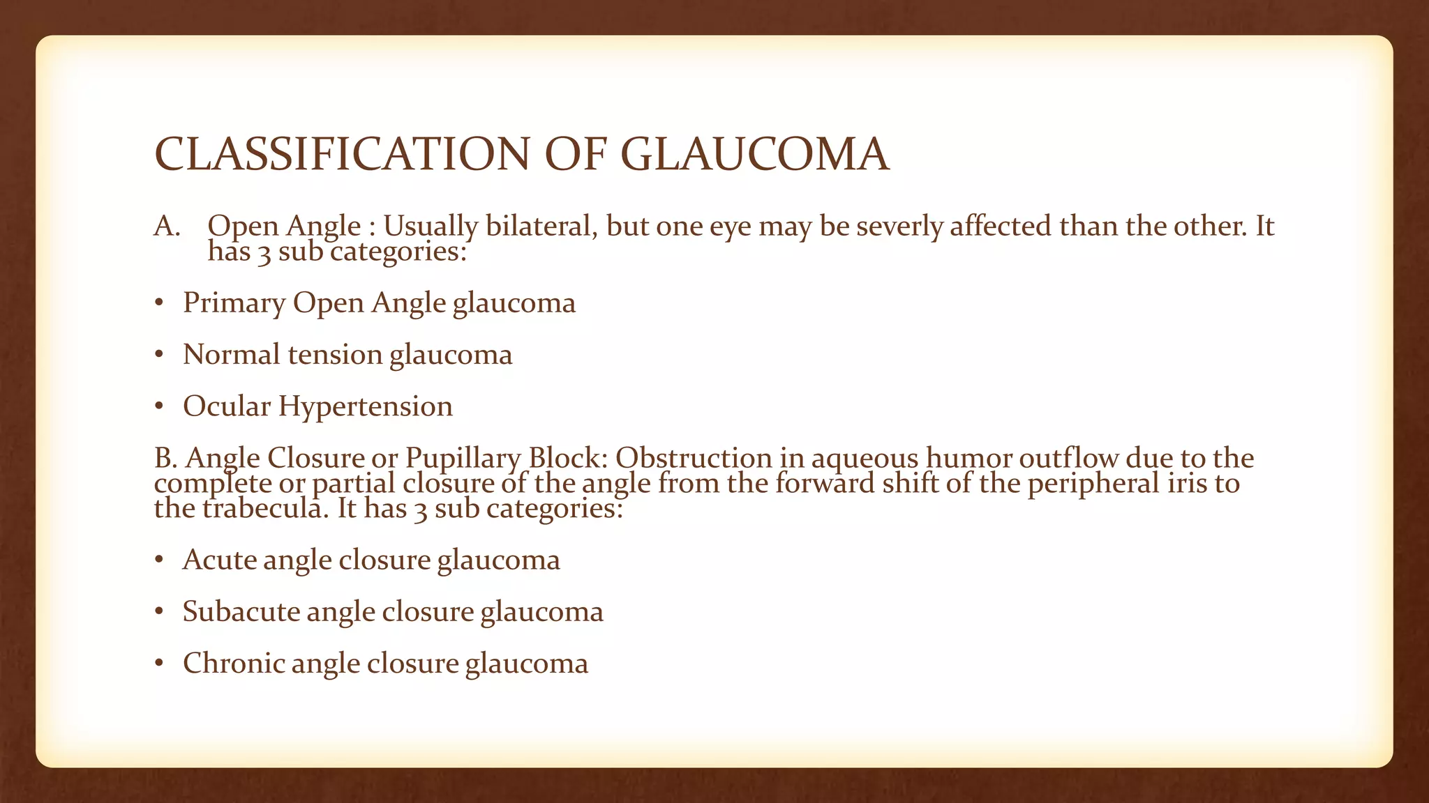
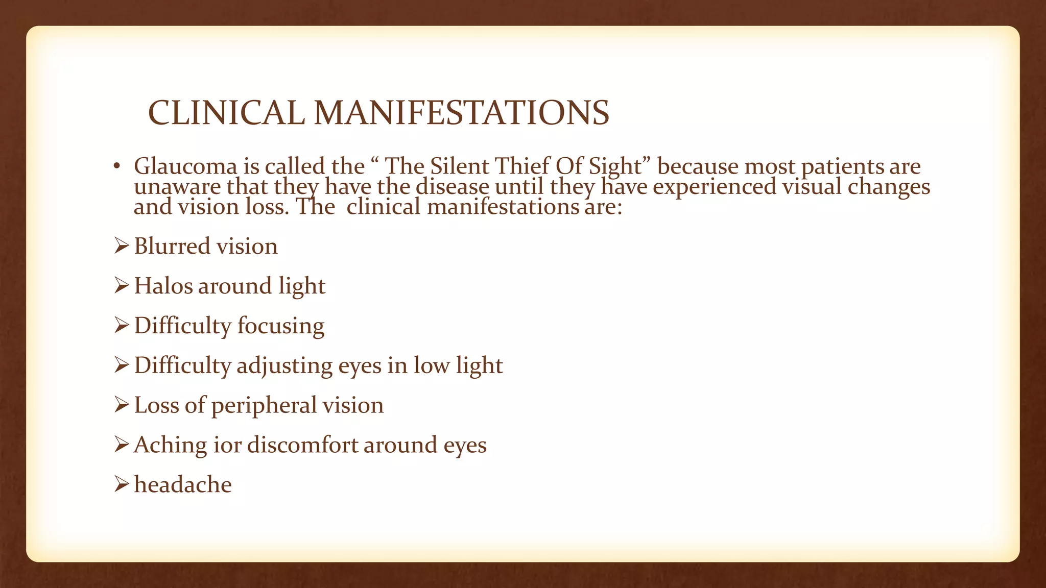
2. INTRODUCTION
• The term ‘Glaucoma’ is used to refer to a group of ocular conditions
characterized by optic nerve damage.
• Increased intra ocular pressure damages the optic nerve and the nerve
fiber layer.
• The degree of harm is highly variable.
• The optic nerve damage is related to the IOP caused by congestion of
aqueous humor in the eye.
• Glaucoma is estimated to affect 2.2 million Americans, and 3 to 6
million more are at risk for the disease.
3. CLASSIFICATION OF GLAUCOMA
A. Open Angle : Usually bilateral, but one eye may be severly affected than the other. It
has 3 sub categories:
• Primary Open Angle glaucoma
• Normal tension glaucoma
• Ocular Hypertension
B. Angle Closure or Pupillary Block: Obstruction in aqueous humor outflow due to the
complete or partial closure of the angle from the forward shift of the peripheral iris to
the trabecula. It has 3 sub categories:
• Acute angle closure glaucoma
• Subacute angle closure glaucoma
• Chronic angle closure glaucoma
4. RISK FACTORS
• Family history of Glaucoma.
• Thin cornea
• African American Race
• Older Age
• Diabetes
• Cardiovascular Disease
• Migraine Syndrome
• Myopia
• Eye Trauma
• Prolonged use of topical corticosteroids
5. PATHOPHYSIOLOGY
There are two theories how increased IOP damages the optic nerve in
glaucoma.
• The Direct Mechanical Theory: It suggests that high IOP damages the
retinal layer as it passes through the optic nerve head.
• The Indirect Ischemic Theory: It suggests that high IOP compresses
the microcirculation in the optic nerve head, resulting in cell injury
and death.
6. CLINICAL MANIFESTATIONS
• Glaucoma is called the “ The Silent Thief Of Sight” because most patients are
unaware that they have the disease until they have experienced visual changes
and vision loss. The clinical manifestations are:
➢Blurred vision
➢Halos around light
➢Difficulty focusing
➢Difficulty adjusting eyes in low light
➢Loss of peripheral vision
➢Aching ior discomfort around eyes
➢headache
7.
8. ASSESSMENT AND DIAGNOSTIC FINDINGS
EXAMINING NAME OF TEST
1) The inner eye pressure Tonometry
2) The shape and color of the optic nerve Ophthalmoscopy
3) The complete field of vision Perimetry
4) The angle in the eye where the iris meets the
cornea
Gonioscopy
5) Thickness of the cornea Pachymetry
9. DIFFERENT TESTS
• Tonometry: Tonometry measures the pressure within your eye. During the
tonometry, eye drops are used to numb the eyes. Then doctor uses a device
called Tonometer to measure the inner pressure of the eye.
• Ophthalmoscopy: This diagnostic procedure helps the doctor examine your
optic nerve for glaucoma damage. Eye drops are used to dilate the pupil so
that the doctor can see through your eye to examine shape and colour of the
optic nerve.
• Perimetry: It is a visual field test that produces a map of your complete field of
vision. This test will help a doctor determine whether your vision has been
affected by Glaucoma.
During this test , you will be asked to look straight ahead a light spot is
presented in different areas of your peripheral vision.
10. • Gonioscopy: This diagnostic test helps determine whether the angle
where the iris meets the cornea is open and wide or narrow and closed.
During the exam, eye drops are used to numb the eye. A hand- held
contact lens is gently placed on the eye. This contact lens has a mirror
which shows angle between the iris and cornea is closed and blocked.
• Pachymetry: It is simple, painless test to measure the thickness of your
cornea.
A probe called pachymeter is gently placed on the front of the eye to
measure its thickness. Pachymetry can help in diagnosis because corneal
thickness has the potential to influence eye pressure readings.
11. MEDICAL MANAGEMENT
• The aim of all the Glaucoma treatment is prevention of optic nerve damage.
PHARMACOLOGIC THERAPY
❑Cholinergics: Increase aqueous fluid outflow by contracting the ciliary muscle and
causing miosis and opening of trabecular meshwork. Example: pilocarpine, carbachol
❑Beta- Blockers- Decreases aqueous humor production. Example: timolol maleate
❑Alpha- adrenergic agonists: Decreases aqueous humor production. Example:
apraclonidine
❑Carbonic anhydrase inhibitors: Decreases aqueous humor production. Example:
acetazolamide and dorzolamide.
❑Prostaglandin analogues: Increases uveoscleral outflow. Example: latanoprost,
bimatoprost.
12. SURGICAL MANAGEMENT
• Laser trabeculoplasty: Laser burns are applied to the inner surface of
the trabecular meshwork to open the intra trabecular spaces and widen
the canal of schlemn, thereby promoting the outflow of aqueous
humor and decreasing IOP.
• Peripheral Iridotomy( for pupillary block glaucoma): An opening is
made in the iris to eliminate the pupillary blockage. Contraindicated in
patient with corneal edema.
• Filtering Procedures(for chronic glaucoma): They are used to create an
opening or fistula in the trabecular meshwork to drain aqueous humor
from the anterior chamber to the subconjunctival space into bleb.
13. • Trabeculectomy: It is the standard filtering technique used to remove
part of the trabecular meshwork.
• Drainage implants or shunts: These are the tubes implanted in the
anterior chamber to shunt aqueous humor to the episcleral plate in the
conjunctival space.
implants are used when failure has occurred with one or more
trabeculectomies in which anti- fibrotic agents were used.
14. NURSING DIAGNOSIS
• Disturbed sensory perception related to altered
status of sense organ.
• Anxiety related to change in health status.
• Deficient knowledge related to lack of exposure with
resources.
15. NURSING INTERVENTION
• Determine type and degree of visual loss.
• Allow expression of feelings about loss and possibility of loss of vision.
• Demonstrate administration of eye drops.
• Evaluate anxiety level, degree of pain experienced.
• Identify helpful resources and people.
• Stress the importance on glaucoma screening.
• Encourage patient to make necessary changes in the lifestyle.
• Discuss dietary considerations.
• Stress importance of routine checkups.