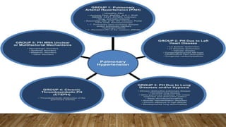
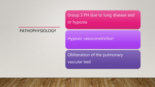
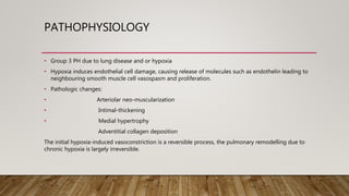
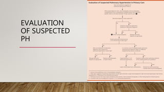
PATHOPHYSIOLOGY
AND DIAGNOSIS OF
PULMONARY
HYPERTENSION
PRESENTER: DR. SOPHIA C.
BROWNE
MODERATOR: DR. A AQUART
DATE: 10TH MAY 2021
PULMONARY
HYPERTENSION
• Complex group of disorders that result from
different pathophysiologic mechanisms
• It is defined by a mPAP of ≥ 25mmHG
• The symptoms are typically non-specific or easily
attributable to comorbid conditions
• Diagnosis can be challenging and requires a
stepwise evaluation
CLINICAL
CLASSIFICATION
OF PULMONARY
HYPERTENSION
PATHOPHYSIOLOGY Pulmonary Circulation
• The role is to carry deoxygenated blood from the
heart to the lungs and returning oxygenated blood
back to the heart for delivery to the systemic
circulation.
• Cardiac output, low pressure and pulmonary
vascular resistance (PVR) is normally maintained
due to the abundance of small pulmonary arteries
and capillaries with high cross-sectional area.
• More capillaries are recruited during exercise to
maintain low PA pressure
• Elevations in PVR with subsequent increases in PA
pressure are observed in the development of PH.
PATHOPHYSIOLOGY
• Ohm’s law change in pressure = flow x resistance
• mPAP = (Right ventricle cardiac output x pulmonary vascular
resistance)+ pulmonary alveolar occlusion pressure
• Primary cause of Pulmonary Hypertension is increase pulmonary
vascular resistance (PVR)
• Increased flow alone i.e. right ventricle output, does not usually
cause significant PH because the pulmonary vascular bed vasodilates
and recruits vessels in response to increased flow.
•
• Increased pulmonary venous pressure which is represented by the
alveolar occlusion pressure, alone does not usually cause significant PH.
•
• Chronic increase of either flow and /or pulmonary venous pressure
can increase pulmonary vascular resistance.
PATHOPHYSIOLOGY
• Group 1- PAH
• Progressive narrowing of the
distal pulmonary arteries
attributed to a variety of
pathologic insults
• Arterial vasoconstriction
• Medial Hypertrophy
• Intimal Proliferation
• Fibrosis
PATHOPHYSIOLOGY
• Gene mutations leading to a predisposition for
the development of PAH.
Mutations in the BMPR2; the gene product binds
members of the TGF-ß superfamily of ligands
Rare mutations in other members of the TGF-ß
• ALK-1
• ENG
• SMAD9
• Other gene mutations without direct relation to
the TGF-ß family CAV1 and KCNK3
PATHOPHYSIOLOGY
The mechanism of anorexigens in PAH is unclear although it is believed to involve
serotonin biology given their interaction with the serotonin transporter to block
serotonin uptake and the ability to induce PASMC growth in PAH.
Dasatinib tyrosine kinase inhibitor, in the treatment of chronic myelogenic leukemia
has been associated with severe PAH development.
Decreases platelet derived growth factor (PDGF), it is a mitogenic and promigratory
stimuli for PASMCs.
Plexiform lesions in HIV associated PAH
PATHOPHYSIOLOGY
Group 2 PH due to Left Heart Disease
A passive backward transmission of filling pressures that increases mPAP
, loss of left atrial compliance, diastolic dysfunction, mitral regurgitation.
A further increase in mPAP, endothelial dysfunction, vasoconstriction
Worsening pulmonary vascular remodeling, right ventricular failure and
death.
PATHOPHYSIOLOGY
Group 3 PH due to lung disease and
or hypoxia
Hypoxic vasoconstriction
Obliteration of the pulmonary
vascular bed
PATHOPHYSIOLOGY
• Group 3 PH due to lung disease and or hypoxia
• Hypoxia induces endothelial cell damage, causing release of molecules such as endothelin leading to
neighbouring smooth muscle cell vasospasm and proliferation.
• Pathologic changes:
• Arteriolar neo-muscularization
• Intimal-thickening
• Medial hypertrophy
Adventitial collagen deposition
The initial hypoxia-induced vasoconstriction is a reversible process, the pulmonary remodelling due to
chronic hypoxia is largely irreversible.
PATHOPHYSIOLOGY
• Group 4 CTEPH
• Abnormal mechanisms of fibrinolysis/underlying haematological disorders or
autoimmune disorders
• Contributing to a hypercoagulable state and poor resolution of thrombi
• Underyling pulmonary arteriopathy or in situ thrombosis likely contributes to
CTEPH.
Vascular remodeling occurs may occur in the small muscular arteries and arterioles
at the site of vessel occlusion, as well as in the spared non-obstructed vessels
secondary to the resulting high shear stress.
PATHOPHYSIOLOGY
• Group 5 PH with unclear multifactorial mechanisms
• Given the multiple etiologies of Group 5 PH, further research is needed to fully
characterize this group of conditions and elucidate the pathophysiologic
mechanisms of disease.
DIAGNOSIS
DIAGNOSIS
• Symptoms
- Exertional dypsnea
- Chest pain
- Dizziness
- Hypersomnolence in Obstructive sleep Apnea
- Skin changes in scleroderma
DIAGNOSIS
• Physical Examination Findings
• Prominent pulmonic component (P2) of the S2
• Elevated JVP
• Left Parasternal Heave ( from a right ventricular hypertrophy)
• Right-sided S3
• Tricuspid Regurgitation Murmur
• Pedal Edema
DIAGNOSIS
• Imaging Studies
• CXR
- Enlarged Pulmonary arteries with rapid tapering of vessels toward the periphery of the lungs
- Right heart enlargement
- Secondary PH caused by parenchymal lung disease, hyperinflation and bulbous disease
suggestive of COPD or increased interstitial lung markings suggestive of ILD.
DIAGNOSIS
DIAGNOSIS
• Echocardiography
- Permits the evaluation for signs of right ventricular pressure overload, including paradoxical
bulging of the septum into the left ventricle during systole and hypertrophy of the right ventricle
wall.
- Doppler estimates of PA pressure based on the velocity of TR can suggest the presence of PH but
do not correlate well with directly measure PA pressure.
- Pulmonary Functions Tests: typically show decreased diffusing capacity
- Ventilation-perfusion scan: helps identify patients with secondary PH caused by chronic
thromboembolic disease, but IPAH can also produce abnormal scans.
- Right-heart catherization : Permits direct measurement of PA pressure, and with angiography, a
definitive diagnosis of chronic thromboembolic disease.
EVALUATION
OF SUSPECTED
PH
REFERENCES
• Dunlap,Beth et al Pulmonary Hypertension: Diagnosis and Treatment www.aafp.org/afp Vol 94, No. 6
Sept,15,2016
• Sysol, J,R et al Classification and Pathophysiology of Hypertension Continuing Cardiology Education, 2018
https://doi.org/10.1002/cce2.71
• Gladwin, Mark et al Pulmonary Hypertension MSD Manuals Sept 2020 https://www.msdmanuals.com
. Ashar, Bimal H. et al The John Hopkins Internal Medicine Board Review 5th Edition: Pulmonary Hypertension
• Pg 183-185
• Elizabeth S Klings, MD Pulmonary Hypertension due to lung disease and/or hypoxemia www.UpToDate.com

Pulmonary hypertension

  • 1.
    PATHOPHYSIOLOGY AND DIAGNOSIS OF PULMONARY HYPERTENSION PRESENTER:DR. SOPHIA C. BROWNE MODERATOR: DR. A AQUART DATE: 10TH MAY 2021
  • 2.
    PULMONARY HYPERTENSION • Complex groupof disorders that result from different pathophysiologic mechanisms • It is defined by a mPAP of ≥ 25mmHG • The symptoms are typically non-specific or easily attributable to comorbid conditions • Diagnosis can be challenging and requires a stepwise evaluation
  • 3.
  • 4.
    PATHOPHYSIOLOGY Pulmonary Circulation •The role is to carry deoxygenated blood from the heart to the lungs and returning oxygenated blood back to the heart for delivery to the systemic circulation. • Cardiac output, low pressure and pulmonary vascular resistance (PVR) is normally maintained due to the abundance of small pulmonary arteries and capillaries with high cross-sectional area. • More capillaries are recruited during exercise to maintain low PA pressure • Elevations in PVR with subsequent increases in PA pressure are observed in the development of PH.
  • 5.
    PATHOPHYSIOLOGY • Ohm’s lawchange in pressure = flow x resistance • mPAP = (Right ventricle cardiac output x pulmonary vascular resistance)+ pulmonary alveolar occlusion pressure • Primary cause of Pulmonary Hypertension is increase pulmonary vascular resistance (PVR) • Increased flow alone i.e. right ventricle output, does not usually cause significant PH because the pulmonary vascular bed vasodilates and recruits vessels in response to increased flow. • • Increased pulmonary venous pressure which is represented by the alveolar occlusion pressure, alone does not usually cause significant PH. • • Chronic increase of either flow and /or pulmonary venous pressure can increase pulmonary vascular resistance.
  • 6.
    PATHOPHYSIOLOGY • Group 1-PAH • Progressive narrowing of the distal pulmonary arteries attributed to a variety of pathologic insults • Arterial vasoconstriction • Medial Hypertrophy • Intimal Proliferation • Fibrosis
  • 7.
    PATHOPHYSIOLOGY • Gene mutationsleading to a predisposition for the development of PAH. Mutations in the BMPR2; the gene product binds members of the TGF-ß superfamily of ligands Rare mutations in other members of the TGF-ß • ALK-1 • ENG • SMAD9 • Other gene mutations without direct relation to the TGF-ß family CAV1 and KCNK3
  • 8.
    PATHOPHYSIOLOGY The mechanism ofanorexigens in PAH is unclear although it is believed to involve serotonin biology given their interaction with the serotonin transporter to block serotonin uptake and the ability to induce PASMC growth in PAH. Dasatinib tyrosine kinase inhibitor, in the treatment of chronic myelogenic leukemia has been associated with severe PAH development. Decreases platelet derived growth factor (PDGF), it is a mitogenic and promigratory stimuli for PASMCs. Plexiform lesions in HIV associated PAH
  • 10.
    PATHOPHYSIOLOGY Group 2 PHdue to Left Heart Disease A passive backward transmission of filling pressures that increases mPAP , loss of left atrial compliance, diastolic dysfunction, mitral regurgitation. A further increase in mPAP, endothelial dysfunction, vasoconstriction Worsening pulmonary vascular remodeling, right ventricular failure and death.
  • 13.
    PATHOPHYSIOLOGY Group 3 PHdue to lung disease and or hypoxia Hypoxic vasoconstriction Obliteration of the pulmonary vascular bed
  • 14.
    PATHOPHYSIOLOGY • Group 3PH due to lung disease and or hypoxia • Hypoxia induces endothelial cell damage, causing release of molecules such as endothelin leading to neighbouring smooth muscle cell vasospasm and proliferation. • Pathologic changes: • Arteriolar neo-muscularization • Intimal-thickening • Medial hypertrophy Adventitial collagen deposition The initial hypoxia-induced vasoconstriction is a reversible process, the pulmonary remodelling due to chronic hypoxia is largely irreversible.
  • 15.
    PATHOPHYSIOLOGY • Group 4CTEPH • Abnormal mechanisms of fibrinolysis/underlying haematological disorders or autoimmune disorders • Contributing to a hypercoagulable state and poor resolution of thrombi • Underyling pulmonary arteriopathy or in situ thrombosis likely contributes to CTEPH. Vascular remodeling occurs may occur in the small muscular arteries and arterioles at the site of vessel occlusion, as well as in the spared non-obstructed vessels secondary to the resulting high shear stress.
  • 16.
    PATHOPHYSIOLOGY • Group 5PH with unclear multifactorial mechanisms • Given the multiple etiologies of Group 5 PH, further research is needed to fully characterize this group of conditions and elucidate the pathophysiologic mechanisms of disease.
  • 17.
  • 18.
    DIAGNOSIS • Symptoms - Exertionaldypsnea - Chest pain - Dizziness - Hypersomnolence in Obstructive sleep Apnea - Skin changes in scleroderma
  • 19.
    DIAGNOSIS • Physical ExaminationFindings • Prominent pulmonic component (P2) of the S2 • Elevated JVP • Left Parasternal Heave ( from a right ventricular hypertrophy) • Right-sided S3 • Tricuspid Regurgitation Murmur • Pedal Edema
  • 20.
    DIAGNOSIS • Imaging Studies •CXR - Enlarged Pulmonary arteries with rapid tapering of vessels toward the periphery of the lungs - Right heart enlargement - Secondary PH caused by parenchymal lung disease, hyperinflation and bulbous disease suggestive of COPD or increased interstitial lung markings suggestive of ILD.
  • 21.
  • 22.
    DIAGNOSIS • Echocardiography - Permitsthe evaluation for signs of right ventricular pressure overload, including paradoxical bulging of the septum into the left ventricle during systole and hypertrophy of the right ventricle wall. - Doppler estimates of PA pressure based on the velocity of TR can suggest the presence of PH but do not correlate well with directly measure PA pressure. - Pulmonary Functions Tests: typically show decreased diffusing capacity - Ventilation-perfusion scan: helps identify patients with secondary PH caused by chronic thromboembolic disease, but IPAH can also produce abnormal scans. - Right-heart catherization : Permits direct measurement of PA pressure, and with angiography, a definitive diagnosis of chronic thromboembolic disease.
  • 23.
  • 24.
    REFERENCES • Dunlap,Beth etal Pulmonary Hypertension: Diagnosis and Treatment www.aafp.org/afp Vol 94, No. 6 Sept,15,2016 • Sysol, J,R et al Classification and Pathophysiology of Hypertension Continuing Cardiology Education, 2018 https://doi.org/10.1002/cce2.71 • Gladwin, Mark et al Pulmonary Hypertension MSD Manuals Sept 2020 https://www.msdmanuals.com . Ashar, Bimal H. et al The John Hopkins Internal Medicine Board Review 5th Edition: Pulmonary Hypertension • Pg 183-185 • Elizabeth S Klings, MD Pulmonary Hypertension due to lung disease and/or hypoxemia www.UpToDate.com