
Recommended
More Related Content
What's hot
What's hot (20)
Similar to Prof. ghabisha saif posterior abdominal wall anatomy
Similar to Prof. ghabisha saif posterior abdominal wall anatomy (20)
Recently uploaded
Recently uploaded (20)
Prof. ghabisha saif posterior abdominal wall anatomy
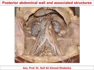
- 1. Posterior abdominal wall and associated structures Ass. Prof. Dr. Saif Ali Ahmed Ghabisha
- 2. • The posterior abdominal wall extends from the 12th rib above to the pelvic brim below. Posterior abdominal wall and associated structures
- 3. Bony part: • In the median plane, it is made up of bodies, intervertebral disc, and transverse processes of the five lumbar vertebrae. • Laterally it is divided into upper and lower parts by the iliac crest. • The part above the iliac crest is made of inner surfaces of the 12th rib and the part below the iliac crest is made of iliac fossa. The posterior abdominal wall is constructed as: Posterior abdominal wall and associated structures
- 4. Muscular part: • Above the iliac crest, from medial to lateral sides, it is made up of psoas major, quadratus lumborum, and transversus abdominis muscles. • Below the iliac crest on either side of the lumbar vertebral column from medial to lateral sides, it is made up of psoas major and iliacus muscles. The posterior abdominal wall is constructed as: Fasciae: • The psoas major and iliacus muscles are covered by fascia iliaca. • The quadratus lumborum is enclosed between the anterior and posterior layers of the thoracolumbar fascia. Posterior abdominal wall and associated structures
- 5. The structures in the posterior abdominal wall: • Muscles and fasciae of the posterior abdominal wall. • Great vessels of the abdomen (abdominal aorta and inferior vena cava). • Azygos and hemiazygos veins. • Lymph nodes and lymphatics of the posterior abdominal wall. • Nerves of the posterior abdominal wall. Posterior abdominal wall and associated structures
- 6. Muscles of the posterior abdominal wall Three muscles: • psoas major • iliacus • quadratus lumborum on each side of the vertebral column form most of the posterior abdominal wall. Posterior abdominal wall and associated structures
- 7. Psoas major • The psoas major is a long fusiform muscle extending from the sides of lumbar vertebrae to the lesser trochanter of the femur. • It is enclosed in a fascial sheath called psoas sheath. Origin: The muscle arises from 14 fleshy slips: • Five slips from intervertebral discs between T12–L5 vertebrae. • Five slips from anterior surfaces and lower borders of the transverse process of five lumbar vertebrae. • Four slips from tendinous arches along the sides of upper four lumbar vertebrae. Insertion: Enters the anterior surface of the tip of the lesser trochanter of the femur. Nerve supply: The nerve supply is by direct branches from ventral rami of L2, L3, L4 spinal nerves. Posterior abdominal wall and associated structures
- 8. Psoas major Posterior abdominal wall and associated structures
- 9. It is formed by the psoas fascia. The attachments of psoas fascia: Above: It is thickened to form medial arcuate ligament, which extends from the body of L1 vertebra to the tip of its transverse process. Laterally: It blends with the anterior layer of the thoracolumbar fascia. Medially: It is attached to the bodies and intervening intervertebral discs of lumbar vertebrae and presents four tendinous arches. Below: It fuses with the arcuate line of the pelvis and the fascia covering the iliacus muscle (iliac fascia). Psoas sheath Posterior abdominal wall and associated structures
- 10. • This muscle is present in about 50% individuals. • When present, it runs downward in front of psoas major. • In form and shape, it resembles the plantaris muscle of the leg and is confined to the abdomen. Origin: It arises from the side of the intervertebral disc between T12 and L1 vertebrae and adjoining parts of their bodies. Insertion: From the site of origin, the muscle runs in front of the psoas major and ends in a long flat tendon, which is inserted into the iliopubic eminence. Nerve supply: It is by a branch of L1 spinal nerve. Action: It is a weak flexor of the trunk. Psoas minor Posterior abdominal wall and associated structures
- 11. • It is a fan-shaped muscle and forms the lateral component of the iliopsoas muscle. • Origin: it arises from the upper two thirds of the floor of iliac fossa, inner lip of iliac crest and upper surface of the lateral part of the sacrum. • Insertion: the fibres converge on and fuse with the lower part of the psoas major laterally and inserted with it on the anterior surface of lesser trochanter and an area (2.5 cm long) below it. • Nerve supply: it is by the femoral nerve. • Actions: along with the psoas major, it causes flexion of the thigh and the lumbar part of the vertebral column. Iliacus Posterior abdominal wall and associated structures
- 12. Quadratus lumborum It is a quadrilateral muscle which fills the medial half of the gap between the 12th rib, the iliac crest, and the tips of transverse processes of lumbar vertebrae. The quadratus lumborum muscle is enclosed between the anterior and middle layers of the thoraco-lumbar fascia. Origin: it arises from: Posterior one-third of the inner lip of the iliac crest and iliolumbar ligament. Lower two to four transverse processes of lumbar vertebrae. Insertion: it is inserted into the medial part of the anterior surface of the 12th rib. It is also inserted into the upper lumbar transverse processes, posterior to its slips of origin. Nerve supply: ventral rami of t12–l3/l4 lumbar spinal n.. Posterior abdominal wall and associated structures
- 13. Great vessels of the abdomen The great vessels of the abdomen are abdominal aorta and inferior vena cava. Posterior abdominal wall and associated structures
- 14. Abdominal aorta • The abdominal aorta begins as the continuation of descending thoracic aorta at the aortic orifice of the diaphragm opposite to the lower border of the T12 vertebra or intervertebral disc between vertebrae T12 and L1. • It descends vertically downward and slightly to the left, in front of the vertebral column, and terminates in front of the lower part of the body of L4 vertebra (about 1.25 cm) to the left of the median plane by dividing into right and left common iliac arteries. • Length: 10–11 cm, Width: 2 cm. Posterior abdominal wall and associated structures
- 15. Relations Posterior: • Bodies of the upper four lumbar vertebrae and intervening intervertebral discs. • Anterior longitudinal ligament. • Third and fourth left lumbar veins. Anterior: From above downward: • Pancreas and splenic vein. • Left renal vein. • Third part of the duodenum. • Root of the mesentery. • Coils of the small intestine separated by parietal peritoneum. Right side: Inferior vena cava. Left side: Left sympathetic trunk. Posterior abdominal wall and associated structures
- 16. Branches • Three unpaired ventral branches to the gut. • Three paired lateral branches to three paired glands (suprarenal glands, kidneys, and gonads). • Paired posterolateral branches to the abdominal wall. • The aorta also gives rise to paired inferior phrenic artery, unpaired median sacral artery, and two terminal branches. Abdominal aorta Posterior abdominal wall and associated structures
- 17. Inferior vena cava (IVC) • The IVC is the largest and widest vein of the body. • It drains most of the blood from the body below the diaphragm into the right atrium of the heart. • The IVC is formed by the union of right and left common iliac veins in front of the body of L5 vertebra, below the aortic bifurcation, and behind the right common iliac artery. • It ascends in front of the vertebral column on the right side of the aorta. • It reaches the groove on the posterior surface of the liver between the right and caudate lobes, just above the groove it pierces the central tendon of the diaphragm at the level of T8 vertebra and terminates by entering the right atrium of the heart. Posterior abdominal wall and associated structures
- 18. Anterior: From below upward: • Root of the mesentery. • Right testicular/ovarian artery. • Third part of the duodenum. • Head of the pancreas and bile duct. • Portal vein (posterior to first of duodenum and in the right free margin of lesser omentum). • Posterior surface of the liver between the right and • caudate lobes. Posterior: From below upward: • Right sympathetic chain and psoas major. • Right renal artery. • Right coeliac ganglion. • Right suprarenal gland (medial part). • Right middle suprarenal vein. • Right inferior phrenic artery. Inferior vena cava (IVC) Relations Posterior abdominal wall and associated structures
- 19. • Three formative veins: two common iliac veins and the median sacral vein. The latter may join the left common iliac vein. Each common iliac vein receives an iliolumbar vein. • Three abdominal wall tributaries: inferior phrenic vein and third and fourth lumbar veins. The first and second lumbar veins end in the ascending lumbar vein. • Three lateral visceral tributaries: right suprarenal vein, renal veins, and right testicular/ovarian vein. The left suprarenal vein and left gonadal veins drain into the left renal vein. • Three anterior visceral tributaries: right, middle, and left hepatic veins. Inferior vena cava (IVC) Tributaries Posterior abdominal wall and associated structures
- 20. Inferior vena cava (IVC) Tributaries Posterior abdominal wall and associated structures
- 21. Lymphatics and lymph nodes of the posterior abdominal wall Lymphatics • The lymph vessels draining the posterior abdominal wall and most of the abdominopelvic organs except part of the liver terminate in the cisterna chyli and thoracic duct. • The lymphatic stream is intercepted by a series of lymph node groups before reaching the cisterna chyli and the thoracic duct. Tributaries of cisterna chyli • Right and left intestinal lymph trunks: from the preaortic lymph nodes, which open in its middle. These trunks drain the lymph from the small intestine, stomach, and liver. • Right and left lumbar lymph trunks: from the paraaortic lymph nodes, which open in it inferiorly. • A pair of lymph vessels: from the lower intercostal lymph nodes, which open in it superiorly. Posterior abdominal wall and associated structures
- 22. Lymphatics and lymph nodes of the posterior abdominal wall Posterior abdominal wall and associated structures
- 23. Lymph nodes Lymphatics and lymph nodes of the posterior abdominal wall External iliac nodes: these are 8–10 in number and lie along the external iliac vessels. Common iliac nodes: these are 4–6 in number and lie along the common iliac vessels (lateral group) and below the bifurcation of the aorta (medial group). Aortic lymph nodes: they are situated along the abdominal aorta and inferior vena cava, and are arranged into two groups: pre-aortic and para-aortic. Pre-aortic nodes: the nodes are located around the Origin of ventral branches to the gut. Para-aortic (lateral aortic) nodes: they are situated on each side of the abdominal aorta, some nodes of this group lie behind the aorta and called retroaortic nodes. Posterior abdominal wall and associated structures
- 24. Lymphatics and lymph nodes of the posterior abdominal wall Posterior abdominal wall and associated structures
- 25. Nerves of the posterior abdominal wall The nerves of the posterior abdominal wall include subcostal nerve, ventral rami of lumbar nerves, and lumbar sympathetic chains. Posterior abdominal wall and associated structures
- 26. • It is the ventral ramus of the 12th thoracic spinal nerve. • It enters the abdomen behind the lateral accurate ligament (lateral lumbocostal arch) and runs downward and laterally in front of the quadratus lumborum beneath the anterior layer of thoraco-lumbar fascia. Subcostal nerve Posterior abdominal wall and associated structures
- 27. Lumbar plexus Posterior abdominal wall and associated structures
- 28. Abdominal part of the autonomic nervous system The abdominal part of the autonomic nervous system: • Lumbar sympathetic chains. • Autonomic plexuses of the posterior abdominal wall. Posterior abdominal wall and associated structures
- 29. Lumbar sympathetic chain • It is a ganglionated chain situated on either side of the lumbar vertebrae. • It commences deep to the medial arcuate ligament of the diaphragm as the continuation of the thoracic sympathetic trunk. • The chain enters the pelvis in front of the ala of sacrum beneath the common iliac vessels, where it continues as the sacral sympathetic chain in front of the sacrum. Branches • White rami communicantes • Gray rami communicantes • Lumbar splanchnic nerves Posterior abdominal wall and associated structures
- 30. Autonomic plexuses of the posterior abdominal wall The preganglionic and postganglionic sympathetic fibres, preganglionic parasympathetic fibres, and visceral afferent fibres form a plexus of nerves around the abdominal aorta. This plexus form two major plexuses: • Coeliac • Superior hypogastric plexuses. Posterior abdominal wall and associated structures
- 31. Coeliac plexus (solar plexus) • The coeliac plexus is located on the front of the abdominal aorta around the coeliac trunk and origin of the superior mesenteric artery. • The coeliac ganglia are two irregular masses of nerve cells situated one on each side of the origin of coeliac artery. • Each ganglion is divided into a large upper part and a lower part. • The lower part is called aorticorenal ganglion. Posterior abdominal wall and associated structures
- 32. Coeliac plexus (solar plexus) Posterior abdominal wall and associated structures
- 33. Branches • Phrenic plexus • Hepatic plexus • Left gastric plexus • Splenic plexus • Suprarenal plexus • Renal plexus • Testicular plexus • Ovarian plexus • Superior mesenteric plexus • Abdominal aortic plexus (intermesenteric plexus) • Inferior mesenteric plexus Posterior abdominal wall and associated structures Coeliac plexus (solar plexus)
- 34. • It lies in front of the bifurcation of the abdominal aorta and body of the fifth lumbar vertebra between the two common iliac arteries. • It is often referred to as presacral nerve but the plexus is never sufficiently condensed to resemble a single nerve and moreover plexus is prelumbar rather than presacral in position. It is formed by the union of: • Descending fibres of the aortic plexus. • Third and fourth lumbar splanchnic nerves. • Ascending filaments of inferior hypogastric plexus. • These fibres supply the parts of gut derived from the hindgut. Superior hypogastric plexus (presacral nerve) Posterior abdominal wall and associated structures
- 35. Superior hypogastric plexus (presacral nerve) Posterior abdominal wall and associated structures
- 36. Posterior abdominal wall and associated structures Superior hypogastric plexus (presacral nerve)