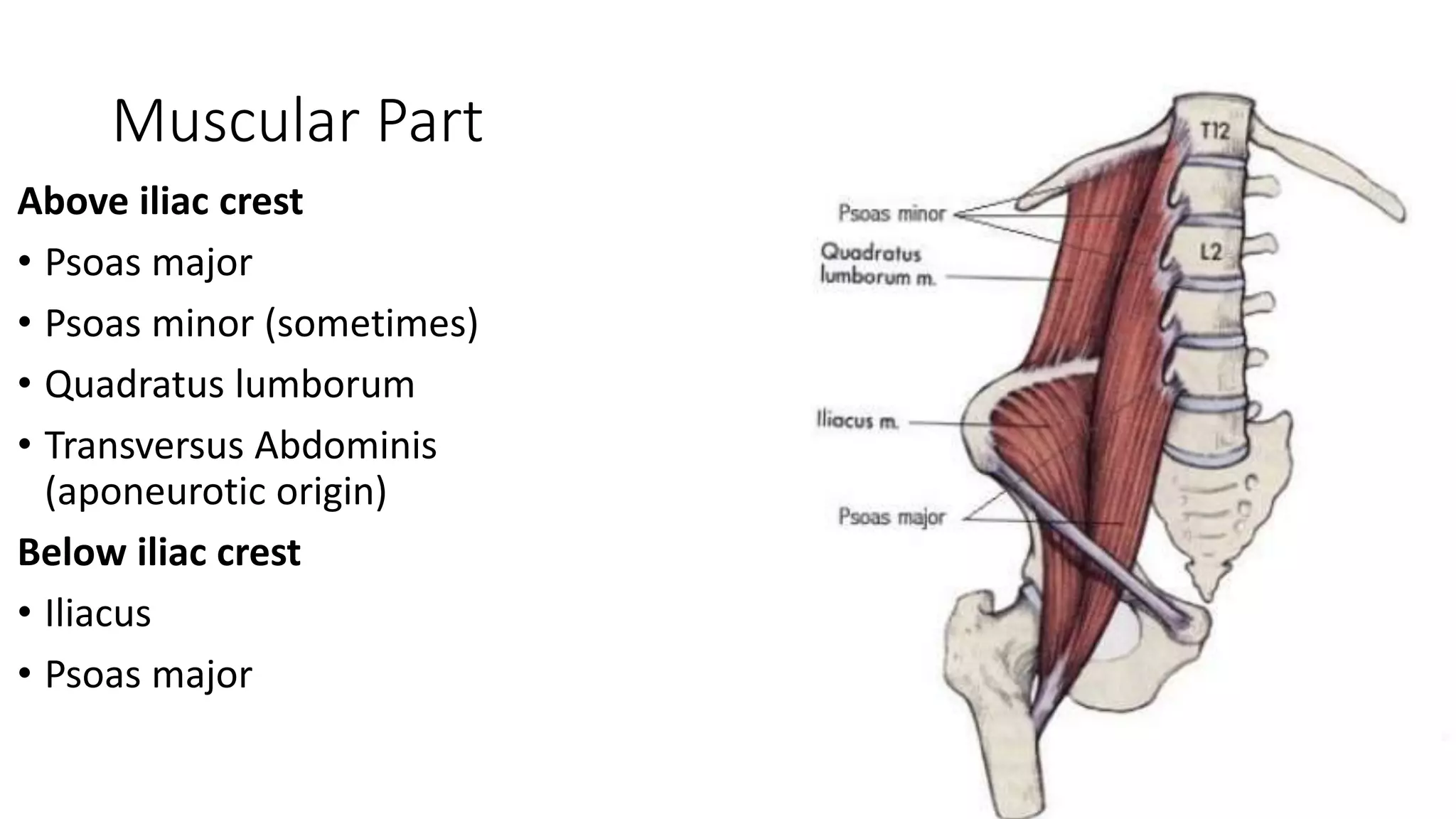
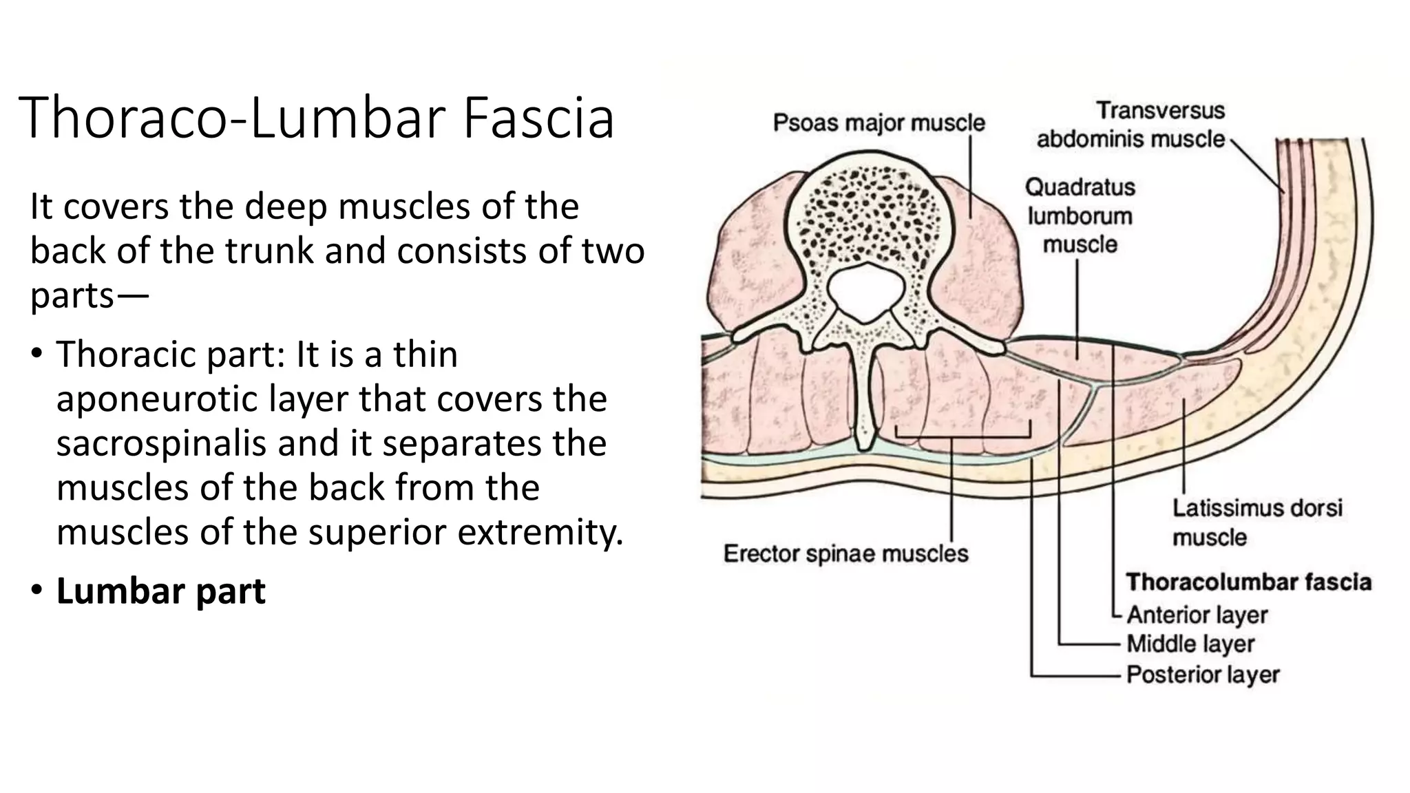
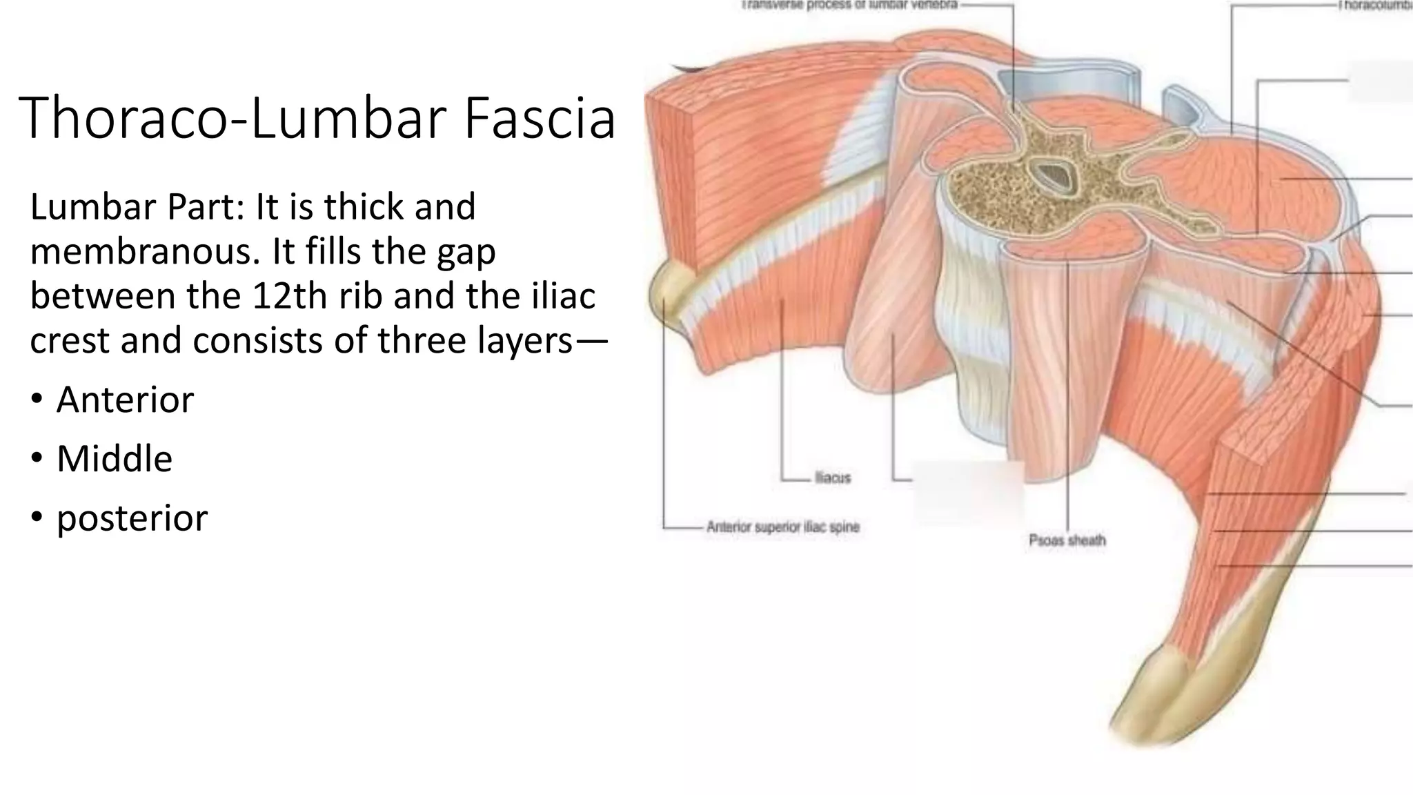
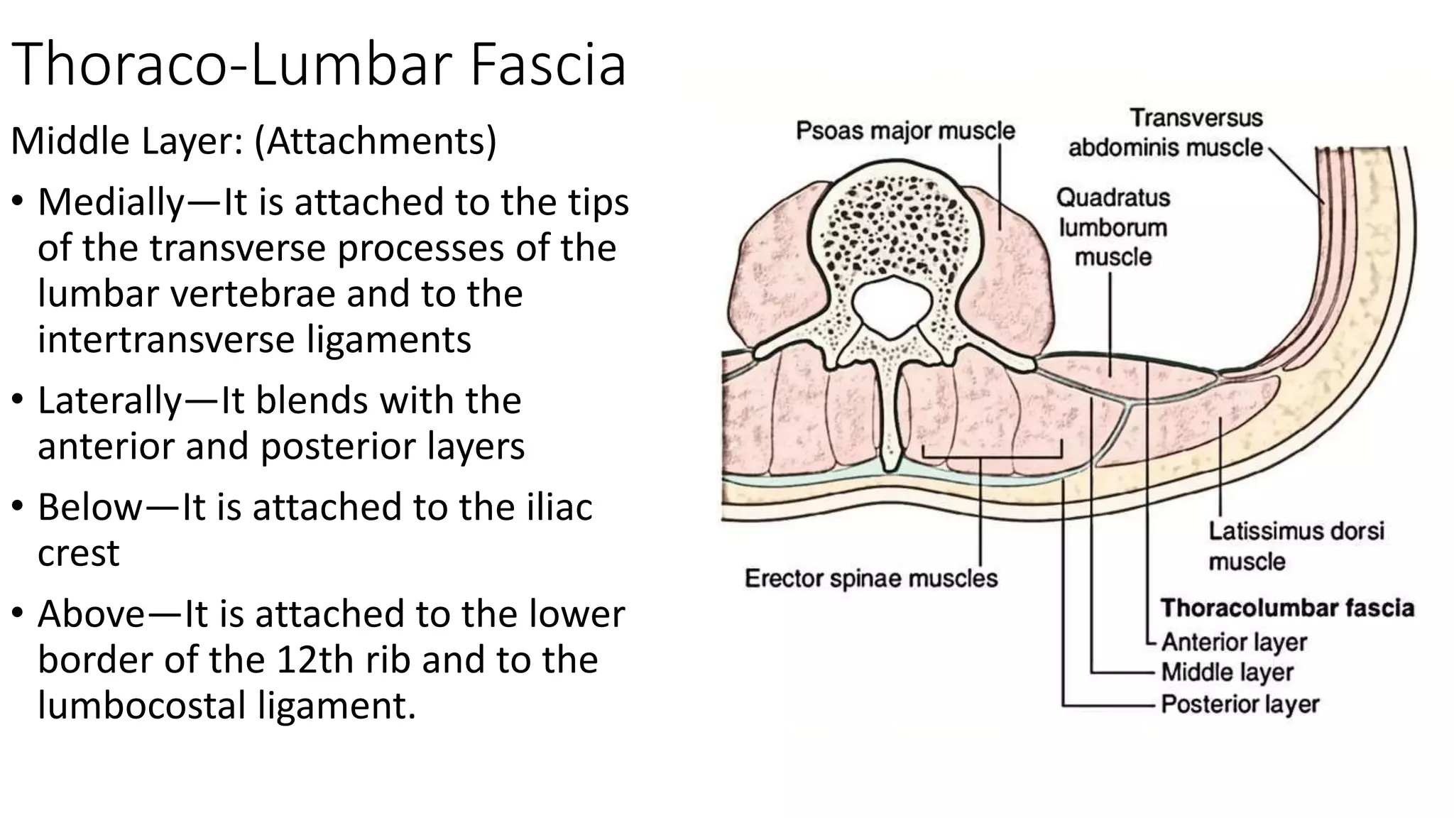
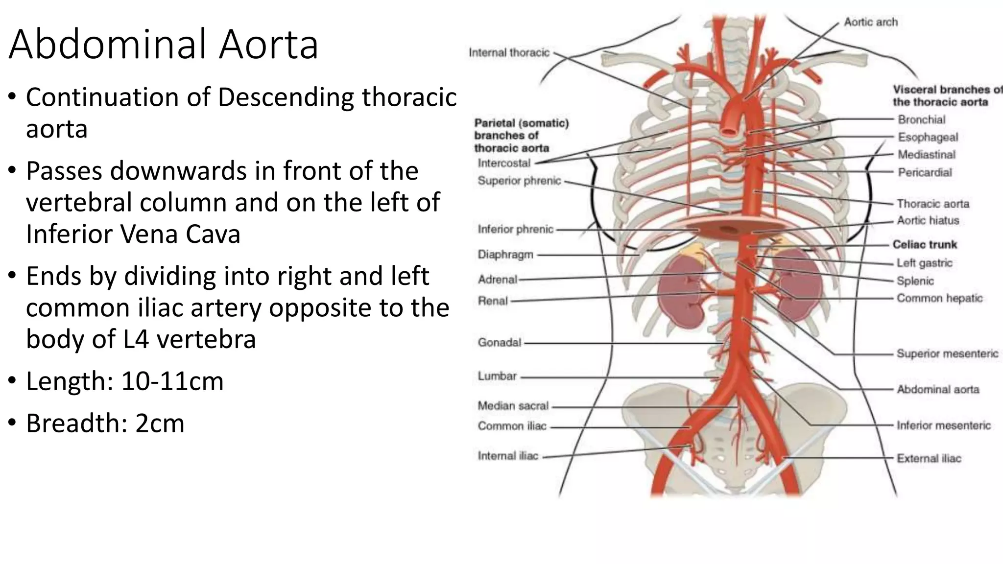
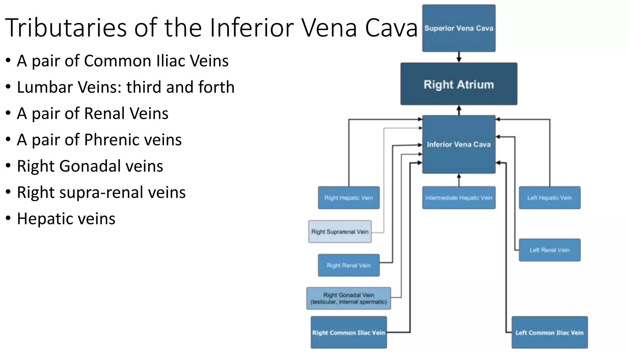
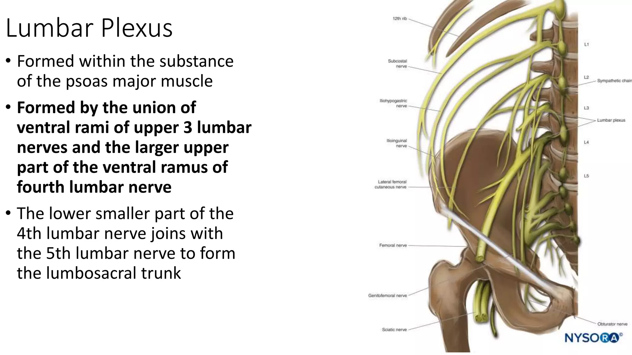
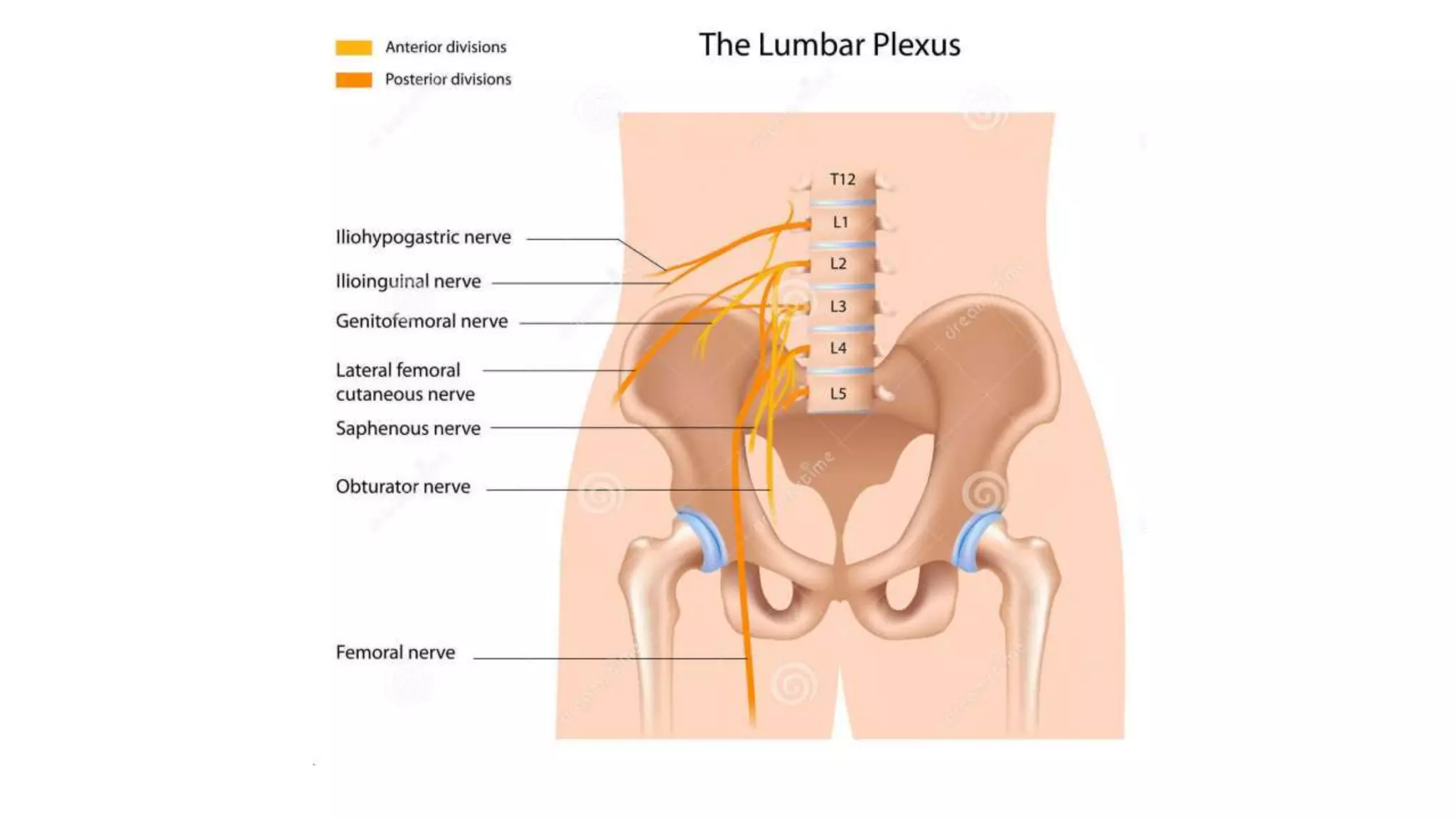
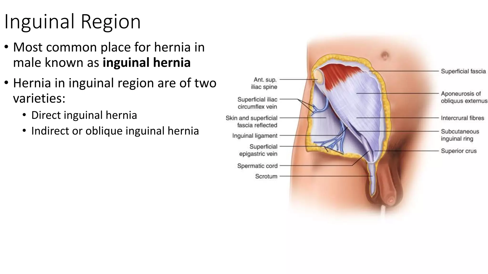
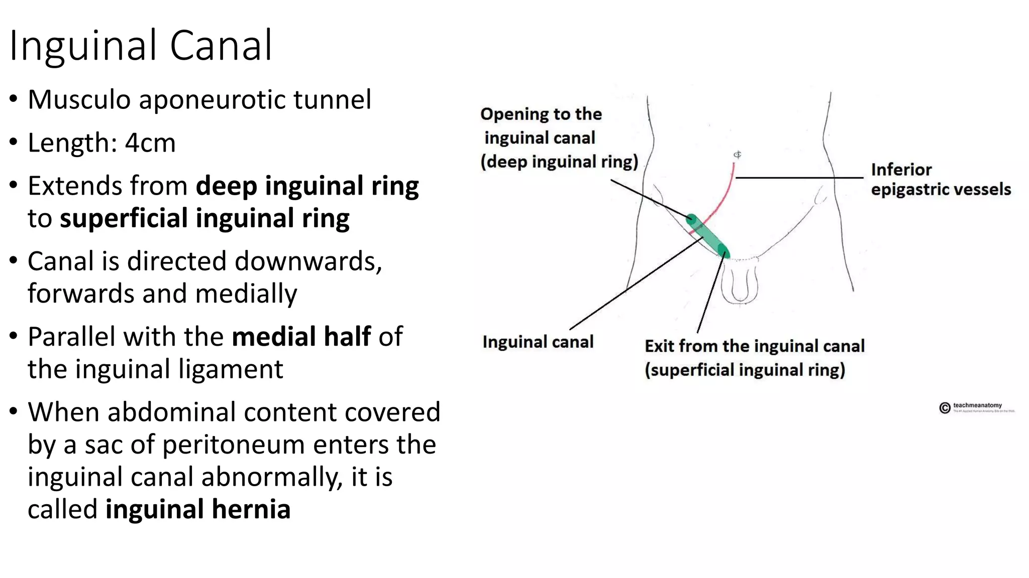
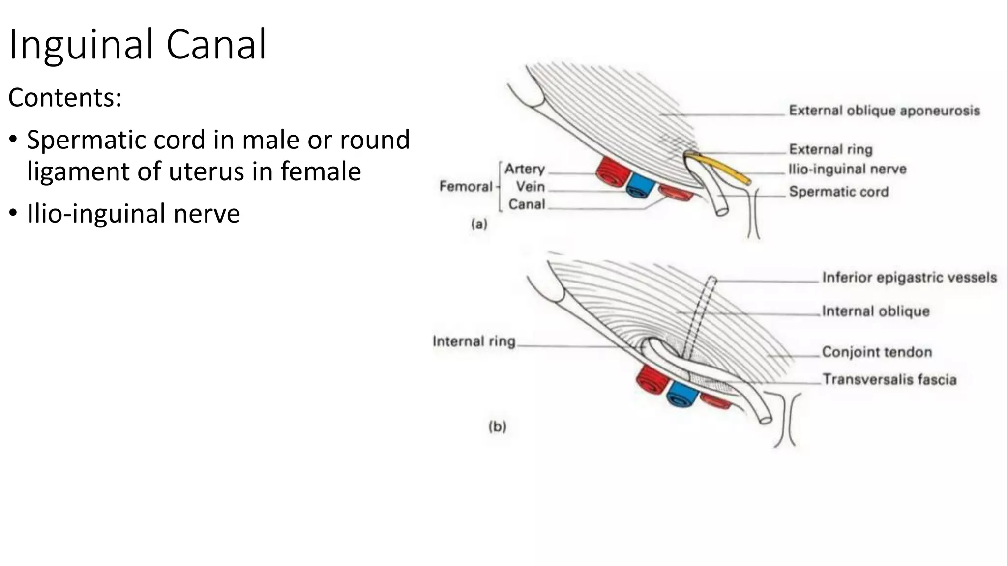
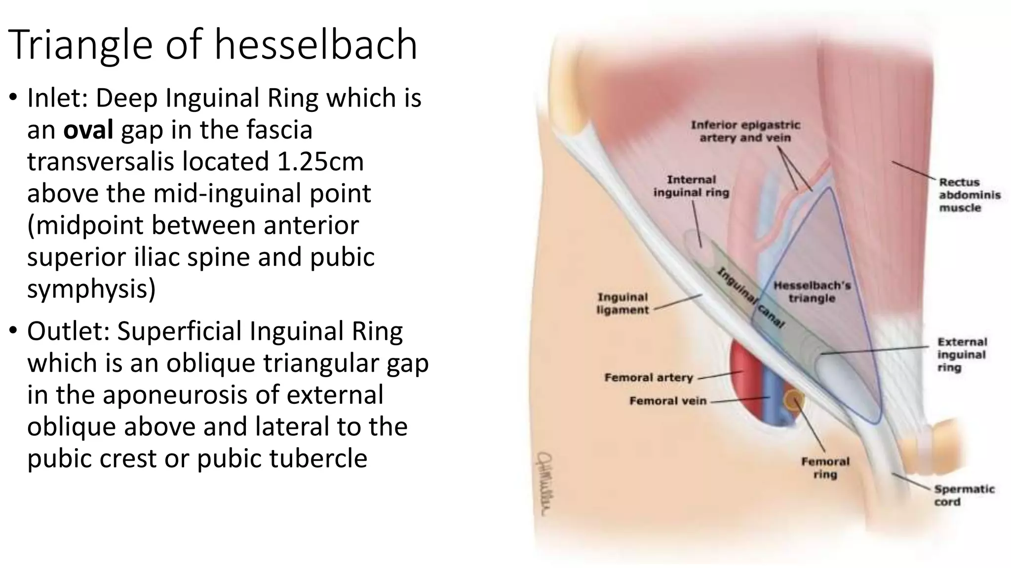
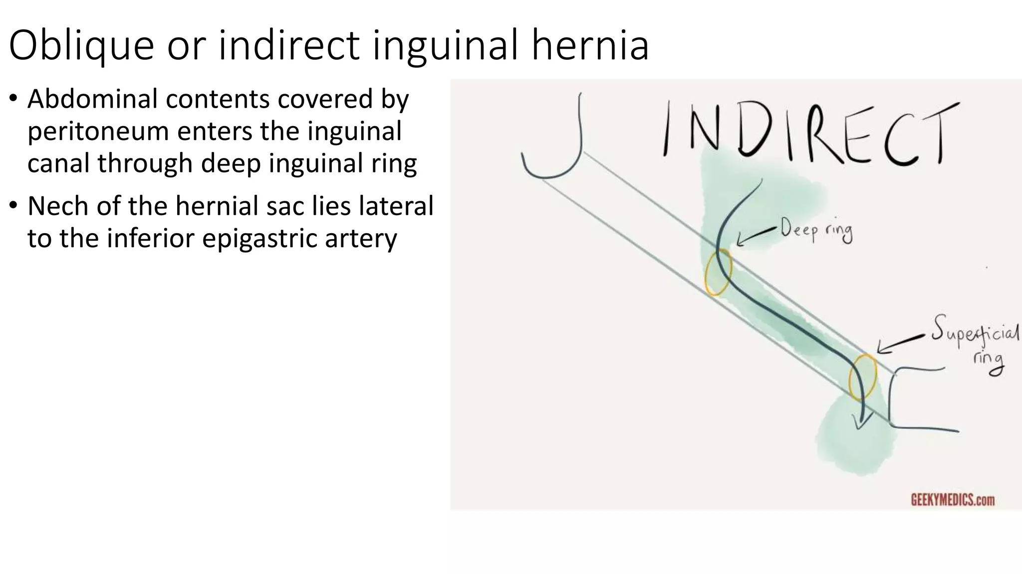
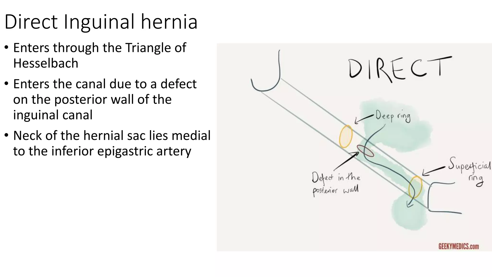
Posterior Abdominal Wall
Dr. Mushfiqul Hoque
Lecturer
Dhaka National Medical College
Posterior Abdominal Wall
• Osseo-musculo-fascial wall
• Extends from the last rib to the pelvic brim
• Consists of 3 parts:
1. Bony part
2. Muscular part
3. Fascial part
Bony Part
• In the midline
• Consists of bodies of the five lumbar
vertebrae
• Inner surface of the lower ribs
• Iliac fossa and alae of the sacrum
Muscular Part
Above iliac crest
• Psoas major
• Psoas minor (sometimes)
• Quadratus lumborum
• Transversus Abdominis
(aponeurotic origin)
Below iliac crest
• Iliacus
• Psoas major
Steps of dissection
Kidney from back
• A longitudinal incision in the middle
line from the eleventh thoracic spine
to the third lumbar spine
• Two transverse incisions, one from
each end of the 1st incision laterally
for about 3½ inches
Layers after skin incision
Above iliac crest
• Skin
• Superficial fascia
• Deep fascia (posterior layer of the thoracolumbar fascia)
• Sacrospinalis (erector spinae)
• Middle layer of the thoracolumbar fascia
• Quadratus lumborum
• Anterior layer of the thoracolumbar fascia
• Subcostal vessels and nerve, iliohypogastric and
ilioinguinal nerves
• Paranephric body (pararenal fat)
• Renal fascia (false capsule)
• Perinephric fat or perirenal fat (fatty capsule)
• True capsule—then kidney.
Thoraco-Lumbar Fascia
It covers the deep muscles of the
back of the trunk and consists of two
parts—
• Thoracic part: It is a thin
aponeurotic layer that covers the
sacrospinalis and it separates the
muscles of the back from the
muscles of the superior extremity.
• Lumbar part
Thoraco-Lumbar Fascia
Lumbar Part: It is thick and
membranous. It fills the gap
between the 12th rib and the iliac
crest and consists of three layers—
• Anterior
• Middle
• posterior
Thoraco-Lumbar Fascia
Posterior Layer:
• Very thick and strong and covers
the sacrospinalis (erector spinae)
muscle
Thoraco-Lumbar Fascia
Posterior Layer: (Attachments)
• Medially—It is attached to the tips
of the spinous processes of the
lumbar and sacral vertebrae and to
the supraspinous ligament.
Abdomen
• Laterally—It fuses with the middle
layer at the lateral border of the
sacrospinalis (erector spinae)
• Below—It is attached to the
posterior fourth of the outer lip of
the iliac crest
• Above—It is continuous with the
thoracic part of the fascia beneath
the serratus posterior inferior.
Thoraco-Lumbar Fascia
Middle Layer:
• Less thicker and covers the posterior
surface of the quadratus lumborum
Thoraco-Lumbar Fascia
Middle Layer: (Attachments)
• Medially—It is attached to the tips
of the transverse processes of the
lumbar vertebrae and to the
intertransverse ligaments
• Laterally—It blends with the
anterior and posterior layers
• Below—It is attached to the iliac
crest
• Above—It is attached to the lower
border of the 12th rib and to the
lumbocostal ligament.
Thoraco-Lumbar Fascia
Anterior Layer:
• Comparatively very thin and covers
the anterior surface of the
quadratus lumborum
Thoraco-Lumbar Fascia
Anterior Layer: (Attachments)
• Medially—It is attached to a vertical ridge on
the anterior surface of the transverse
processes of the lumbar vertebra and it
separates the quadratus lumborum from the
psoas major
• Laterally—It fuses with the middle layer at
the lateral border of the quadratus
lumborum and is then continuous with the
fascia transversalis
• Below—It is attached to the iliolumbar
ligament and to the adjoining part of the
inner lip of the iliac crest
• Above—It is thickened to form the lateral
arcuate ligament which is attached medially
to the tip of the transverse process of the 1st
lumbar vertebra and laterally to the free end
and lower border of the last rib.
Abdominal Aorta
• Continuation of Descending thoracic
aorta
• Passes downwards in front of the
vertebral column and on the left of
Inferior Vena Cava
• Ends by dividing into right and left
common iliac artery opposite to the
body of L4 vertebra
• Length: 10-11cm
• Breadth: 2cm
Inferior Vena Cava
• Drains blood from the parts of the
body below the diaphragm into the
right atrium of the heart
• Formed by the union of right and
left common iliac veins in front of
the body of L5 vertebra about 2.5
cm to the right of the midline
• Length: 20-23cm
• Breadth: 2.5cm
Tributaries of the Inferior Vena Cava
• A pair of Common Iliac Veins
• Lumbar Veins: third and forth
• A pair of Renal Veins
• A pair of Phrenic veins
• Right Gonadal veins
• Right supra-renal veins
• Hepatic veins
Lumbar Plexus
• Formed within the substance
of the psoas major muscle
• Formed by the union of
ventral rami of upper 3 lumbar
nerves and the larger upper
part of the ventral ramus of
fourth lumbar nerve
• The lower smaller part of the
4th lumbar nerve joins with
the 5th lumbar nerve to form
the lumbosacral trunk
Hernial Region
Inguinal Hernia
Hernia
• Definition: Protrusion of a viscus or a part of a viscus from its normal
position to an abnormal position through an abnormal opening on the
wall of its containing cavity due to weakness on the wall.
Inguinal Region
• Most common place for hernia in
male known as inguinal hernia
• Hernia in inguinal region are of two
varieties:
• Direct inguinal hernia
• Indirect or oblique inguinal hernia
Inguinal Canal
• Musculo aponeurotic tunnel
• Length: 4cm
• Extends from deep inguinal ring
to superficial inguinal ring
• Canal is directed downwards,
forwards and medially
• Parallel with the medial half of
the inguinal ligament
• When abdominal content covered
by a sac of peritoneum enters the
inguinal canal abnormally, it is
called inguinal hernia
Inguinal Canal
Contents:
• Spermatic cord in male or round
ligament of uterus in female
• Ilio-inguinal nerve
Inguinal Canal
Boundaries:
• Anterior wall: skin, superficial
fascia and aponeurosis of external
oblique muscle
• Posterior wall: Fascia Transversalis
• Roof: Arched fibres of the internal
oblique and transversus
abdominis muscles
• Floor: grooved upper surface of
Inguinal Ligament, on the medial
side floor is formed by upper
surface of lacunar ligament.
Inguinal Canal
• Inlet: Deep Inguinal Ring which is
an oval gap in the fascia
transversalis located 1.25cm
above the mid-inguinal point
(midpoint between anterior
superior iliac spine and pubic
symphysis)
• Outlet: Superficial Inguinal Ring
which is an oblique triangular gap
in the aponeurosis of external
oblique above and lateral to the
pubic crest or pubic tubercle
Triangle of hesselbach
• Inlet: Deep Inguinal Ring which is
an oval gap in the fascia
transversalis located 1.25cm
above the mid-inguinal point
(midpoint between anterior
superior iliac spine and pubic
symphysis)
• Outlet: Superficial Inguinal Ring
which is an oblique triangular gap
in the aponeurosis of external
oblique above and lateral to the
pubic crest or pubic tubercle
Oblique or indirect inguinal hernia
• Abdominal contents covered by
peritoneum enters the inguinal
canal through deep inguinal ring
• Nech of the hernial sac lies lateral
to the inferior epigastric artery
Direct Inguinal hernia
• Enters through the Triangle of
Hesselbach
• Enters the canal due to a defect
on the posterior wall of the
inguinal canal
• Neck of the hernial sac lies medial
to the inferior epigastric artery
End of class

Posterior abdominal wall and hernia

  • 1.
    Posterior Abdominal Wall Dr.Mushfiqul Hoque Lecturer Dhaka National Medical College
  • 2.
    Posterior Abdominal Wall •Osseo-musculo-fascial wall • Extends from the last rib to the pelvic brim • Consists of 3 parts: 1. Bony part 2. Muscular part 3. Fascial part
  • 3.
    Bony Part • Inthe midline • Consists of bodies of the five lumbar vertebrae • Inner surface of the lower ribs • Iliac fossa and alae of the sacrum
  • 4.
    Muscular Part Above iliaccrest • Psoas major • Psoas minor (sometimes) • Quadratus lumborum • Transversus Abdominis (aponeurotic origin) Below iliac crest • Iliacus • Psoas major
  • 5.
    Steps of dissection Kidneyfrom back • A longitudinal incision in the middle line from the eleventh thoracic spine to the third lumbar spine • Two transverse incisions, one from each end of the 1st incision laterally for about 3½ inches
  • 6.
    Layers after skinincision Above iliac crest • Skin • Superficial fascia • Deep fascia (posterior layer of the thoracolumbar fascia) • Sacrospinalis (erector spinae) • Middle layer of the thoracolumbar fascia • Quadratus lumborum • Anterior layer of the thoracolumbar fascia • Subcostal vessels and nerve, iliohypogastric and ilioinguinal nerves • Paranephric body (pararenal fat) • Renal fascia (false capsule) • Perinephric fat or perirenal fat (fatty capsule) • True capsule—then kidney.
  • 7.
    Thoraco-Lumbar Fascia It coversthe deep muscles of the back of the trunk and consists of two parts— • Thoracic part: It is a thin aponeurotic layer that covers the sacrospinalis and it separates the muscles of the back from the muscles of the superior extremity. • Lumbar part
  • 8.
    Thoraco-Lumbar Fascia Lumbar Part:It is thick and membranous. It fills the gap between the 12th rib and the iliac crest and consists of three layers— • Anterior • Middle • posterior
  • 9.
    Thoraco-Lumbar Fascia Posterior Layer: •Very thick and strong and covers the sacrospinalis (erector spinae) muscle
  • 10.
    Thoraco-Lumbar Fascia Posterior Layer:(Attachments) • Medially—It is attached to the tips of the spinous processes of the lumbar and sacral vertebrae and to the supraspinous ligament. Abdomen • Laterally—It fuses with the middle layer at the lateral border of the sacrospinalis (erector spinae) • Below—It is attached to the posterior fourth of the outer lip of the iliac crest • Above—It is continuous with the thoracic part of the fascia beneath the serratus posterior inferior.
  • 11.
    Thoraco-Lumbar Fascia Middle Layer: •Less thicker and covers the posterior surface of the quadratus lumborum
  • 12.
    Thoraco-Lumbar Fascia Middle Layer:(Attachments) • Medially—It is attached to the tips of the transverse processes of the lumbar vertebrae and to the intertransverse ligaments • Laterally—It blends with the anterior and posterior layers • Below—It is attached to the iliac crest • Above—It is attached to the lower border of the 12th rib and to the lumbocostal ligament.
  • 13.
    Thoraco-Lumbar Fascia Anterior Layer: •Comparatively very thin and covers the anterior surface of the quadratus lumborum
  • 14.
    Thoraco-Lumbar Fascia Anterior Layer:(Attachments) • Medially—It is attached to a vertical ridge on the anterior surface of the transverse processes of the lumbar vertebra and it separates the quadratus lumborum from the psoas major • Laterally—It fuses with the middle layer at the lateral border of the quadratus lumborum and is then continuous with the fascia transversalis • Below—It is attached to the iliolumbar ligament and to the adjoining part of the inner lip of the iliac crest • Above—It is thickened to form the lateral arcuate ligament which is attached medially to the tip of the transverse process of the 1st lumbar vertebra and laterally to the free end and lower border of the last rib.
  • 15.
    Abdominal Aorta • Continuationof Descending thoracic aorta • Passes downwards in front of the vertebral column and on the left of Inferior Vena Cava • Ends by dividing into right and left common iliac artery opposite to the body of L4 vertebra • Length: 10-11cm • Breadth: 2cm
  • 18.
    Inferior Vena Cava •Drains blood from the parts of the body below the diaphragm into the right atrium of the heart • Formed by the union of right and left common iliac veins in front of the body of L5 vertebra about 2.5 cm to the right of the midline • Length: 20-23cm • Breadth: 2.5cm
  • 19.
    Tributaries of theInferior Vena Cava • A pair of Common Iliac Veins • Lumbar Veins: third and forth • A pair of Renal Veins • A pair of Phrenic veins • Right Gonadal veins • Right supra-renal veins • Hepatic veins
  • 20.
    Lumbar Plexus • Formedwithin the substance of the psoas major muscle • Formed by the union of ventral rami of upper 3 lumbar nerves and the larger upper part of the ventral ramus of fourth lumbar nerve • The lower smaller part of the 4th lumbar nerve joins with the 5th lumbar nerve to form the lumbosacral trunk
  • 23.
  • 24.
    Hernia • Definition: Protrusionof a viscus or a part of a viscus from its normal position to an abnormal position through an abnormal opening on the wall of its containing cavity due to weakness on the wall.
  • 25.
    Inguinal Region • Mostcommon place for hernia in male known as inguinal hernia • Hernia in inguinal region are of two varieties: • Direct inguinal hernia • Indirect or oblique inguinal hernia
  • 26.
    Inguinal Canal • Musculoaponeurotic tunnel • Length: 4cm • Extends from deep inguinal ring to superficial inguinal ring • Canal is directed downwards, forwards and medially • Parallel with the medial half of the inguinal ligament • When abdominal content covered by a sac of peritoneum enters the inguinal canal abnormally, it is called inguinal hernia
  • 27.
    Inguinal Canal Contents: • Spermaticcord in male or round ligament of uterus in female • Ilio-inguinal nerve
  • 28.
    Inguinal Canal Boundaries: • Anteriorwall: skin, superficial fascia and aponeurosis of external oblique muscle • Posterior wall: Fascia Transversalis • Roof: Arched fibres of the internal oblique and transversus abdominis muscles • Floor: grooved upper surface of Inguinal Ligament, on the medial side floor is formed by upper surface of lacunar ligament.
  • 29.
    Inguinal Canal • Inlet:Deep Inguinal Ring which is an oval gap in the fascia transversalis located 1.25cm above the mid-inguinal point (midpoint between anterior superior iliac spine and pubic symphysis) • Outlet: Superficial Inguinal Ring which is an oblique triangular gap in the aponeurosis of external oblique above and lateral to the pubic crest or pubic tubercle
  • 30.
    Triangle of hesselbach •Inlet: Deep Inguinal Ring which is an oval gap in the fascia transversalis located 1.25cm above the mid-inguinal point (midpoint between anterior superior iliac spine and pubic symphysis) • Outlet: Superficial Inguinal Ring which is an oblique triangular gap in the aponeurosis of external oblique above and lateral to the pubic crest or pubic tubercle
  • 31.
    Oblique or indirectinguinal hernia • Abdominal contents covered by peritoneum enters the inguinal canal through deep inguinal ring • Nech of the hernial sac lies lateral to the inferior epigastric artery
  • 32.
    Direct Inguinal hernia •Enters through the Triangle of Hesselbach • Enters the canal due to a defect on the posterior wall of the inguinal canal • Neck of the hernial sac lies medial to the inferior epigastric artery
  • 34.