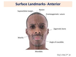
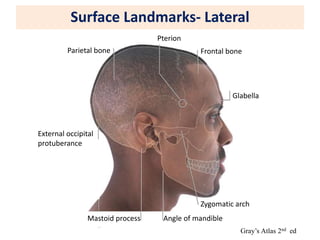
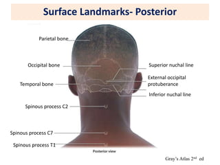
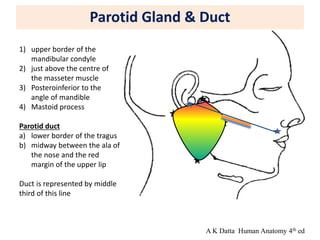
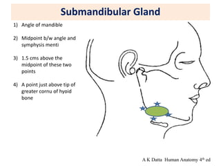
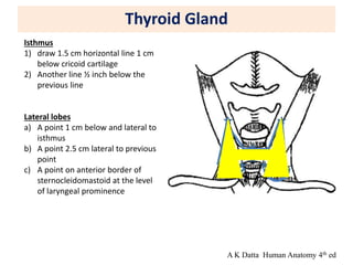
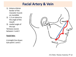
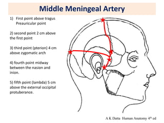
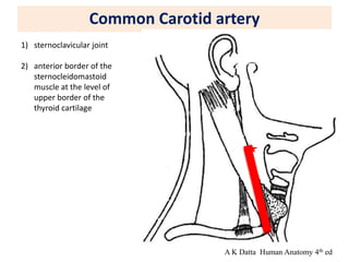
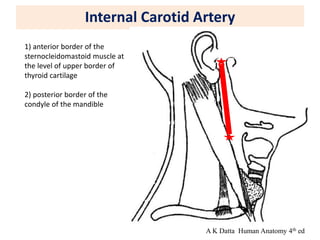
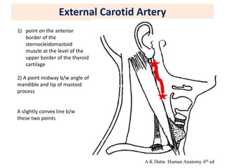
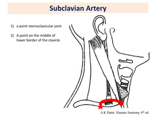
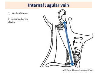
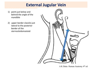
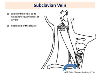
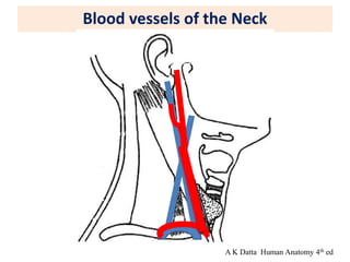
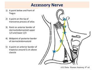
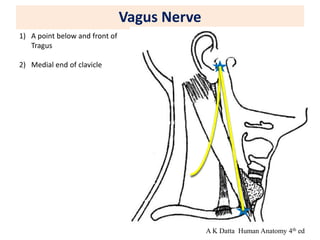
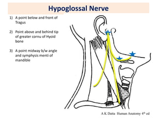
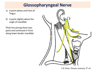
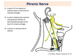
Surface Marking of
Head & Neck region
Dr Shivshanker S Patil
Assistant Professor
Department of Anatomy
ESIC MCH Hyderabad
Objectives
1) To study the bony and soft tissue landmarks in head and neck region
2) Surface marking of important structures in Head and neck region
Glands:-
Parotid gland, Submandibular gland, Parotid duct, Thyroid gland
Artery and Veins
Common carotid, External carotid, Internal carotid, Subclavian, Facial, Middle
meningeal artery
Internal Jugular, External jugular veins, Subclavian, Facial vein
Nerves
Cervical sympathetic, Vagus, Glossopharyngeal, Hypoglossal, Accessory, Phrenic
nerve
Surface Landmarks- Anterior
Gray’s Atlas 2nd ed
Frontozygomatic suture
Nasion
Supraorbital margin
Zygomatic bone
Mandible
Maxilla
Angle of mandible
Surface Landmarks- Lateral
Gray’s Atlas 2nd ed
Glabella
Frontal bone
Pterion
Parietal bone
External occipital
protuberance
Zygomatic arch
Angle of mandible
Mastoid process
Surface Landmarks- Posterior
Gray’s Atlas 2nd ed
Parietal bone
External occipital
protuberance
Superior nuchal line
Inferior nuchal line
Spinous process C2
Spinous process C7
Spinous process T1
Temporal bone
Occipital bone
Skull : Anatomical position
( Image: Vishram singh)
Frankfurt’s plane - Infraorbital margins and upper margins of external
acoustic meatuses lie in the same horizontal plane.
Orbital cavities are directed forwards, cranium superiorly, mandible inferiorly.
Parotid Gland & Duct
1) upper border of the
mandibular condyle
2) just above the centre of
the masseter muscle
3) Posteroinferior to the
angle of mandible
4) Mastoid process
Parotid duct
a) lower border of the tragus
b) midway between the ala of
the nose and the red
margin of the upper lip
Duct is represented by middle
third of this line
A K Datta Human Anatomy 4th ed
Submandibular Gland
1) Angle of mandible
2) Midpoint b/w angle and
symphysis menti
3) 1.5 cms above the
midpoint of these two
points
4) A point just above tip of
greater cornu of hyoid
bone
A K Datta Human Anatomy 4th ed
Thyroid Gland
Isthmus
1) draw 1.5 cm horizontal line 1 cm
below cricoid cartilage
2) Another line ½ inch below the
previous line
Lateral lobes
a) A point 1 cm below and lateral to
isthmus
b) A point 2.5 cm lateral to previous
point
c) A point on anterior border of
sternocleidomastoid at the level
of laryngeal prominence
A K Datta Human Anatomy 4th ed
Facial Artery & Vein
1) Antero-inferior
border of the
masseter muscle
on mandible
2) 1.2 cm lateral to
the angle of the
mouth
3) medial angle of
the eye
Tortuous course
between 1 and 2
Facial Vein:
A straight line drawn
b/w point 1 and 3
A K Datta Human Anatomy 4th ed
Middle Meningeal Artery
1) First point above tragus
Preauricular point
2) second point 2 cm above
the first point
3) third point (pterion) 4 cm
above zygomatic arch
4) fourth point midway
between the nasion and
inion.
5) fifth point (lambda) 5 cm
above the external occipital
protuberance.
A K Datta Human Anatomy 4th ed
Common Carotid artery
1) sternoclavicular joint
2) anterior border of the
sternocleidomastoid
muscle at the level of
upper border of the
thyroid cartilage
A K Datta Human Anatomy 4th ed
Internal Carotid Artery
1) anterior border of the
sternocleidomastoid muscle at
the level of upper border of
thyroid cartilage
2) posterior border of the
condyle of the mandible
A K Datta Human Anatomy 4th ed
External Carotid Artery
1) point on the anterior
border of the
sternocleidomastoid
muscle at the level of the
upper border of the thyroid
cartilage
2) A point midway b/w angle of
mandible and tip of mastoid
process
A slightly convex line b/w
these two points
A K Datta Human Anatomy 4th ed
Subclavian Artery
1) a point sternoclavicular joint
2) A point on the middle of
lower border of the clavicle
A K Datta Human Anatomy 4th ed
Internal Jugular vein
1) lobule of the ear
2) medial end of the
clavicle
A K Datta Human Anatomy 4th ed
External Jugular Vein
1) point just below and
behind the angle of the
mandible
2) upper border clavicle just
lateral to the posterior
border of the
sternocleidomastoid
A K Datta Human Anatomy 4th ed
Subclavian Vein
1) a point little medial to its
midpoint to lower border of
clavicle
2) medial end of the clavicle
A K Datta Human Anatomy 4th ed
Blood vessels of the Neck
A K Datta Human Anatomy 4th ed
Accessory Nerve
1) A point below and front of
Tragus
2) A point on the tip of
transverse process of atlas
3) Point on anterior border of
sternocleidomastoid upper
1/3 and lower 2/3
4) Midpoint of posterior border
of sternocleidomastoid
5) A point on anterior border of
trapezius around 5 cm above
clavicle
A K Datta Human Anatomy 4th ed
Vagus Nerve
1) A point below and front of
Tragus
2) Medial end of clavicle
A K Datta Human Anatomy 4th ed
Hypoglossal Nerve
1) A point below and front of
Tragus
2) Point above and behind tip
of greater cornu of Hyoid
bone
3) A point midway b/w angle
and symphysis menti of
mandible
A K Datta Human Anatomy 4th ed
Glossopharyngeal Nerve
1) A point below and front of
Tragus
2) A point slightly above the
angle of mandible
Thick line joining these two
point and continued in front
along lower border mandible
A K Datta Human Anatomy 4th ed
Phrenic Nerve
1) A point 3.5 cm lateral to
median plane at the level of
Thyroid cartilage
2) A point midway b/w anterior
and posterior border of
sternocleidomastoid at the
level of cricoid cartilage
3) A point on sternal end of
clavicle
A K Datta Human Anatomy 4th ed
Sympathetic trunk
1) a point posterior to condyle
of mandible
2) a point on the
sternoclavicular joint
Superior cervical ganglion
Spindle from transverse process
of atlas to level of greater cornu
of hyoid bone
Middle cervical ganglion
Small circle opposite the level
of cricoid cartilage
Inferior cervical ganglion
A small circle 3cm above the
sternoclavicular joint
A K Datta Human Anatomy 4th ed
Brachial plexus
1) a point midway b/w
anterior and posterior
border of
sternocleidomastoid at the
level of cricoid cartilage
2) A point at the junction of
clavicle and posterior
border of
sternocleidomastoid
3) A point just lateral to
midpoint of clavicle
A K Datta Human Anatomy 4th ed
Facial nerve
1) First point on the
anterior border of
mastoid process
2) A point behind the neck
of mandible
A K Datta Human Anatomy 4th ed
Surface marking Head Neck.ppt

Surface marking Head Neck.ppt

  • 1.
    Surface Marking of Head& Neck region Dr Shivshanker S Patil Assistant Professor Department of Anatomy ESIC MCH Hyderabad
  • 2.
    Objectives 1) To studythe bony and soft tissue landmarks in head and neck region 2) Surface marking of important structures in Head and neck region Glands:- Parotid gland, Submandibular gland, Parotid duct, Thyroid gland Artery and Veins Common carotid, External carotid, Internal carotid, Subclavian, Facial, Middle meningeal artery Internal Jugular, External jugular veins, Subclavian, Facial vein Nerves Cervical sympathetic, Vagus, Glossopharyngeal, Hypoglossal, Accessory, Phrenic nerve
  • 3.
    Surface Landmarks- Anterior Gray’sAtlas 2nd ed Frontozygomatic suture Nasion Supraorbital margin Zygomatic bone Mandible Maxilla Angle of mandible
  • 4.
    Surface Landmarks- Lateral Gray’sAtlas 2nd ed Glabella Frontal bone Pterion Parietal bone External occipital protuberance Zygomatic arch Angle of mandible Mastoid process
  • 5.
    Surface Landmarks- Posterior Gray’sAtlas 2nd ed Parietal bone External occipital protuberance Superior nuchal line Inferior nuchal line Spinous process C2 Spinous process C7 Spinous process T1 Temporal bone Occipital bone
  • 6.
    Skull : Anatomicalposition ( Image: Vishram singh) Frankfurt’s plane - Infraorbital margins and upper margins of external acoustic meatuses lie in the same horizontal plane. Orbital cavities are directed forwards, cranium superiorly, mandible inferiorly.
  • 7.
    Parotid Gland &Duct 1) upper border of the mandibular condyle 2) just above the centre of the masseter muscle 3) Posteroinferior to the angle of mandible 4) Mastoid process Parotid duct a) lower border of the tragus b) midway between the ala of the nose and the red margin of the upper lip Duct is represented by middle third of this line A K Datta Human Anatomy 4th ed
  • 8.
    Submandibular Gland 1) Angleof mandible 2) Midpoint b/w angle and symphysis menti 3) 1.5 cms above the midpoint of these two points 4) A point just above tip of greater cornu of hyoid bone A K Datta Human Anatomy 4th ed
  • 9.
    Thyroid Gland Isthmus 1) draw1.5 cm horizontal line 1 cm below cricoid cartilage 2) Another line ½ inch below the previous line Lateral lobes a) A point 1 cm below and lateral to isthmus b) A point 2.5 cm lateral to previous point c) A point on anterior border of sternocleidomastoid at the level of laryngeal prominence A K Datta Human Anatomy 4th ed
  • 10.
    Facial Artery &Vein 1) Antero-inferior border of the masseter muscle on mandible 2) 1.2 cm lateral to the angle of the mouth 3) medial angle of the eye Tortuous course between 1 and 2 Facial Vein: A straight line drawn b/w point 1 and 3 A K Datta Human Anatomy 4th ed
  • 11.
    Middle Meningeal Artery 1)First point above tragus Preauricular point 2) second point 2 cm above the first point 3) third point (pterion) 4 cm above zygomatic arch 4) fourth point midway between the nasion and inion. 5) fifth point (lambda) 5 cm above the external occipital protuberance. A K Datta Human Anatomy 4th ed
  • 12.
    Common Carotid artery 1)sternoclavicular joint 2) anterior border of the sternocleidomastoid muscle at the level of upper border of the thyroid cartilage A K Datta Human Anatomy 4th ed
  • 13.
    Internal Carotid Artery 1)anterior border of the sternocleidomastoid muscle at the level of upper border of thyroid cartilage 2) posterior border of the condyle of the mandible A K Datta Human Anatomy 4th ed
  • 14.
    External Carotid Artery 1)point on the anterior border of the sternocleidomastoid muscle at the level of the upper border of the thyroid cartilage 2) A point midway b/w angle of mandible and tip of mastoid process A slightly convex line b/w these two points A K Datta Human Anatomy 4th ed
  • 15.
    Subclavian Artery 1) apoint sternoclavicular joint 2) A point on the middle of lower border of the clavicle A K Datta Human Anatomy 4th ed
  • 16.
    Internal Jugular vein 1)lobule of the ear 2) medial end of the clavicle A K Datta Human Anatomy 4th ed
  • 17.
    External Jugular Vein 1)point just below and behind the angle of the mandible 2) upper border clavicle just lateral to the posterior border of the sternocleidomastoid A K Datta Human Anatomy 4th ed
  • 18.
    Subclavian Vein 1) apoint little medial to its midpoint to lower border of clavicle 2) medial end of the clavicle A K Datta Human Anatomy 4th ed
  • 19.
    Blood vessels ofthe Neck A K Datta Human Anatomy 4th ed
  • 20.
    Accessory Nerve 1) Apoint below and front of Tragus 2) A point on the tip of transverse process of atlas 3) Point on anterior border of sternocleidomastoid upper 1/3 and lower 2/3 4) Midpoint of posterior border of sternocleidomastoid 5) A point on anterior border of trapezius around 5 cm above clavicle A K Datta Human Anatomy 4th ed
  • 21.
    Vagus Nerve 1) Apoint below and front of Tragus 2) Medial end of clavicle A K Datta Human Anatomy 4th ed
  • 22.
    Hypoglossal Nerve 1) Apoint below and front of Tragus 2) Point above and behind tip of greater cornu of Hyoid bone 3) A point midway b/w angle and symphysis menti of mandible A K Datta Human Anatomy 4th ed
  • 23.
    Glossopharyngeal Nerve 1) Apoint below and front of Tragus 2) A point slightly above the angle of mandible Thick line joining these two point and continued in front along lower border mandible A K Datta Human Anatomy 4th ed
  • 24.
    Phrenic Nerve 1) Apoint 3.5 cm lateral to median plane at the level of Thyroid cartilage 2) A point midway b/w anterior and posterior border of sternocleidomastoid at the level of cricoid cartilage 3) A point on sternal end of clavicle A K Datta Human Anatomy 4th ed
  • 25.
    Sympathetic trunk 1) apoint posterior to condyle of mandible 2) a point on the sternoclavicular joint Superior cervical ganglion Spindle from transverse process of atlas to level of greater cornu of hyoid bone Middle cervical ganglion Small circle opposite the level of cricoid cartilage Inferior cervical ganglion A small circle 3cm above the sternoclavicular joint A K Datta Human Anatomy 4th ed
  • 26.
    Brachial plexus 1) apoint midway b/w anterior and posterior border of sternocleidomastoid at the level of cricoid cartilage 2) A point at the junction of clavicle and posterior border of sternocleidomastoid 3) A point just lateral to midpoint of clavicle A K Datta Human Anatomy 4th ed
  • 27.
    Facial nerve 1) Firstpoint on the anterior border of mastoid process 2) A point behind the neck of mandible A K Datta Human Anatomy 4th ed