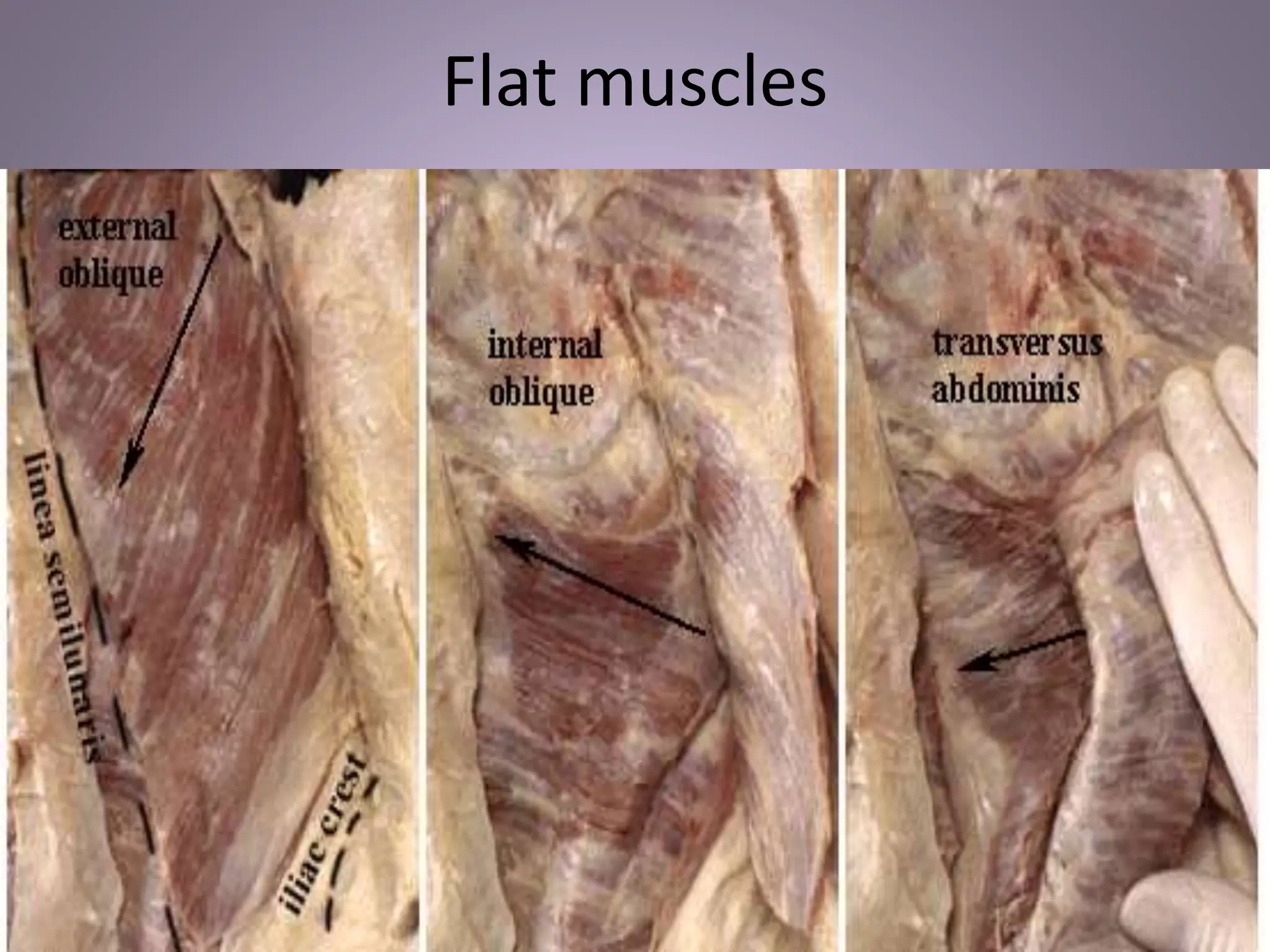
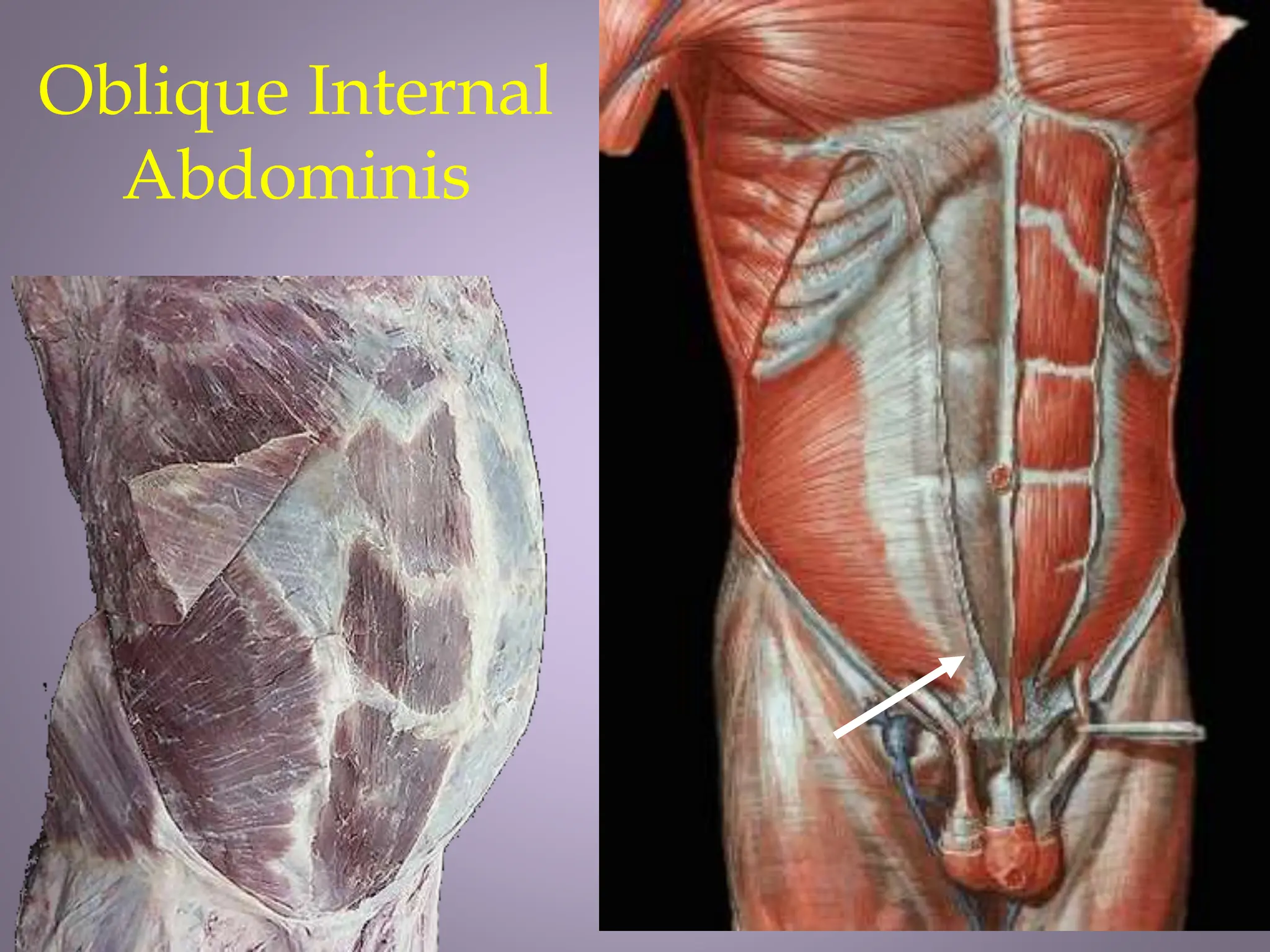
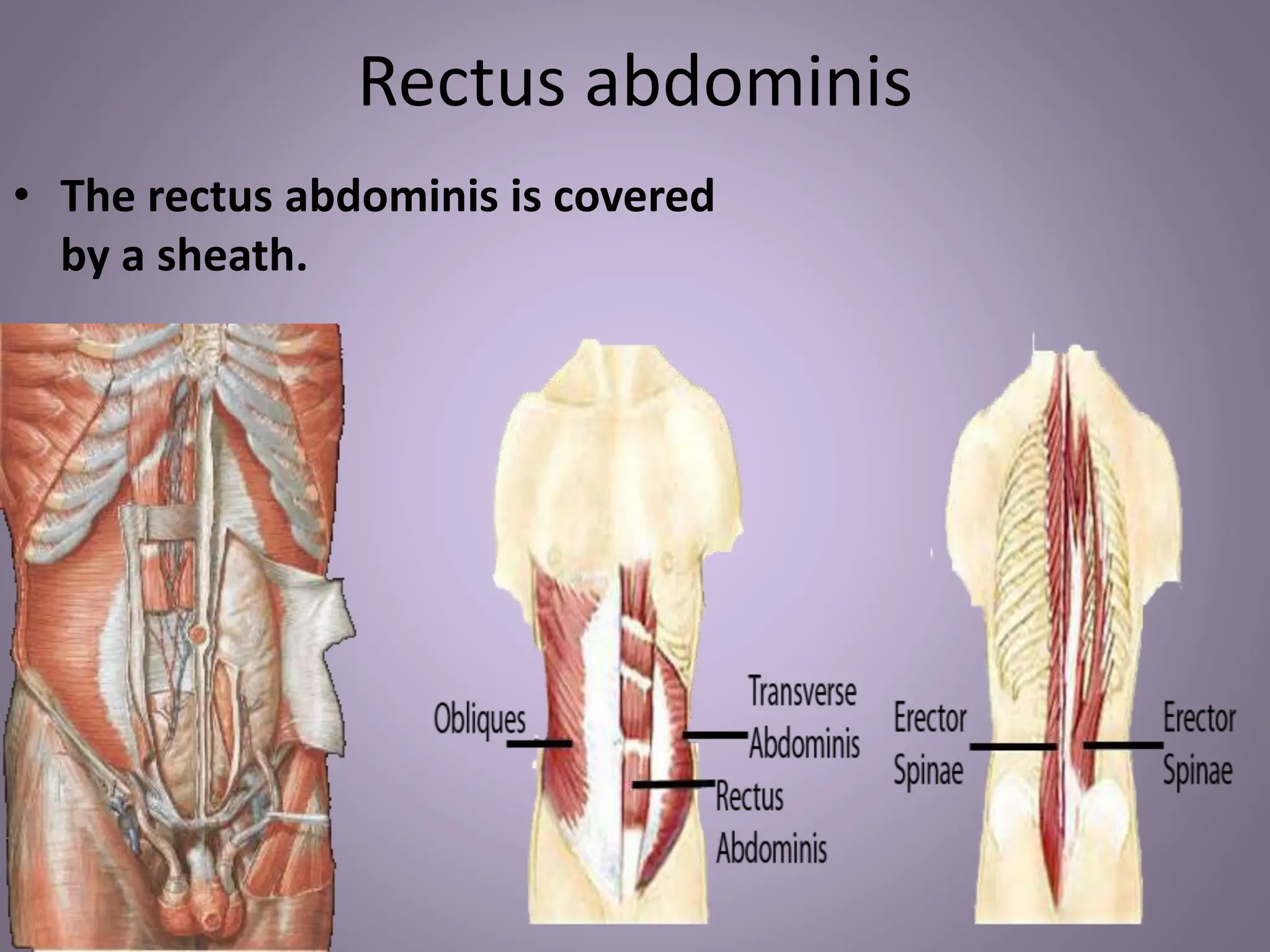
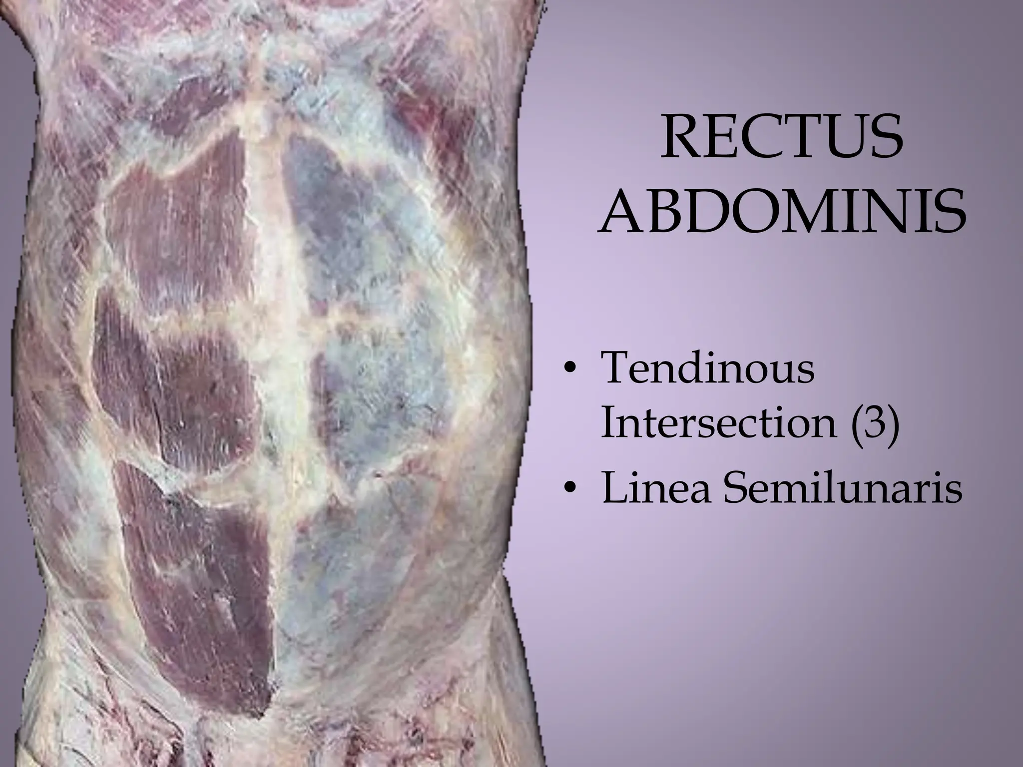
ANATOMY OF
ANTERIOR ABDOMEN
AND PERITONEUM
Organization of the antero-lateral
abdominal wall
BOUNDARIES
Superiorly:
• Xiphoid process.(Xiphisternum)
• Costal cartilages of the 7th and 10th
ribs.
Inferiorly:
• Iliac crest.
• Anterior superior iliac spine.
• Inguinal ligament.
• Pubic tubercle, pubic crest and
pubic symphysis.
Divided into 4 quadrants i.e
1. Right and left upper quadrants
2. Right and left lower quadrants
• Transpyloric plane(L1, L2)
• Transtubercular plane
• Mid-clavicular/ mid-inguinal
line
Abdominal Regions
Divisions of the abdomen
Can be Divided into 9 regions.
• Two vertical planes- midclavicular
• Two horizontal planes:
– Subcostal plane – joining the most inferior
points of the costal margins, and passing at
L3.
– Transtubercular plane-joining the
tubercles of the iliac crest.
Note the 9 regions.
• Right and left hypochondrial regions (1and 3)
• Middle epigastric region (2)
• Right and left lumbar regions (6 and 4)
• A middle umbilical region (5)
• Right and left iliac (inguinal) region (7and 9)
• A middle hypogastric / suprapubic region (8)
Abdominal wall
Anterolateral
abdominal wall
Posterior
abdominal wall
Layers of the abdominal wall through L1
1. skin
2. superficial fascia:
fatty and
menbaraneous
3. deep fascia
4. muscle
5. Extra peritoneal
tissue-
endoabdominal
fascia
6. peritoneum
LAYERS OF ANTERIOR ABDOMINAL WALL
1. Skin
Shows ‘creases' which represent the lines of orientation of collagen
fibres in the dermis- Langer's lines.
These lines are surgically important – incisions along them heal better
leaving a thin scar; while those across them leave big scars.
In pregnant women, obese people and those with abdominal distention
from whatever cause, there are dark elongate lines called striae
gravidara.
The skin is very sensitive to touch, and quickly when touched, the
muscles contract.
2.Superficial fascia
Consists of two layers below umbilicus;
a. Fatty layer (Camper's fascia) containing variable amounts
of fat, more in females and in the lower abdomen.
b. Membranous layer (Scarpa's fascia).
(+) Contains fibrous tissue and very little fat.
(+) Fuses with fascia lata below inguinal ligament)
(+) Continuous with the superficial perineal fascia
(Colle's fascia) and with that investing the scrotum and
penis.
- boundaries, Holdensline, Fournier’s gangrene
3. Deep fascia
Deep fascia is thin and invests the muscles/ bone
4. The muscles
There are 4 main muscles
to note:
– External oblique
– Internal oblique
– Transversus abdominis
– Rectus abdominis
• All muscles are derived
from the hypomere
MUSCLES
Anterior Group Lateral Group
•Rectus Abdominis
•Pyramidalis
•External Oblique
•Internal Oblique
•Transversus
Linea Alba
• Fibrous band that extends from symphysis
pubis to the xiphoid process and lies in the
midline.
• Fusion of the aponeuroses of the muscles of
the anterior abdominal wall and is
represented on the surface by a slight median
groove
LINEA ALBA
Linea Semilunaris
• Lateral edge of the rectus abdominis muscle,
crosses the costal margin at the tip of the
ninth costal cartilage
• To accentuate, the patient is asked to lie on his
back and raise his shoulders off the couch
without using his arms
Flat muscles
External Oblique
• Origin – lower eight ribs
• Insertion – xiphoid process, linea alba, pubic crest, iliac
crest, the pubic tubercle and the anterior superior iliac
spine as the inguinal ligament
– fibers run anterior and inferior (your hands in outside
pockets).
• Nerve supply – lower 6 thoracic nerves
• Action – supports abdominal contents, assists in
forced expiration, micturition, defecation,
partuition, vomiting
Oblique Internal
Abdominis
Internal Oblique
• Origin – thoracolumbar fascia, iliac crest, lateral two
thirds of the inguinal ligament
• Insertion – Lower three ribs and costal cartilages,
xiphoid process, linea alba, symphysis pubis, pecten
pubis via the conjoint tendon
– fibers run at right angles to the external oblique (hands
in inside pockets),
• Nerve supply – lower six thoracic nerves
• Action – same with external obliques
1. Inguinal ligament
2. Muscular part of transversus abdominis
3. Transversus abdominis aponeurosis
4. Muscular part of internal oblique
5. Internal oblique aponeurosis
6. Transversalis fascia
7. Cremasteric fascia forming middle coating of spermatic cord
8. Pubic tubercle
Transversus
Abdominis
Transversus Abdominis
• Origin – lower six costal cartilages, thoracolumbar
fascia, iliac crest, lateral third of inguinal ligament
• Insertion – xiphoid process, linea alba, symphysis
pubis,pecten pubis via the conjoint tendon
– fibers run transversely
• Nerve supply – lower six thoracic nerves
• Action – compresses abdominal contents
Rectus abdominis
• The rectus abdominis is covered
by a sheath.
RECTUS
ABDOMINIS
• Tendinous
Intersection (3)
• Linea Semilunaris
Rectus Abdominis
• Origin – symphysis pubis and pubic crest
• Insertion – fifth, sixth and seventh costal cartilages
and xiphoid process
• Nerve supply – lower six thoracic nerves
• Action – compresses abdominal contents and flexes
vertebral column
Pyramidalis
• Origin – anterior surface of pubis
• Insertion – Linea alba
• Nerve supply – Twelfth thoracic nerve
• Action – tenses the linea alba
• It is often absent in approximately 20% of people
• Surgeons use the attachment of the pyramidalis to
the linea alba as a landmark for an accurate median
abdominal incision
PYRAMIDALIS
Functions of the muscles
• Support and protection for abdominal viscera
• Movement of the trunk – flexion, extension, twisting
and lateral bending.
• Maintenance of posture
• Increase intra abdominal pressure in functions such as
defecation, micturition and parturition etc.
THE RECTUS SHEATH
Location
Fibrous compartment for rectus abdominis muscle in the paramedian abdominal wall.s
Formation
Formed of the aponeurosis of abdominal muscles.
Proximal 1/3rd
The anterior layer joins the aponeurosis of the external oblique to form the anterior wall of the
rectus sheath.
The posterior layer joins with the aponeurosis of the transversus abdominis to form the
posterior wall of the rectus sheath.
Middle 1/3 rd
Aponeurosis of internal oblique joins external oblique aponeurosis to form anterior wall.
Posterior wall is formed by aponeurosis of transversus abdominis muscle
Distal 1/3 rd
Mid way between umbilicus and pubic crest all three aponeurosis form the anterior layer
The posterior layer is formed only by fascia transversalis
Rectus sheath
• Anterior layer- derived from the External oblique
abdominis and anterior layer of the Internal Oblique
Abdominis
• Posterior layer- formed from the aponeurosis of the
Internal Oblique Abdominis and Transverse Abdominis.
• Below the arcuate line, all three flat muscles contribute
to the anterior portion of the rectus sheath and there is
no posterior component.
• At and below the arcuate line, Rectus abdominis
muscle lies entirely on the transversalis fascia.
• The rectus sheath is connected by tendinous
intersections representing embryologic segmentation.
• Antagonist- erector spinae complex
• Strong extensors of the vertebral column.
Note:
1. The anterior and posterior
layers fuse in the midline to
form the linear alba, a
fibrous intersection
extending from the xiphoid
process to the pubic
symphysis.
2. The inferior ¼ of the rectus
sheath is deficient
posteriorly. The limit of the
posterior wall is marked by
the arcuate line
The superior ¾ , posterior
wallis covered by
aponeurotic sheath
3. The lateral margin of rectus
sheath is called linea
semilunaris
Rectus sheath
• Above the arcuate line the rectus
sheath has two layers
– an anterior layer
– a posterior layer
Rectus
Sheath
Arcuate
line
Arteries
• 5 intercostal arteries
• subcostal arteries
• 4 lumbar arteries
• Superior epigastric artery—
internal thoracic artery
• Inferior epigastric artery -
external iliac artery
• Deep iliac circumflex artery-
external iliac artery
Contents of Rectus Sheath
• Rectus abdominis muscle
• Inferior and superior epigastric vessels
• Terminal parts of the lower five intercostal
nerves, and the Subcostal nerve.
• Fibro fatty connective tissue
• Occasionally lymph node(s)
Extra peritoneal fascia
• Transparent ‘membrane' which lines the inside of
the abdominal wall,lying between the parietal
peritoneum and the transversalis fascia
• Its parts are named according to what it lines e.g.
(+) diaphragmatic fascia;
(+) iliac fascia;
(+) Psoas fascia.
(+) fascia transversalis ( part covering the muscle
transversus abdominis ).
Extraperitoneal
Fascia
Blood supply to the anterior abdominal
wall
ARTERIES
• Inferior epigastric : External iliac
• Superficial circumflex iliac : Femoral arterty
• Deep circumflex iliac : femoral artery
• Superior epigastric : internal thoracic
• Lower intercostal : Abdominal Aorta
• Subcostal arteries : Abdominal aorta
VEINS
• The veins correspond to the arteries, but:
– Inferior epigastric vein anastomoses with lateral thoracic vein.
– Superficial epigastric vein anastomoses with lateral thoracic vein.
• These two unite the veins of the upper and lower halves of the body (of
the azygous system).
SUPERFICIAL
ARTERIES
• Lateral
– Posterior intercostal a.
– Subcostal a.
– Lumbar a.
• Median
– Epigastric a.
– hypogastric a.
• Inferior
– Superficial epigastric a.
– Superficial iliac a.
Arteries
• 5 intercostal arteries
• subcostal arteries
• 4 lumbar arteries
• Superior epigastric
artery—internal thoracic
artery
• Inferior epigastric artery -
external iliac artery
• Deep iliac circumflex
artery- external iliac artery
Arteries of the Anterior and Lateral
Abdominal Walls
• Superior epigastric artery – terminal branch of
the internal thoracic artery; supplies the
upper central part of the anterior abdominal
wall
• Inferior epigastric artery – branch of the
external iliac artery, just above the inguinal
ligament; supplies the lower central part of
the anterior abdominal wall
• Deep circumflex iliac artery – branch of the
external iliac artery; supplies the lower lateral
part of the abdominal wall.
• Posterior Intercostal arteries – 2, branches of
the descending thoracic aorta; supply the
lateral part of the abdominal wall.
Superficial veins
subclavian
femoral
paraumbilical
S epigastric
S circumflex iliac
thoracoepigastric
lateral thoracic
portal
Lymphatic Drainage
Anterior →
Intercostal Lymphatic Nodes
Parasternal Lymphatic Nodes
Middle → Lumbar Lymphatic Nodes
Lower → External Iliac Lymphatic Nodes
Lymphatic Drainage
• Superior to the umbilical level:
– Axillary nodes
– Parasternal nodes
• Inferior to the umbilical level:
– Superficial Inguinal lymph nodes
– Deep inguinal nodes
– External iliac nodes
– Lumbar nodes
Innervation
• Ventral rami of lower 6 thoracic nerves.
• T7-T9 supply region above umbilicus
• T11-L1 below umbilicus
• T10 skin around umbilicus
• The lower intercostals and subcostal
• Segmental with T10 Para umbilical.
Innervation
 T7-12 thoracic n.
 Iliohypogastric n.
 Ilioinguinal n.
 Genitofemoral n.
Innervation
• Intercostal n.
– Anterior
cutaneous branch
– Lateral cutaneous
branch
The internal surface of anterior abdominal
wall
• In the midline, there are elevations of peritoneum with free edges,
called folds.
• Superior to the umbilicus:
– A median fold, the falciform ligament. This contains the ligamentum teres,
the obliterated umbilical vein. Note that the umbilical vein is patent for
sometime after birth and may be used for exchange transfusion.
• Inferior to the umbilicus, there are 5 folds:
– The median umbilical fold is due to median umbilical ligament, the
remnant of the urachus, which develops from the allantois. It attaches to
the urinary bladder.
– 2 medial umbilical folds formed by medial umbilical ligaments – the
obliterated umbilical arteries.
– 2 lateral umbilical folds – formed by the inferior epigastric vessels
Abdominal wall
Dr.G.Bhanu Prakash
www.gims-org.com
Inguinal Ligament
• Rolled-under inferior margin of
the external oblique muscle
• Attached laterally to the ASIS,
curves downward and medially to
be attached to the pubic tubercle
Superficial Inguinal Ring
• Triangular aperture in the aponeurosis of the
external oblique muscle situated above and
medial to the pubic tubercle
• In males – the margins can be felt by
invaginating the skin of the upper part of the
scrotum with the tip of the little finger
• In females – smaller and difficult to palpate
Cremaster
Conjoint Tendon
Conjoint Tendon
Cremaster
Transverse Abdominal Fascia
abdominal inguinal ring
(deep inguinal ring)
Superficial Inguinal Ring
Medial Crus
Lateral Crus
Intercrural Fibers
Reflected Ligament
The inguinal canal
• Canal represents path taken by testis out of the
abdomen.
• BOUNDARIES
• Floor: Inguinal ligament and lacunar ligament
Roof: Arching fibres of internal oblique and
transversus abdominis.
• Antero lateral: Aponeurosis of external oblique
Posterior: Fascia transversalis laterally and
conjoint tendon medially (of transversus and
internal oblique abdominal muscles)
• CONTENTS
• Spermatic cord in male
• Round ligament of uterus in female and its artery
• Ilioinguinal nerve
It may contain:
• Iliohypogastric nerve
• Genitofemoral nerve (genital branch)
• Subcostal (in the upper part)
• Inguinal Rings
Testis
Ductus deferens
Pampiniform plexus
Testicular artery
Cremasteric artery
Spermatic cord
Aponeurosis of internal
oblique
External oblique
Inguinal Rings
• Superficial ring is a triangular
aperture in the aponeurosis of
the external oblique muscle.
(+) Base: pubic crest
(+) Sides: crura of the external
oblique aponeurosis.
• The deep ring is a deficit in
transversalis fascia, lateral to
inferior epigastric artery.
•
Inguinal triangle
Bounded by:
• Medially: Linear semilunaris (lateral edge of
Rectus abdomis)
• Inferiorly: Inguinal ligament
• Supero-laterally: Inferior epigastric artery
Boundaries
Inguinal Triangle
(Hesselbach's triangle )
Direct Hernia
Spermatic Cord
• Comprises of structures running to and from the
testis, surrounded by structures derived from the
anterior abdominal wall. Traverses entire inguinal
canal
Coverings
• Internal spermatic fascia - from transversalis fascia
• Cremasteric fascia - from fascia of internal oblique
• Cremaster muscle - from internal oblique abdomen
• External spermatic fascia - from external oblique
Contents
1. Autonomic nerve plexus
2. Testicular artery; artery to ductus deferens,
cremasteric artery
3. Pampiniform plexus of veins
4. Ductus deferens
• Lymph vessels
• Remnants of processus vaginalis
Cremasteric reflex
• Genital branch of genitofemoral nerve supply
cremasteric muscle.
• Stroking the area of supply of the femoral branch
(superomedial thigh) stimulates the muscle, pulling up
the testis – this is the cremasteric reflex.
• Easy to demonstrate in children, but gets weaker with
age.
Incisions
PARAMEDIAN
• Usually, the rectus abdominis is retracted
laterally, to avoid detaching its nerve and vessels.
It is unwise to cut the muscle longitudinally.
• These incisions, do not meet many blood vessels.
The epigastric vessels (inferior and superior) are
usually easy to identify.
HORIZONTAL INCISION
• Especially in the lower abdomen (Pfanesteil) may
encounter the inferior and superficial epigastric
vessels. If these can be guarded, handled, the
incision heals rapidly, leaving thin scars.
GRIDIRON INCISION
• It is mentioned under appendix.
Review Questions
1. Describe the muscles, blood supply, lymphatic drainage and sensory innervation of the
anterior abdominal wall
2. Discuss the formation and contents of the inguinal canal. Add notes on the distinction
between direct and indirect inguinal hernias. List six structures that must be safeguarded
during hernial repair
3. Describe the formation and contents of the rectus sheath
4. Outline the general organization of the superficial fascia of the anterior abdominal wall,
and the perineum. Add clinical notes on the implication of this organization
5. Describe in detail the pattern and clinical significance of the blood supply of the anterior
abdominal wall.
6. State the advantages and disadvantages of the various incisions in the anterior abdominal
wall.
Abdominal wall herniae

lecture 2.pptx on abdomen internal external

  • 1.
  • 3.
    Organization of theantero-lateral abdominal wall BOUNDARIES Superiorly: • Xiphoid process.(Xiphisternum) • Costal cartilages of the 7th and 10th ribs. Inferiorly: • Iliac crest. • Anterior superior iliac spine. • Inguinal ligament. • Pubic tubercle, pubic crest and pubic symphysis. Divided into 4 quadrants i.e 1. Right and left upper quadrants 2. Right and left lower quadrants
  • 4.
    • Transpyloric plane(L1,L2) • Transtubercular plane • Mid-clavicular/ mid-inguinal line
  • 5.
  • 9.
    Divisions of theabdomen Can be Divided into 9 regions. • Two vertical planes- midclavicular • Two horizontal planes: – Subcostal plane – joining the most inferior points of the costal margins, and passing at L3. – Transtubercular plane-joining the tubercles of the iliac crest. Note the 9 regions. • Right and left hypochondrial regions (1and 3) • Middle epigastric region (2) • Right and left lumbar regions (6 and 4) • A middle umbilical region (5) • Right and left iliac (inguinal) region (7and 9) • A middle hypogastric / suprapubic region (8)
  • 11.
  • 12.
    Layers of theabdominal wall through L1 1. skin 2. superficial fascia: fatty and menbaraneous 3. deep fascia 4. muscle 5. Extra peritoneal tissue- endoabdominal fascia 6. peritoneum
  • 13.
    LAYERS OF ANTERIORABDOMINAL WALL 1. Skin Shows ‘creases' which represent the lines of orientation of collagen fibres in the dermis- Langer's lines. These lines are surgically important – incisions along them heal better leaving a thin scar; while those across them leave big scars. In pregnant women, obese people and those with abdominal distention from whatever cause, there are dark elongate lines called striae gravidara. The skin is very sensitive to touch, and quickly when touched, the muscles contract.
  • 14.
    2.Superficial fascia Consists oftwo layers below umbilicus; a. Fatty layer (Camper's fascia) containing variable amounts of fat, more in females and in the lower abdomen. b. Membranous layer (Scarpa's fascia). (+) Contains fibrous tissue and very little fat. (+) Fuses with fascia lata below inguinal ligament) (+) Continuous with the superficial perineal fascia (Colle's fascia) and with that investing the scrotum and penis. - boundaries, Holdensline, Fournier’s gangrene 3. Deep fascia Deep fascia is thin and invests the muscles/ bone
  • 16.
    4. The muscles Thereare 4 main muscles to note: – External oblique – Internal oblique – Transversus abdominis – Rectus abdominis • All muscles are derived from the hypomere
  • 17.
    MUSCLES Anterior Group LateralGroup •Rectus Abdominis •Pyramidalis •External Oblique •Internal Oblique •Transversus
  • 18.
    Linea Alba • Fibrousband that extends from symphysis pubis to the xiphoid process and lies in the midline. • Fusion of the aponeuroses of the muscles of the anterior abdominal wall and is represented on the surface by a slight median groove
  • 20.
  • 21.
    Linea Semilunaris • Lateraledge of the rectus abdominis muscle, crosses the costal margin at the tip of the ninth costal cartilage • To accentuate, the patient is asked to lie on his back and raise his shoulders off the couch without using his arms
  • 24.
  • 27.
    External Oblique • Origin– lower eight ribs • Insertion – xiphoid process, linea alba, pubic crest, iliac crest, the pubic tubercle and the anterior superior iliac spine as the inguinal ligament – fibers run anterior and inferior (your hands in outside pockets). • Nerve supply – lower 6 thoracic nerves • Action – supports abdominal contents, assists in forced expiration, micturition, defecation, partuition, vomiting
  • 29.
  • 31.
    Internal Oblique • Origin– thoracolumbar fascia, iliac crest, lateral two thirds of the inguinal ligament • Insertion – Lower three ribs and costal cartilages, xiphoid process, linea alba, symphysis pubis, pecten pubis via the conjoint tendon – fibers run at right angles to the external oblique (hands in inside pockets), • Nerve supply – lower six thoracic nerves • Action – same with external obliques
  • 32.
    1. Inguinal ligament 2.Muscular part of transversus abdominis 3. Transversus abdominis aponeurosis 4. Muscular part of internal oblique 5. Internal oblique aponeurosis 6. Transversalis fascia 7. Cremasteric fascia forming middle coating of spermatic cord 8. Pubic tubercle
  • 33.
  • 35.
    Transversus Abdominis • Origin– lower six costal cartilages, thoracolumbar fascia, iliac crest, lateral third of inguinal ligament • Insertion – xiphoid process, linea alba, symphysis pubis,pecten pubis via the conjoint tendon – fibers run transversely • Nerve supply – lower six thoracic nerves • Action – compresses abdominal contents
  • 36.
    Rectus abdominis • Therectus abdominis is covered by a sheath.
  • 37.
  • 38.
    Rectus Abdominis • Origin– symphysis pubis and pubic crest • Insertion – fifth, sixth and seventh costal cartilages and xiphoid process • Nerve supply – lower six thoracic nerves • Action – compresses abdominal contents and flexes vertebral column
  • 39.
    Pyramidalis • Origin –anterior surface of pubis • Insertion – Linea alba • Nerve supply – Twelfth thoracic nerve • Action – tenses the linea alba • It is often absent in approximately 20% of people • Surgeons use the attachment of the pyramidalis to the linea alba as a landmark for an accurate median abdominal incision
  • 40.
  • 41.
    Functions of themuscles • Support and protection for abdominal viscera • Movement of the trunk – flexion, extension, twisting and lateral bending. • Maintenance of posture • Increase intra abdominal pressure in functions such as defecation, micturition and parturition etc.
  • 42.
    THE RECTUS SHEATH Location Fibrouscompartment for rectus abdominis muscle in the paramedian abdominal wall.s Formation Formed of the aponeurosis of abdominal muscles. Proximal 1/3rd The anterior layer joins the aponeurosis of the external oblique to form the anterior wall of the rectus sheath. The posterior layer joins with the aponeurosis of the transversus abdominis to form the posterior wall of the rectus sheath. Middle 1/3 rd Aponeurosis of internal oblique joins external oblique aponeurosis to form anterior wall. Posterior wall is formed by aponeurosis of transversus abdominis muscle Distal 1/3 rd Mid way between umbilicus and pubic crest all three aponeurosis form the anterior layer The posterior layer is formed only by fascia transversalis
  • 43.
    Rectus sheath • Anteriorlayer- derived from the External oblique abdominis and anterior layer of the Internal Oblique Abdominis • Posterior layer- formed from the aponeurosis of the Internal Oblique Abdominis and Transverse Abdominis. • Below the arcuate line, all three flat muscles contribute to the anterior portion of the rectus sheath and there is no posterior component. • At and below the arcuate line, Rectus abdominis muscle lies entirely on the transversalis fascia. • The rectus sheath is connected by tendinous intersections representing embryologic segmentation. • Antagonist- erector spinae complex • Strong extensors of the vertebral column.
  • 44.
    Note: 1. The anteriorand posterior layers fuse in the midline to form the linear alba, a fibrous intersection extending from the xiphoid process to the pubic symphysis. 2. The inferior ¼ of the rectus sheath is deficient posteriorly. The limit of the posterior wall is marked by the arcuate line The superior ¾ , posterior wallis covered by aponeurotic sheath 3. The lateral margin of rectus sheath is called linea semilunaris
  • 45.
    Rectus sheath • Abovethe arcuate line the rectus sheath has two layers – an anterior layer – a posterior layer
  • 46.
  • 47.
  • 48.
    Arteries • 5 intercostalarteries • subcostal arteries • 4 lumbar arteries • Superior epigastric artery— internal thoracic artery • Inferior epigastric artery - external iliac artery • Deep iliac circumflex artery- external iliac artery
  • 52.
    Contents of RectusSheath • Rectus abdominis muscle • Inferior and superior epigastric vessels • Terminal parts of the lower five intercostal nerves, and the Subcostal nerve. • Fibro fatty connective tissue • Occasionally lymph node(s)
  • 53.
    Extra peritoneal fascia •Transparent ‘membrane' which lines the inside of the abdominal wall,lying between the parietal peritoneum and the transversalis fascia • Its parts are named according to what it lines e.g. (+) diaphragmatic fascia; (+) iliac fascia; (+) Psoas fascia. (+) fascia transversalis ( part covering the muscle transversus abdominis ).
  • 54.
  • 55.
    Blood supply tothe anterior abdominal wall ARTERIES • Inferior epigastric : External iliac • Superficial circumflex iliac : Femoral arterty • Deep circumflex iliac : femoral artery • Superior epigastric : internal thoracic • Lower intercostal : Abdominal Aorta • Subcostal arteries : Abdominal aorta VEINS • The veins correspond to the arteries, but: – Inferior epigastric vein anastomoses with lateral thoracic vein. – Superficial epigastric vein anastomoses with lateral thoracic vein. • These two unite the veins of the upper and lower halves of the body (of the azygous system).
  • 56.
    SUPERFICIAL ARTERIES • Lateral – Posteriorintercostal a. – Subcostal a. – Lumbar a. • Median – Epigastric a. – hypogastric a. • Inferior – Superficial epigastric a. – Superficial iliac a.
  • 57.
    Arteries • 5 intercostalarteries • subcostal arteries • 4 lumbar arteries • Superior epigastric artery—internal thoracic artery • Inferior epigastric artery - external iliac artery • Deep iliac circumflex artery- external iliac artery
  • 59.
    Arteries of theAnterior and Lateral Abdominal Walls • Superior epigastric artery – terminal branch of the internal thoracic artery; supplies the upper central part of the anterior abdominal wall • Inferior epigastric artery – branch of the external iliac artery, just above the inguinal ligament; supplies the lower central part of the anterior abdominal wall
  • 60.
    • Deep circumflexiliac artery – branch of the external iliac artery; supplies the lower lateral part of the abdominal wall. • Posterior Intercostal arteries – 2, branches of the descending thoracic aorta; supply the lateral part of the abdominal wall.
  • 61.
    Superficial veins subclavian femoral paraumbilical S epigastric Scircumflex iliac thoracoepigastric lateral thoracic portal
  • 62.
    Lymphatic Drainage Anterior → IntercostalLymphatic Nodes Parasternal Lymphatic Nodes Middle → Lumbar Lymphatic Nodes Lower → External Iliac Lymphatic Nodes
  • 63.
    Lymphatic Drainage • Superiorto the umbilical level: – Axillary nodes – Parasternal nodes • Inferior to the umbilical level: – Superficial Inguinal lymph nodes – Deep inguinal nodes – External iliac nodes – Lumbar nodes Innervation • Ventral rami of lower 6 thoracic nerves. • T7-T9 supply region above umbilicus • T11-L1 below umbilicus • T10 skin around umbilicus • The lower intercostals and subcostal • Segmental with T10 Para umbilical.
  • 64.
    Innervation  T7-12 thoracicn.  Iliohypogastric n.  Ilioinguinal n.  Genitofemoral n.
  • 65.
    Innervation • Intercostal n. –Anterior cutaneous branch – Lateral cutaneous branch
  • 66.
    The internal surfaceof anterior abdominal wall • In the midline, there are elevations of peritoneum with free edges, called folds. • Superior to the umbilicus: – A median fold, the falciform ligament. This contains the ligamentum teres, the obliterated umbilical vein. Note that the umbilical vein is patent for sometime after birth and may be used for exchange transfusion. • Inferior to the umbilicus, there are 5 folds: – The median umbilical fold is due to median umbilical ligament, the remnant of the urachus, which develops from the allantois. It attaches to the urinary bladder. – 2 medial umbilical folds formed by medial umbilical ligaments – the obliterated umbilical arteries. – 2 lateral umbilical folds – formed by the inferior epigastric vessels
  • 67.
  • 68.
    Inguinal Ligament • Rolled-underinferior margin of the external oblique muscle • Attached laterally to the ASIS, curves downward and medially to be attached to the pubic tubercle
  • 69.
    Superficial Inguinal Ring •Triangular aperture in the aponeurosis of the external oblique muscle situated above and medial to the pubic tubercle • In males – the margins can be felt by invaginating the skin of the upper part of the scrotum with the tip of the little finger • In females – smaller and difficult to palpate
  • 70.
  • 71.
  • 72.
    Transverse Abdominal Fascia abdominalinguinal ring (deep inguinal ring)
  • 73.
    Superficial Inguinal Ring MedialCrus Lateral Crus Intercrural Fibers Reflected Ligament
  • 74.
    The inguinal canal •Canal represents path taken by testis out of the abdomen. • BOUNDARIES • Floor: Inguinal ligament and lacunar ligament Roof: Arching fibres of internal oblique and transversus abdominis. • Antero lateral: Aponeurosis of external oblique Posterior: Fascia transversalis laterally and conjoint tendon medially (of transversus and internal oblique abdominal muscles) • CONTENTS • Spermatic cord in male • Round ligament of uterus in female and its artery • Ilioinguinal nerve It may contain: • Iliohypogastric nerve • Genitofemoral nerve (genital branch) • Subcostal (in the upper part) • Inguinal Rings Testis Ductus deferens Pampiniform plexus Testicular artery Cremasteric artery Spermatic cord Aponeurosis of internal oblique External oblique
  • 75.
    Inguinal Rings • Superficialring is a triangular aperture in the aponeurosis of the external oblique muscle. (+) Base: pubic crest (+) Sides: crura of the external oblique aponeurosis. • The deep ring is a deficit in transversalis fascia, lateral to inferior epigastric artery. •
  • 76.
    Inguinal triangle Bounded by: •Medially: Linear semilunaris (lateral edge of Rectus abdomis) • Inferiorly: Inguinal ligament • Supero-laterally: Inferior epigastric artery
  • 77.
  • 78.
  • 79.
    Spermatic Cord • Comprisesof structures running to and from the testis, surrounded by structures derived from the anterior abdominal wall. Traverses entire inguinal canal Coverings • Internal spermatic fascia - from transversalis fascia • Cremasteric fascia - from fascia of internal oblique • Cremaster muscle - from internal oblique abdomen • External spermatic fascia - from external oblique Contents 1. Autonomic nerve plexus 2. Testicular artery; artery to ductus deferens, cremasteric artery 3. Pampiniform plexus of veins 4. Ductus deferens • Lymph vessels • Remnants of processus vaginalis
  • 80.
    Cremasteric reflex • Genitalbranch of genitofemoral nerve supply cremasteric muscle. • Stroking the area of supply of the femoral branch (superomedial thigh) stimulates the muscle, pulling up the testis – this is the cremasteric reflex. • Easy to demonstrate in children, but gets weaker with age.
  • 81.
  • 82.
    PARAMEDIAN • Usually, therectus abdominis is retracted laterally, to avoid detaching its nerve and vessels. It is unwise to cut the muscle longitudinally. • These incisions, do not meet many blood vessels. The epigastric vessels (inferior and superior) are usually easy to identify.
  • 83.
    HORIZONTAL INCISION • Especiallyin the lower abdomen (Pfanesteil) may encounter the inferior and superficial epigastric vessels. If these can be guarded, handled, the incision heals rapidly, leaving thin scars. GRIDIRON INCISION • It is mentioned under appendix.
  • 84.
    Review Questions 1. Describethe muscles, blood supply, lymphatic drainage and sensory innervation of the anterior abdominal wall 2. Discuss the formation and contents of the inguinal canal. Add notes on the distinction between direct and indirect inguinal hernias. List six structures that must be safeguarded during hernial repair 3. Describe the formation and contents of the rectus sheath 4. Outline the general organization of the superficial fascia of the anterior abdominal wall, and the perineum. Add clinical notes on the implication of this organization 5. Describe in detail the pattern and clinical significance of the blood supply of the anterior abdominal wall. 6. State the advantages and disadvantages of the various incisions in the anterior abdominal wall.
  • 85.