Downloaded 89 times


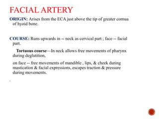
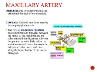
![SECOND PART – Deep to
hyoglossus, runs horizontally
forward along the upper border of
hyoid bone between hyoglossus
laterally and middle constrictor,
stylohyoid ligament medially.
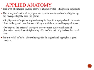
THIRD PART [ ‘arteria profunda
linguae’ ]—Also called as deep
lingual artery.
-It runs upwards along the anterior
Border of hyoglossus, then
horizontally forwards on the
undersurface of tongue on each side
of frenum linguae.
-In vertical course,it lies b/t the
genioglossus medially & inferior
longitudinal muscle of tongue
laterally. Horizontal part is
accompanied by lingual nerve.](https://image.slidesharecdn.com/mohit-triangle-of-neck11-170331154747/85/Triangle-of-Neck-by-Mohit-41-320.jpg)
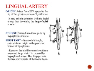
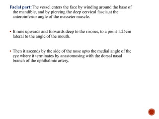
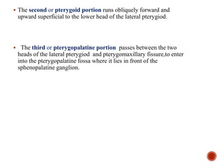
![Cervical part : Cervical part Runs
upwards on superior constrictor
of pharynx deep to the posterior
belly of digastric.
-It grooves the posterior border
of submandibular gland, makes
S-bend [2 loops] 1st winding
down over submandibular gland
& then up over the base of
mandible.](https://image.slidesharecdn.com/mohit-triangle-of-neck11-170331154747/85/Triangle-of-Neck-by-Mohit-45-320.jpg)


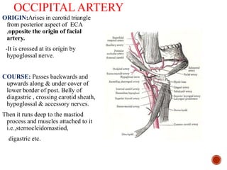

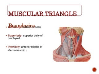

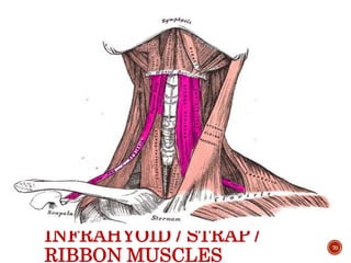

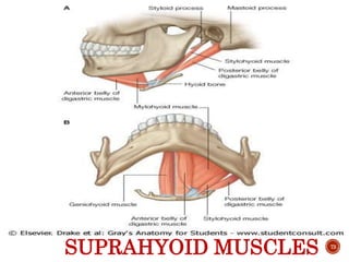

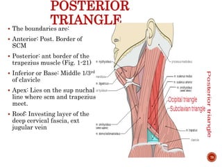
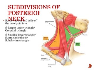
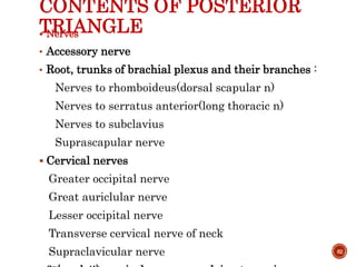


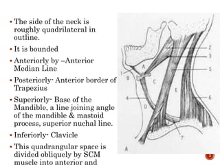
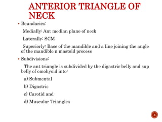
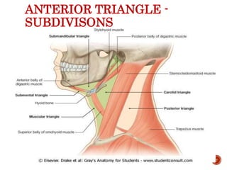
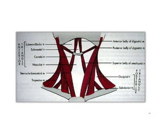
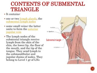
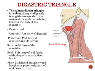
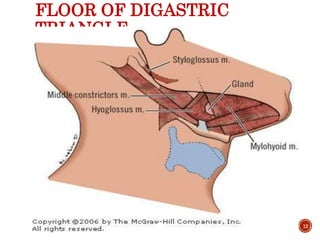
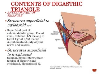
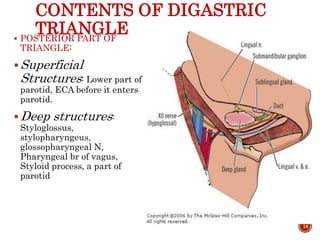
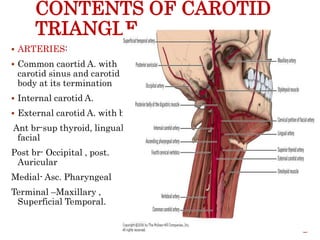
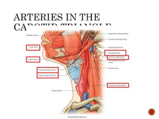
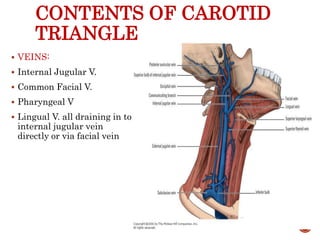
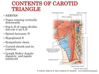
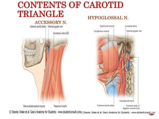
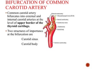
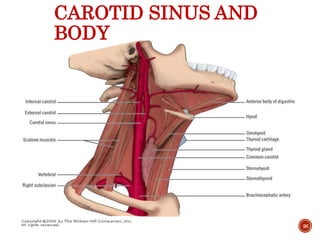
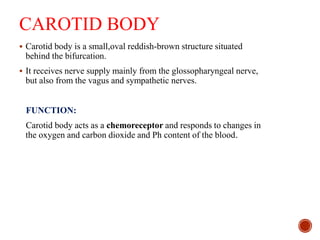
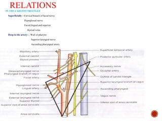
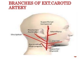
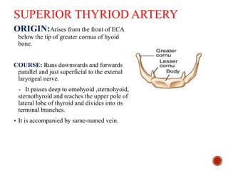
The sternocleidomastoid muscle divides the neck into anterior and posterior triangles. The anterior triangle contains several important structures and is further divided. The submental triangle contains lymph nodes and veins. The digastric triangle contains the submandibular gland, vessels, and nerves. The carotid triangle contains the common carotid artery and its branches, the internal and external carotid arteries. The external carotid artery gives off branches that supply the face and neck structures including the superior thyroid, lingual, and facial arteries.


![Polymer [ बहुलक ] Chemistry Notes PDF - Irfanullah Mehar - JJ Sir Chemistry.pdf](https://cdn.slidesharecdn.com/ss_thumbnails/polymerchemistrynotespdf-irfanullahmehar-jjsirchemistry-260210172118-3f9b37f7-thumbnail.jpg?width=640&height=640&fit=bounds)


