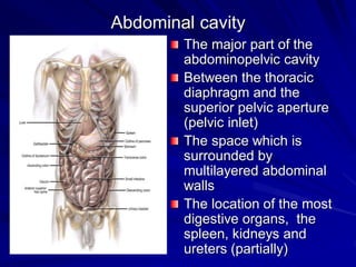
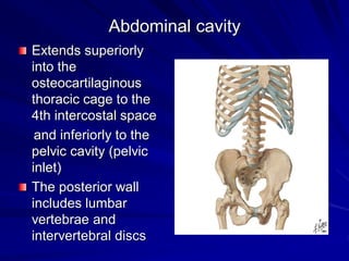
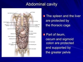
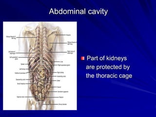
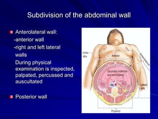
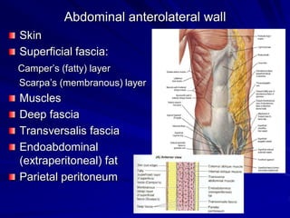
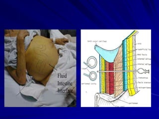
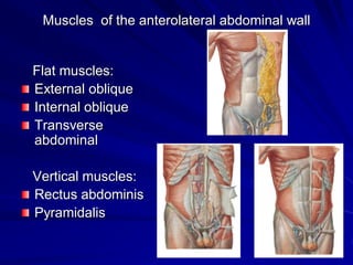
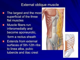
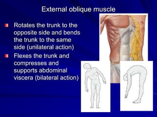
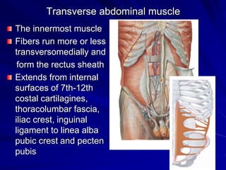
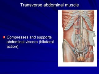
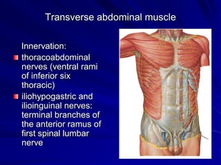
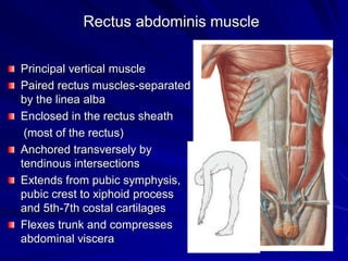
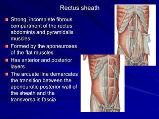
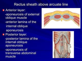
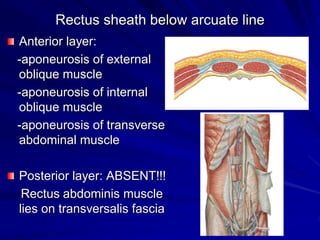
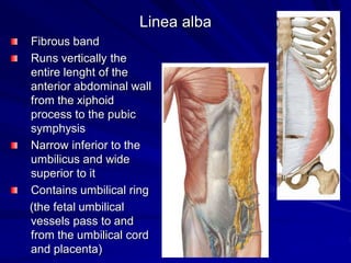
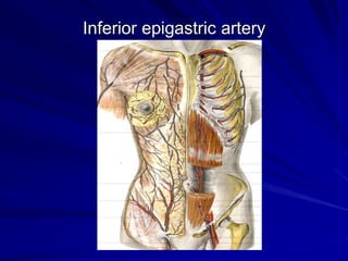
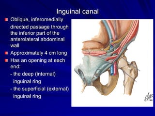
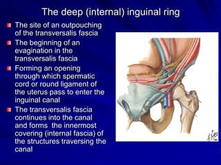
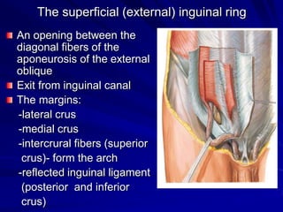
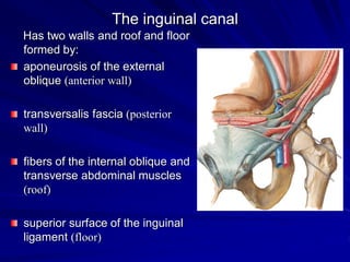
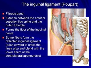
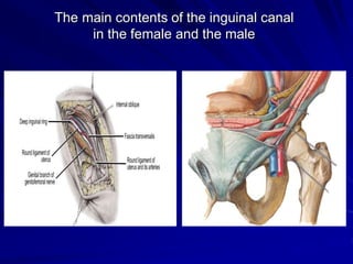
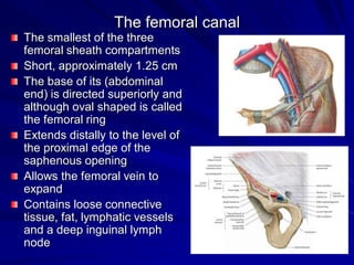
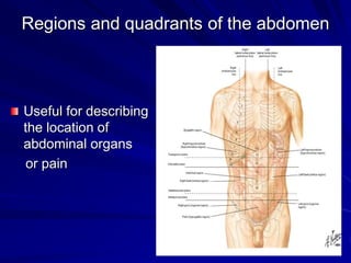
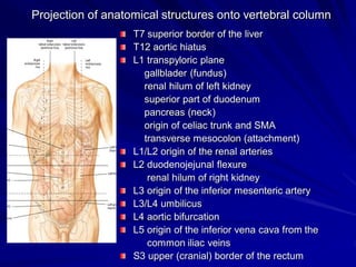
ABDOMINAL CAVITY
the first lecture
Abdominal cavity
Body walls
Rectus sheath
Inguinal canal
Sites of hernias
Arteries and veins of abdominal walls
Planes and regions
Abdominal cavity
The major part of the
abdominopelvic cavity
Between the thoracic
diaphragm and the
superior pelvic aperture
(pelvic inlet)
The space which is
surrounded by
multilayered abdominal
walls
The location of the most
digestive organs, the
spleen, kidneys and
ureters (partially)
Abdominal cavity
Extends superiorly
into the
osteocartilaginous
thoracic cage to the
4th intercostal space
and inferiorly to the
pelvic cavity (pelvic
inlet)
The posterior wall
includes lumbar
vertebrae and
intervertebral discs
Abdominal cavity
The spleen and the liver
are protected by
the thoracic cage
Part of ileum,
cecum and sigmoid
colon are protected
and supported by
the greater pelvis
Abdominal cavity
Part of kidneys
are protected by
the thoracic cage
Subdivision of the abdominal wall
Anterolateral wall:
-anterior wall
-right and left lateral
walls
During physical
examination is inspected,
palpated, percussed and
auscultated
Posterior wall
Abdominal anterolateral wall
Skin
Superficial fascia:
Camper’s (fatty) layer
Scarpa’s (membranous) layer
Muscles
Deep fascia
Transversalis fascia
Endoabdominal
(extraperitoneal) fat
Parietal peritoneum
Muscles of the anterolateral abdominal wall
Flat muscles:
External oblique
Internal oblique
Transverse
abdominal
Vertical muscles:
Rectus abdominis
Pyramidalis
External oblique muscle
The largest and the most
superficial of the three
flat muscles
Muscle fibers run
inferomedially and
become aponeurotic,
form a rectus sheath
Extends from external
surfaces of 5th-12th ribs
to linea alba, pubic
tubercle and iliac crest
External oblique muscle
Rotates the trunk to the
opposite side and bends
the trunk to the same
side (unilateral action)
Flexes the trunk and
compresses and
supports abdominal
viscera (bilateral action)
External oblique muscle
Innervation:
thoracoabdominal
nerves (ventral rami
of inferior 5 thoracic
nerves)
subcostal nerve
Internal oblique muscle
Intermediate of the three flat
muscles
Aponeurotic fibers form a
rectus sheath
The superior 2/3 of
aponeurosis splits into two
laminae
Extends from thoracolumbar
fascia, iliac crest and inguinal
ligament to inferior borders of
10th-12th ribs, linea alba and
pecten pubis
Internal oblique muscle
Bends and rotates the trunk to
the same side (unilateral
action)
Flexes the trunk and
compresses and supports
abdominal viscera (bilateral
action)
Internal oblique muscle
Innervation:
thoracoabdominal
nerves (ventral rami
of inferior six thoracic
nerves)
iliohypogastric and
ilioinguinal nerves:
terminal branches of
the anterior ramus of
first spinal lumbar
nerve
Transverse abdominal muscle
The innermost muscle
Fibers run more or less
transversomedially and
form the rectus sheath
Extends from internal
surfaces of 7th-12th
costal cartilagines,
thoracolumbar fascia,
iliac crest, inguinal
ligament to linea alba
pubic crest and pecten
pubis
Transverse abdominal muscle
Compresses and supports
abdominal viscera (bilateral
action)
Transverse abdominal muscle
Innervation:
thoracoabdominal
nerves (ventral rami
of inferior six
thoracic)
iliohypogastric and
ilioinguinal nerves:
terminal branches of
the anterior ramus of
first spinal lumbar
nerve
Rectus abdominis muscle
Principal vertical muscle
Paired rectus muscles-separated
by the linea alba
Enclosed in the rectus sheath
(most of the rectus)
Anchored transversely by
tendinous intersections
Extends from pubic symphysis,
pubic crest to xiphoid process
and 5th-7th costal cartilages
Flexes trunk and compresses
abdominal viscera
Rectus abdominis muscle
Innervation:
thoracoabdominal
nerves (ventral rami of
inferior six thoracic)
Pyramidalis muscle
Absent in approximately 20%
of people
Located in the rectus sheath
anterior to the most inferior
part of the rectus abdominis
muscle
Extends from the pubic crest
of the hip bone to the linea
alba
Draws down on the linea alba
(tenses the linea alba)
The attachment is a
landmark for an accurate
median abdominal incision
Dermatomes
Function and action of the anterolateral abdominal muscles
Form a strong expandable support for
the anterolateral abdominal wall
Protect the abdominal viscera from
injury
Compress the abdominal contents
Help to maintain or increase the intra-
abdominal pressure oppose the
diaphragm (produce expiration and
produce the force required for
defecation, vomiting, micturition and
parturition-maternal pushing during
childbirth)
Move the trunk and help to maintain
posture
Rectus sheath
Strong, incomplete fibrous
compartment of the rectus
abdominis and pyramidalis
muscles
Formed by the aponeuroses
of the flat muscles
Has anterior and posterior
layers
The arcuate line demarcates
the transition between the
aponeurotic posterior wall of
the sheath and the
transversalis fascia
Rectus sheath above arcuate line
Anterior layer:
-aponeurosis of external
oblique muscle
-anterior lamina of the
internal oblique
aponeurosis
Posterior layer:
-posterior lamina of the
internal oblique
aponeurosis
-aponeurosis of
transverse abdominal
muscle
Rectus sheath below arcuate line
Anterior layer:
-aponeurosis of external
oblique muscle
-aponeurosis of internal
oblique muscle
-aponeurosis of transverse
abdominal muscle
Posterior layer: ABSENT!!!
Rectus abdominis muscle
lies on transversalis fascia
Linea alba
Fibrous band
Runs vertically the
entire lenght of the
anterior abdominal wall
from the xiphoid
process to the pubic
symphysis
Narrow inferior to the
umbilicus and wide
superior to it
Contains umbilical ring
(the fetal umbilical
vessels pass to and
from the umbilical cord
and placenta)
Linea alba
Formed by the fibers
of the anterior and
posterior layers of the
sheath
peritoneal fold and peritoneal recess
Peritoneal fold- a reflection
of peritoneum that is raised
from the body wall by
underlying blood vessels,
ducts and obliterated fetal
vessels
Peritoneal recess (fossa)-
a pouch of peritoneum that
is formed by a peritoneal
fold
Internal surface of the anterolateral wall:
Internal surface of the anterolateral wall- peritoneal folds
Umbilical peritoneal
folds:
-the median umbilical fold
(covers the median
umbilical ligament- the
remnant of urachus, in
fetus runs within the
umbilical cord)
-two medial umbilical
folds (cover the medial
umbilical ligaments- the
remnants of the
occluded fetal umbilical
arteries)
-two lateral umbilical
folds
(cover the inferior
epigastric vessels)
Internal surface of the anterolateral wall – peritoneal
recesses
Peritoneal fossae (depressions
lateral to the umbilical folds):
-supravesical fossae
-medial inguinal fossae (called
inguinal triangles)
-lateral inguinal fossae
(include the deep inguinal
rings)
The fossae are potential sites for
hernias
The falciform ligament:
a sagittally oriented peritoneal
reflection in the supraumbilical
part of the internal surface
Inferior epigastric artery
Inguinal canal
Oblique, inferomedially
directed passage through
the inferior part of the
anterolateral abdominal
wall
Approximately 4 cm long
Has an opening at each
end:
- the deep (internal)
inguinal ring
- the superficial (external)
inguinal ring
The deep (internal) inguinal ring
The site of an outpouching
of the transversalis fascia
The beginning of an
evagination in the
transversalis fascia
Forming an opening
through which spermatic
cord or round ligament of
the uterus pass to enter the
inguinal canal
The transversalis fascia
continues into the canal
and forms the innermost
covering (internal fascia) of
the structures traversing the
canal
The superficial (external) inguinal ring
An opening between the
diagonal fibers of the
aponeurosis of the external
oblique
Exit from inguinal canal
The margins:
-lateral crus
-medial crus
-intercrural fibers (superior
crus)- form the arch
-reflected inguinal ligament
(posterior and inferior
crus)
The inguinal canal
Has two walls and roof and floor
formed by:
aponeurosis of the external
oblique (anterior wall)
transversalis fascia (posterior
wall)
fibers of the internal oblique and
transverse abdominal muscles
(roof)
superior surface of the inguinal
ligament (floor)
The inguinal ligament (Poupart)
Fibrous band
Extends between the anterior
superior iliac spine and the
pubic tubercle
Forms the floor of the inguinal
canal
Some fibers form the
reflected inguinal ligament
(pass upward to cross the
linea alba and blend with the
lower fibers of the
contralateral aponeurosis)
The main contents of the inguinal canal
in the female and the male
The femoral canal
The smallest of the three
femoral sheath compartments
Short, approximately 1.25 cm
The base of its (abdominal
end) is directed superiorly and
although oval shaped is called
the femoral ring
Extends distally to the level of
the proximal edge of the
saphenous opening
Allows the femoral vein to
expand
Contains loose connective
tissue, fat, lymphatic vessels
and a deep inguinal lymph
node
Abdominal hernias
Hernia= „rupture”
The result from increased
intra-abdominal pressure in
the presence of weakness
The protrusion of parietal
peritoneum through an
anatomical opening or a
secondary defect
Hernial opening- the orifice or
defect
The hernial sac- the pouch,
generally lined by parietal
peritoneum
Hernial contents- usually
consist of greater omentum
or loops of small bowel
Indirect inguinal hernias
Pass through the deep inguinal
ring (it is lateral to the inferior
epigastric artery) with the sac
following the course of the
spermatic cord
Strikes the pad of the finger
Direct inguinal hernias
Bulge directly through the
abdominal wall, medial to
the inferior epigastric artery
Strikes the distal tip of the
finger during palpation from
the scrotum
The „three finger rule”
Usefull to appreciate
the topographic
anatomy of inguinal or
femoral hernias and
differentiate among
direct and indirect
inguinal hernias and
femoral hernias
The examiner places
thenar eminence on the
anterior superior iliac
spine- fingers point to
hernias
Posterior abdominal wall
Five lumbar vertebrae
and IV discs
Posterior abdominal
wall muscles
Lumbar plexus
Fascia, including
thoracolumbar fascia
Diaphragm
Fat, nerves, vessels
and lymph nodes
Thoracolumbar fascia
The lumbar part is
extending between
the 12th rib and the
iliac crest
Attaches laterally to
the internal oblique
and transverse
abdominal muscles
Main muscles of the posterior abdominal wall
Psoas major
Iliacus
Quadratus lumborum
Quadratus lumborum muscle
Extends from the medial
half of the inferior border of
12th rib and tips of lumbar
transverse processes
to the iliolumbar ligament
and internal lip of iliac crest
Extends and laterally flexes
vertebral column, fixes 12th
rib during inspiration
Quadratus lumborum muscle
The subcostal nerve
runs inferolaterally on it
Branches of the lumbar
plexus run inferiorly on
the anterior surface
Ventral branches of
T12 and L1-L4 nerves
supply it
Arteries of the anterolateral abdominal wall
Superior epigastrics from the
internal thoracic arteries
Inferior epigastrics and deep
circumflex iliacs from external
iliac arteries
Superficial circumflex iliacs
and superficial epigastrics
from the femoral artery
Branches of the posterior
intercostal arteries
Branches of the
musculophrenic arteries
(from internal thoracic
arteries)
Inferior epigastric artery
Regions and quadrants of the abdomen
Useful for describing
the location of
abdominal organs
or pain
Regions and planes of abdomen
Four quadrants are
defined by two planes:
Horizontal
(transumbilical)
passing through the
umbilicus and the IV disc
between L3 and L4
vertebrae
Vertical (median)
passing longitudinally
through the body
Regions and planes of the abdomen
Nine regions are
defined by four planes:
Subcostal
passing through the inferior
border of the 10th costal
cartilage on each side
Transtubercular
passing through the iliac
tubercules and the body of
L5 vertebra
Two midclavicular
passing from midpoint of the
clavicles to the midinguinal points
Abdominal regions
Right hypochondriac
Epigastric
Left hypochondriac
Right lateral
Umbilical
Left lateral
Right inguinal
Hypogastric (pubic)
Left inguinal
Transpyloric plane:
- midway between the jugular notch of
the sternum and the superior border
of the pubic symphysis
- at the level of the L1 vertebra
- the pylorus is generally located
slightly below this plane
Semilunar line
Tip of 9th
costal cartilage
to the pubic
tubercle
The digestive tract
Extends from the lips to
the anus.
Consists of oral cavity,
pharynx, esophagus,
stomach, small
intestine and large
intestine.
Associated organs:
liver and pancreas
(major glands of the
digestive tract)
The viscera of the abdomen in situ
Projection of anatomical structures onto vertebral column
T7 superior border of the liver
T12 aortic hiatus
L1 transpyloric plane
gallblader (fundus)
renal hilum of left kidney
superior part of duodenum
pancreas (neck)
origin of celiac trunk and SMA
transverse mesocolon (attachment)
L1/L2 origin of the renal arteries
L2 duodenojejunal flexure
renal hilum of right kidney
L3 origin of the inferior mesenteric artery
L3/L4 umbilicus
L4 aortic bifurcation
L5 origin of the inferior vena cava from the
common iliac veins
S3 upper (cranial) border of the rectum
THANK YOU !!!

01._abdominal_cavity.pdf

  • 1.
  • 2.
    Abdominal cavity Body walls Rectussheath Inguinal canal Sites of hernias Arteries and veins of abdominal walls Planes and regions
  • 3.
    Abdominal cavity The majorpart of the abdominopelvic cavity Between the thoracic diaphragm and the superior pelvic aperture (pelvic inlet) The space which is surrounded by multilayered abdominal walls The location of the most digestive organs, the spleen, kidneys and ureters (partially)
  • 4.
    Abdominal cavity Extends superiorly intothe osteocartilaginous thoracic cage to the 4th intercostal space and inferiorly to the pelvic cavity (pelvic inlet) The posterior wall includes lumbar vertebrae and intervertebral discs
  • 5.
    Abdominal cavity The spleenand the liver are protected by the thoracic cage Part of ileum, cecum and sigmoid colon are protected and supported by the greater pelvis
  • 6.
    Abdominal cavity Part ofkidneys are protected by the thoracic cage
  • 7.
    Subdivision of theabdominal wall Anterolateral wall: -anterior wall -right and left lateral walls During physical examination is inspected, palpated, percussed and auscultated Posterior wall
  • 8.
    Abdominal anterolateral wall Skin Superficialfascia: Camper’s (fatty) layer Scarpa’s (membranous) layer Muscles Deep fascia Transversalis fascia Endoabdominal (extraperitoneal) fat Parietal peritoneum
  • 10.
    Muscles of theanterolateral abdominal wall Flat muscles: External oblique Internal oblique Transverse abdominal Vertical muscles: Rectus abdominis Pyramidalis
  • 11.
    External oblique muscle Thelargest and the most superficial of the three flat muscles Muscle fibers run inferomedially and become aponeurotic, form a rectus sheath Extends from external surfaces of 5th-12th ribs to linea alba, pubic tubercle and iliac crest
  • 12.
    External oblique muscle Rotatesthe trunk to the opposite side and bends the trunk to the same side (unilateral action) Flexes the trunk and compresses and supports abdominal viscera (bilateral action)
  • 13.
    External oblique muscle Innervation: thoracoabdominal nerves(ventral rami of inferior 5 thoracic nerves) subcostal nerve
  • 14.
    Internal oblique muscle Intermediateof the three flat muscles Aponeurotic fibers form a rectus sheath The superior 2/3 of aponeurosis splits into two laminae Extends from thoracolumbar fascia, iliac crest and inguinal ligament to inferior borders of 10th-12th ribs, linea alba and pecten pubis
  • 15.
    Internal oblique muscle Bendsand rotates the trunk to the same side (unilateral action) Flexes the trunk and compresses and supports abdominal viscera (bilateral action)
  • 16.
    Internal oblique muscle Innervation: thoracoabdominal nerves(ventral rami of inferior six thoracic nerves) iliohypogastric and ilioinguinal nerves: terminal branches of the anterior ramus of first spinal lumbar nerve
  • 17.
    Transverse abdominal muscle Theinnermost muscle Fibers run more or less transversomedially and form the rectus sheath Extends from internal surfaces of 7th-12th costal cartilagines, thoracolumbar fascia, iliac crest, inguinal ligament to linea alba pubic crest and pecten pubis
  • 18.
    Transverse abdominal muscle Compressesand supports abdominal viscera (bilateral action)
  • 19.
    Transverse abdominal muscle Innervation: thoracoabdominal nerves(ventral rami of inferior six thoracic) iliohypogastric and ilioinguinal nerves: terminal branches of the anterior ramus of first spinal lumbar nerve
  • 20.
    Rectus abdominis muscle Principalvertical muscle Paired rectus muscles-separated by the linea alba Enclosed in the rectus sheath (most of the rectus) Anchored transversely by tendinous intersections Extends from pubic symphysis, pubic crest to xiphoid process and 5th-7th costal cartilages Flexes trunk and compresses abdominal viscera
  • 21.
    Rectus abdominis muscle Innervation: thoracoabdominal nerves(ventral rami of inferior six thoracic)
  • 22.
    Pyramidalis muscle Absent inapproximately 20% of people Located in the rectus sheath anterior to the most inferior part of the rectus abdominis muscle Extends from the pubic crest of the hip bone to the linea alba Draws down on the linea alba (tenses the linea alba) The attachment is a landmark for an accurate median abdominal incision
  • 23.
  • 24.
    Function and actionof the anterolateral abdominal muscles Form a strong expandable support for the anterolateral abdominal wall Protect the abdominal viscera from injury Compress the abdominal contents Help to maintain or increase the intra- abdominal pressure oppose the diaphragm (produce expiration and produce the force required for defecation, vomiting, micturition and parturition-maternal pushing during childbirth) Move the trunk and help to maintain posture
  • 25.
    Rectus sheath Strong, incompletefibrous compartment of the rectus abdominis and pyramidalis muscles Formed by the aponeuroses of the flat muscles Has anterior and posterior layers The arcuate line demarcates the transition between the aponeurotic posterior wall of the sheath and the transversalis fascia
  • 26.
    Rectus sheath abovearcuate line Anterior layer: -aponeurosis of external oblique muscle -anterior lamina of the internal oblique aponeurosis Posterior layer: -posterior lamina of the internal oblique aponeurosis -aponeurosis of transverse abdominal muscle
  • 27.
    Rectus sheath belowarcuate line Anterior layer: -aponeurosis of external oblique muscle -aponeurosis of internal oblique muscle -aponeurosis of transverse abdominal muscle Posterior layer: ABSENT!!! Rectus abdominis muscle lies on transversalis fascia
  • 28.
    Linea alba Fibrous band Runsvertically the entire lenght of the anterior abdominal wall from the xiphoid process to the pubic symphysis Narrow inferior to the umbilicus and wide superior to it Contains umbilical ring (the fetal umbilical vessels pass to and from the umbilical cord and placenta)
  • 29.
    Linea alba Formed bythe fibers of the anterior and posterior layers of the sheath
  • 30.
    peritoneal fold andperitoneal recess Peritoneal fold- a reflection of peritoneum that is raised from the body wall by underlying blood vessels, ducts and obliterated fetal vessels Peritoneal recess (fossa)- a pouch of peritoneum that is formed by a peritoneal fold Internal surface of the anterolateral wall:
  • 31.
    Internal surface ofthe anterolateral wall- peritoneal folds Umbilical peritoneal folds: -the median umbilical fold (covers the median umbilical ligament- the remnant of urachus, in fetus runs within the umbilical cord) -two medial umbilical folds (cover the medial umbilical ligaments- the remnants of the occluded fetal umbilical arteries) -two lateral umbilical folds (cover the inferior epigastric vessels)
  • 32.
    Internal surface ofthe anterolateral wall – peritoneal recesses Peritoneal fossae (depressions lateral to the umbilical folds): -supravesical fossae -medial inguinal fossae (called inguinal triangles) -lateral inguinal fossae (include the deep inguinal rings) The fossae are potential sites for hernias The falciform ligament: a sagittally oriented peritoneal reflection in the supraumbilical part of the internal surface
  • 33.
  • 34.
    Inguinal canal Oblique, inferomedially directedpassage through the inferior part of the anterolateral abdominal wall Approximately 4 cm long Has an opening at each end: - the deep (internal) inguinal ring - the superficial (external) inguinal ring
  • 35.
    The deep (internal)inguinal ring The site of an outpouching of the transversalis fascia The beginning of an evagination in the transversalis fascia Forming an opening through which spermatic cord or round ligament of the uterus pass to enter the inguinal canal The transversalis fascia continues into the canal and forms the innermost covering (internal fascia) of the structures traversing the canal
  • 36.
    The superficial (external)inguinal ring An opening between the diagonal fibers of the aponeurosis of the external oblique Exit from inguinal canal The margins: -lateral crus -medial crus -intercrural fibers (superior crus)- form the arch -reflected inguinal ligament (posterior and inferior crus)
  • 37.
    The inguinal canal Hastwo walls and roof and floor formed by: aponeurosis of the external oblique (anterior wall) transversalis fascia (posterior wall) fibers of the internal oblique and transverse abdominal muscles (roof) superior surface of the inguinal ligament (floor)
  • 38.
    The inguinal ligament(Poupart) Fibrous band Extends between the anterior superior iliac spine and the pubic tubercle Forms the floor of the inguinal canal Some fibers form the reflected inguinal ligament (pass upward to cross the linea alba and blend with the lower fibers of the contralateral aponeurosis)
  • 39.
    The main contentsof the inguinal canal in the female and the male
  • 40.
    The femoral canal Thesmallest of the three femoral sheath compartments Short, approximately 1.25 cm The base of its (abdominal end) is directed superiorly and although oval shaped is called the femoral ring Extends distally to the level of the proximal edge of the saphenous opening Allows the femoral vein to expand Contains loose connective tissue, fat, lymphatic vessels and a deep inguinal lymph node
  • 41.
    Abdominal hernias Hernia= „rupture” Theresult from increased intra-abdominal pressure in the presence of weakness The protrusion of parietal peritoneum through an anatomical opening or a secondary defect Hernial opening- the orifice or defect The hernial sac- the pouch, generally lined by parietal peritoneum Hernial contents- usually consist of greater omentum or loops of small bowel
  • 42.
    Indirect inguinal hernias Passthrough the deep inguinal ring (it is lateral to the inferior epigastric artery) with the sac following the course of the spermatic cord Strikes the pad of the finger
  • 43.
    Direct inguinal hernias Bulgedirectly through the abdominal wall, medial to the inferior epigastric artery Strikes the distal tip of the finger during palpation from the scrotum
  • 44.
    The „three fingerrule” Usefull to appreciate the topographic anatomy of inguinal or femoral hernias and differentiate among direct and indirect inguinal hernias and femoral hernias The examiner places thenar eminence on the anterior superior iliac spine- fingers point to hernias
  • 45.
    Posterior abdominal wall Fivelumbar vertebrae and IV discs Posterior abdominal wall muscles Lumbar plexus Fascia, including thoracolumbar fascia Diaphragm Fat, nerves, vessels and lymph nodes
  • 46.
    Thoracolumbar fascia The lumbarpart is extending between the 12th rib and the iliac crest Attaches laterally to the internal oblique and transverse abdominal muscles
  • 47.
    Main muscles ofthe posterior abdominal wall Psoas major Iliacus Quadratus lumborum
  • 48.
    Quadratus lumborum muscle Extendsfrom the medial half of the inferior border of 12th rib and tips of lumbar transverse processes to the iliolumbar ligament and internal lip of iliac crest Extends and laterally flexes vertebral column, fixes 12th rib during inspiration
  • 49.
    Quadratus lumborum muscle Thesubcostal nerve runs inferolaterally on it Branches of the lumbar plexus run inferiorly on the anterior surface Ventral branches of T12 and L1-L4 nerves supply it
  • 50.
    Arteries of theanterolateral abdominal wall Superior epigastrics from the internal thoracic arteries Inferior epigastrics and deep circumflex iliacs from external iliac arteries Superficial circumflex iliacs and superficial epigastrics from the femoral artery Branches of the posterior intercostal arteries Branches of the musculophrenic arteries (from internal thoracic arteries)
  • 51.
  • 52.
    Regions and quadrantsof the abdomen Useful for describing the location of abdominal organs or pain
  • 53.
    Regions and planesof abdomen Four quadrants are defined by two planes: Horizontal (transumbilical) passing through the umbilicus and the IV disc between L3 and L4 vertebrae Vertical (median) passing longitudinally through the body
  • 54.
    Regions and planesof the abdomen Nine regions are defined by four planes: Subcostal passing through the inferior border of the 10th costal cartilage on each side Transtubercular passing through the iliac tubercules and the body of L5 vertebra Two midclavicular passing from midpoint of the clavicles to the midinguinal points
  • 55.
    Abdominal regions Right hypochondriac Epigastric Lefthypochondriac Right lateral Umbilical Left lateral Right inguinal Hypogastric (pubic) Left inguinal
  • 56.
    Transpyloric plane: - midwaybetween the jugular notch of the sternum and the superior border of the pubic symphysis - at the level of the L1 vertebra - the pylorus is generally located slightly below this plane
  • 57.
    Semilunar line Tip of9th costal cartilage to the pubic tubercle
  • 58.
    The digestive tract Extendsfrom the lips to the anus. Consists of oral cavity, pharynx, esophagus, stomach, small intestine and large intestine. Associated organs: liver and pancreas (major glands of the digestive tract)
  • 59.
    The viscera ofthe abdomen in situ
  • 60.
    The viscera ofthe abdomen in situ
  • 61.
    Projection of anatomicalstructures onto vertebral column T7 superior border of the liver T12 aortic hiatus L1 transpyloric plane gallblader (fundus) renal hilum of left kidney superior part of duodenum pancreas (neck) origin of celiac trunk and SMA transverse mesocolon (attachment) L1/L2 origin of the renal arteries L2 duodenojejunal flexure renal hilum of right kidney L3 origin of the inferior mesenteric artery L3/L4 umbilicus L4 aortic bifurcation L5 origin of the inferior vena cava from the common iliac veins S3 upper (cranial) border of the rectum
  • 62.