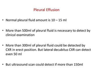
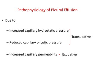
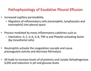
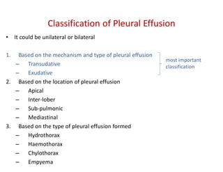
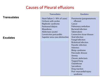
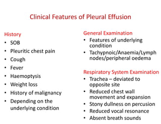
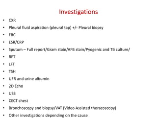
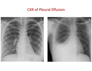
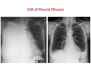
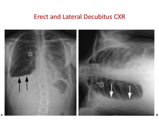
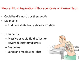
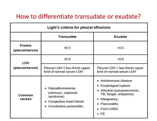
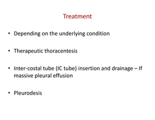
Pleural Effusion
Dr P Mayurathan
MBBS (Jaffna), MD in Medicine (Col), MRCP (UK), FRCP (Lond), FRCP (Edin)
Senior Lecturer in Medicine, FHCS
Honorary Consultant Physician, THB
Pleural Effusion
• Abnormally excessive fluid accumulation in the pleural
space
• Due to the imbalance between formation and absorption
of pleural fluid
• In response to injury, inflammation or both locally and
systemically
Pleural Effusion
• Normal pleural fluid amount is 10 – 15 ml
• More than 500ml of pleural fluid is necessary to detect by
clinical examination
• More than 300ml of pleural fluid could be detected by
CXR in erect position. But lateral decubitus CXR can detect
even 50 ml
• But ultrasound scan could detect if more than 150ml
Pleural Effusion
Pathophysiology of Pleural Effusion
• Due to
– Increased capillary hydrostatic pressure
– Reduced capillary oncotic pressure
– Increased capillary permeability -
Transudative
Exudative
Pathophysiology of Exudative Pleural Effusion
• Increased capillary permeability
– Migration of inflammatory cells (neutrophils, lymphocytes and
eosinophils) into pleural space
• Process mediated by many inflammatory cytokines such as
– Interlukins: IL-1, IL-6, IL-8, TNF-α and Platelet activating factor
(by mesothelial cells)
• Neutrophils activate the coagulation cascade and cause
procoagulant activity and decrease fibrinolysis
• All leads to increase levels of of proteins and Lactate Dehydrogenase
(LDH) and reduction in pH and glucose levels
• It could be unilateral or bilateral
1. Based on the mechanism and type of pleural effusion
– Transudative
– Exudative
2. Based on the location of pleural effusion
– Apical
– Inter-lober
– Sub-pulmonic
– Mediastinal
3. Based on the type of pleural effusion formed
– Hydrothorax
– Haemothorax
– Chylothorax
– Empyema
Classification of Pleural Effusion
most important
classification
Causes of Pleural effusions
History
• SOB
• Pleuritic chest pain
• Cough
• Fever
• Haemoptysis
• Weight loss
• History of malignancy
• Depending on the
underlying condition
General Examination
• Features of underlying
condition
• Tachypnoic/Anaemia/Lymph
nodes/peripheral oedema
Respiratory System Examination
• Trachea – deviated to
opposite site
• Reduced chest wall
movement and expansion
• Stony dullness on percusion
• Reduced vocal resonance
• Absent breath sounds
Clinical Features of Pleural Effusion
• CXR
• Pleural fluid aspiration (pleural tap) +/- Pleural biopsy
• FBC
• ESR/CRP
• Sputum – Full report/Gram stain/AFB stain/Pyogenic and TB culture/
• RFT
• LFT
• TSH
• UFR and urine albumin
• 2D Echo
• USS
• CECT chest
• Bronchoscopy and biopsy/VAT (Video Assisted thoracoscopy)
• Other investigations depending on the cause
Investigations
CXR of Pleural Effusion
L L
CXR of Pleural Effusion
L L
Erect and Lateral Decubitus CXR
Pleural Fluid Aspiration (Thoracentesis or Pleural Tap)
• Could be diagnostic or therapeutic
• Diagnostic
– to differentiate transudate or exudate
• Therapeutic
– Massive or rapid fluid collection
– Severe respiratory distress
– Empyema
– Large and mediastinal shift
Abram’s Pleural Biopsy Needle
Other investigations
• Adenosine Deaminase (ADA)
• pH
• Amylase
• Cholesterol
• Fungal studies
Investigations of Pleural Fluid
• Cytology (including for
malignant cells)
• Protein level
• Glucose
• LDH
• For AFB/TB PCR/TB culture
• Gram stain
• Pyogenic culture
How to differentiate transudate or exudate?
What are the causes for blood stained pleural
effusion (Haemothorax)?
1. TB
2. Malignancy – Primary or Secondary
3. Chest wall injuries/trauma
4. Pulmonary embolism
5. Pulmonary infarction
Treatment
• Depending on the underlying condition
• Therapeutic thoracentesis
• Inter-costal tube (IC tube) insertion and drainage – If
massive pleural effusion
• Pleurodesis
Pleural Effusion and its pathophysiology

Pleural Effusion and its pathophysiology

  • 1.
    Pleural Effusion Dr PMayurathan MBBS (Jaffna), MD in Medicine (Col), MRCP (UK), FRCP (Lond), FRCP (Edin) Senior Lecturer in Medicine, FHCS Honorary Consultant Physician, THB
  • 2.
    Pleural Effusion • Abnormallyexcessive fluid accumulation in the pleural space • Due to the imbalance between formation and absorption of pleural fluid • In response to injury, inflammation or both locally and systemically
  • 3.
  • 5.
    • Normal pleuralfluid amount is 10 – 15 ml • More than 500ml of pleural fluid is necessary to detect by clinical examination • More than 300ml of pleural fluid could be detected by CXR in erect position. But lateral decubitus CXR can detect even 50 ml • But ultrasound scan could detect if more than 150ml Pleural Effusion
  • 6.
    Pathophysiology of PleuralEffusion • Due to – Increased capillary hydrostatic pressure – Reduced capillary oncotic pressure – Increased capillary permeability - Transudative Exudative
  • 7.
    Pathophysiology of ExudativePleural Effusion • Increased capillary permeability – Migration of inflammatory cells (neutrophils, lymphocytes and eosinophils) into pleural space • Process mediated by many inflammatory cytokines such as – Interlukins: IL-1, IL-6, IL-8, TNF-α and Platelet activating factor (by mesothelial cells) • Neutrophils activate the coagulation cascade and cause procoagulant activity and decrease fibrinolysis • All leads to increase levels of of proteins and Lactate Dehydrogenase (LDH) and reduction in pH and glucose levels
  • 8.
    • It couldbe unilateral or bilateral 1. Based on the mechanism and type of pleural effusion – Transudative – Exudative 2. Based on the location of pleural effusion – Apical – Inter-lober – Sub-pulmonic – Mediastinal 3. Based on the type of pleural effusion formed – Hydrothorax – Haemothorax – Chylothorax – Empyema Classification of Pleural Effusion most important classification
  • 9.
  • 10.
    History • SOB • Pleuriticchest pain • Cough • Fever • Haemoptysis • Weight loss • History of malignancy • Depending on the underlying condition General Examination • Features of underlying condition • Tachypnoic/Anaemia/Lymph nodes/peripheral oedema Respiratory System Examination • Trachea – deviated to opposite site • Reduced chest wall movement and expansion • Stony dullness on percusion • Reduced vocal resonance • Absent breath sounds Clinical Features of Pleural Effusion
  • 11.
    • CXR • Pleuralfluid aspiration (pleural tap) +/- Pleural biopsy • FBC • ESR/CRP • Sputum – Full report/Gram stain/AFB stain/Pyogenic and TB culture/ • RFT • LFT • TSH • UFR and urine albumin • 2D Echo • USS • CECT chest • Bronchoscopy and biopsy/VAT (Video Assisted thoracoscopy) • Other investigations depending on the cause Investigations
  • 12.
    CXR of PleuralEffusion L L
  • 13.
    CXR of PleuralEffusion L L
  • 14.
    Erect and LateralDecubitus CXR
  • 15.
    Pleural Fluid Aspiration(Thoracentesis or Pleural Tap) • Could be diagnostic or therapeutic • Diagnostic – to differentiate transudate or exudate • Therapeutic – Massive or rapid fluid collection – Severe respiratory distress – Empyema – Large and mediastinal shift
  • 17.
  • 18.
    Other investigations • AdenosineDeaminase (ADA) • pH • Amylase • Cholesterol • Fungal studies Investigations of Pleural Fluid • Cytology (including for malignant cells) • Protein level • Glucose • LDH • For AFB/TB PCR/TB culture • Gram stain • Pyogenic culture
  • 19.
    How to differentiatetransudate or exudate?
  • 20.
    What are thecauses for blood stained pleural effusion (Haemothorax)? 1. TB 2. Malignancy – Primary or Secondary 3. Chest wall injuries/trauma 4. Pulmonary embolism 5. Pulmonary infarction
  • 21.
    Treatment • Depending onthe underlying condition • Therapeutic thoracentesis • Inter-costal tube (IC tube) insertion and drainage – If massive pleural effusion • Pleurodesis