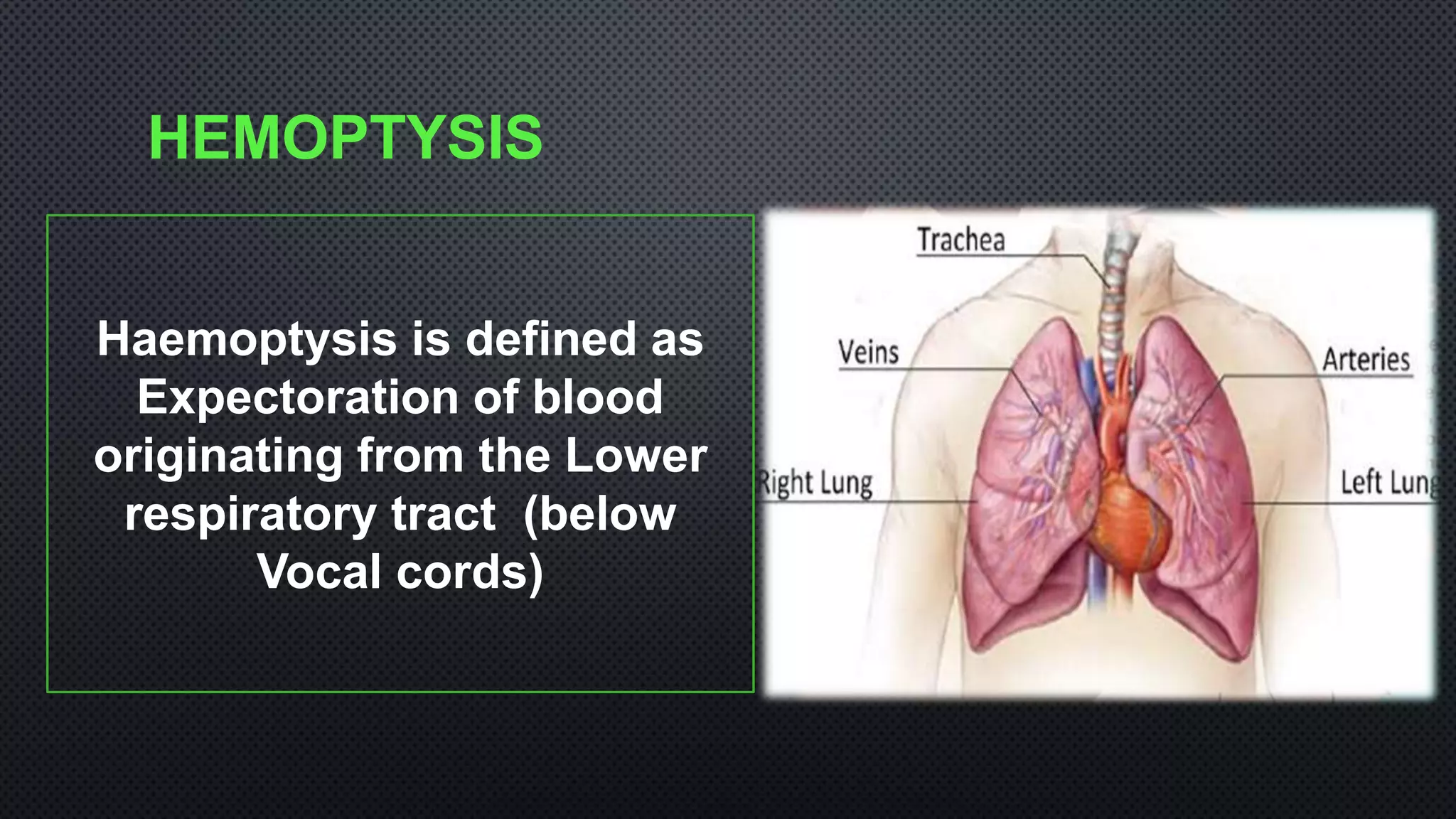
HAEMOPTYSIS
DR MUSTAFA BASHIR
CASE HISTORY
80 Year female
Hypertension (on Telmisartan 40 mg OD), Non diabetic
Presented with 2 day history of
• Coughing out of blood
• Multiple episodes, approx. ½ Cup each episode.
• Associated with mild left sided chest discomfort and shortness of
breath.
• No History of
 Bleeding from other sites
 Fever , weight loss, Cough, purulent sputum, Wheezing
 Orthopnea,PND
 Swelling of Lower limbs
 Skin rash, arthritis
 Reduced urine output
 PAST History: No history of Pulm-tuberculosis
Similar History 4 weeks back
HRCT CHEST
CTPA
MANAGEMENT
• PATIENT WAS MANAGED WITH BRONCHIAL ARTERY
EMBOLIZATION (BAE)
• HAEMOPTYSIS SETTLED AND PATIENT WAS DISCHARGED HOME
• REPORTED TO EMERGENCY SKIMS WITH RECURRENT
EPISODES OF MASSIVE HAEMOPTYSIS
MANAGEMENT
• IN VIEW OF HIGH RISK FOR SURGERY, PATIENT WAS
ATTEMPTED FOR SECOND SESSION OF BAE BUT THAT
FAILED.
• AFTER THE CASE WAS DISCUSSED BY EMERGENCY TEAM
WITH CTVS AND INTERVENTIONAL RADIOLOGIST
COURSE IN THE HOSPITAL
• Patient was Shifted to Internal medicine ward
• Had persistent haemoptysis
• Next day had another massive episode of
Haemoptysis
CONTD…
In view of Massive haemoptysis
1. Case was discussed CTVS for surgical intervention
2. Radiotherapist for Haemostatic dose of RT
3. SICU for selective Intubation or DLT placement
EXAMINATION
PR-110/MIN. GENERAL PHYSICAL EXAM.
BP-140/80 MMHG P +
RR-22/M I
SP02-93%ON ROOM AIR C NIL JVP-N
AFEBRILE TO TOUCH O NO CLUBBING
NO TELANGIECTASIA
EXAM. CONT……
CHEST: FINE CREPTS ISA(BILATERAL)
CVS: S1,S2
NO MURMUR.
P/A: SOFT, NT, ND.
CNS: GCS 15/15
NAD.
INVESTIGATIONS
FURTHER MANAGEMENT
1. LEFT Lateral decubitus position
2. P-RBC support
3. I/V Tranexamic acid
4. Oxygen supplementation
HAEMOPTYSIS SETTLED with conservative management
AFTER 4 days with no episode of haemoptysis patient was discharged
home with advise to follow CTVS in case of Haemoptysis
Recurrent Massive Haemoptysis
with left sided Pulmonary AVM
with failed BAE
DISCUSSION
HEMOPTYSIS
Haemoptysis is defined as
Expectoration of blood
originating from the Lower
respiratory tract (below
Vocal cords)
HAEMOPTYSIS SHOULD BE DIFFERENTIATING
FROM
DIFFERENCE BETWEEN
HAEMOPTYSIS AND HEMATEMESIS
FEATURES HEMOPTYSIS HEMATEMESIS
Definition Coughing out of blood Vomiting out of blood
Colour & Content Bright red ,mixed with sputum Coffee-ground in colour , with
food particles
Premonitory symptoms Respiratory symptoms GI symptoms
Melena Does not occur Usually with melena
Amount Relatively less High in amount
Reaction Alkaline Acidic
SOURCE OF BLEEDING
~90%
Bronchial artery
~10%
Pulmonary artery
SITES OF HAEMOPTYSIS
Airways
Parenchyma
Vasculature
CAUSES OF HAEMOPTYSIS
SEVERITY OF HEMOPTYSIS
Mild • < 100 ml in 24 h
Moderate • 100 -600 ml in 24 h
Massive
• > 600 ml in 24 h or 30 ml/h, with
respiratory failure/Haemodynamic
instability.
Fernando Luiz Cavalcanti J.Bras Pneumol 2010
RISK FACTORS FOR MORTALITY
Malignancy
Aspergillosis
Aspirationintocontralaterallung*
Bleedingfromthepulmonaryartery
Early prediction of in-hospital mortality of patients with hemoptysis: an approach to defining severe
hemoptysis.Fartoukh M, Khoshnood B, Parrot A, Khalil A, Carette MF, Stoclin A, Mayaud C, Cadranel J, Ancel PY -
Respiration. 2012;83(2):106.
DIAGNOSIS
History
Examination
BronchoscopyImaging
Lab
parameters
Clinical features suggestive of cardiac, pulmonary, renal , hepatic or immunological cause
MANAGEMENT
Air way
Breathing
Circulation
PROVIDE SUCTION.
Provide O2
crystalloid solutions
AND blood products
INITIAL STEPS
Management
Resuscitation
Identify
Bleeding
Side
Position of
patient
Airway Patency
Control Of
Bleeding
PROTECTION OF NON BLEEDING LUNG
Ifbleedingsideisknown
Rest
Lateraldecubitus
-Bleeding side down.
SELECTIVE INTUBATION
SINGLELUMENETT
Selectivelyintubate the non
bleeding lung
Selective intubation of L Main bronchus
in R sided massive hemoptysis
SELECTIVE INTUBATION
Speciallydesignedforselectiveintub
ationoftherightorleftmainbronchi
• Last option in an
asphyxiatingpt.
CONTROL THE BLEEDING
Non
Surgical
• Bloodproduc
ts
(PRP,FFP)
• Bronchosco
picmeasures
• BAE
Surgical
• Lobectomy/
• Pneumonect
omy
• Other
surgical
management
directed to
cause
BRONCHOSCOPY
Massive hemoptysis. Assessment and management. Cahill BC, Ingbar DH Clin Chest
Med. 1994;15(1):147.
Flexiblebronchoscopyistheinitialdiagnosticprocedureofchoice:
Performedbedside,andishighlysuccessfulatlocalizi
ng the bleedingsite.
Intubationshouldbeconsidered
BRONCHOSPIC MEASURES
Localisation of bleeding
Guide Intubation:
single limen /double lumen intubation
Control of bleeding:
Cold saline lavage
Topical vasoconstrictors like adrenaline,ornipressin
APC/ Electro cautery
BalloonTamponade
Spigot
• ADVANTAGES:
• Performedatbedside
• Access: UL/distalorifices
• Can do Lavage
• Topicalanaesthesia.
• DISADVANTAGES:
• Poorsuction
• Airwaypatencyisnotgood
• Merits of rigid bronchoscopy
over flexible:-
• Better airway control
• Larger field of view
• Better suctioning
• Better for therapeutic Interventions
• DISADVANTAGES:
• PoorvisibilityofperipherallesionsandU
L
• GA
COLD-SALINE LAVAGE
*Conlan AA, Hurwitz SS, Krige L, Nicolaou N, Pool R: Massive hemoptysis: review of 123 cases. J Thorac Cardiovasc Surg
1983; 85: 120–124.
Lavage:Normalsalineat4°C
ItStopsthebleedingwithmassivehemoptysis
obviatingtheneedforemergencythoracotomy.*
RigidscopeisbetteroverFOB
Topical VasoconstrictiveAgents
Topical epinephrine (1:20,000)
Effective :mild to moderate.
Side effects
-Tachyarrythmias
- HTN
• Newer agents: ADH
derivative
- ornipressin
* Cahill BC, Ingbar DH: Massive hemoptysis. Assessment and management. Clin Chest Med 1994; 15: 147–167.
OTHER
TRANEXAMIC ACID
•ANTIFIBRINOLYTIC DRUG
•ROUTE : PO ,IV & TOPICAL (RECENTLY)
•ENDOBRONCHIAL :*
•DOSE: 500–1,000 MG
•RESPONSE TIME: SECONDS
* Solomonov A, Fruchter O, Zuckerman T,Brenner B, Yigla M: Pulmonary hemorrhage: a novel
mode of therapy. Respir Med 2009; 103: 1196–1200.
BalloonTamponade
• Life threatening hemoptysis.
• 4 Fr 100 cm Fogarthy balloon
catheter by FOB.
• Inflated for 24-48 hrs
* Hiebert C: Balloon catheter control of lifethreatening hemoptysis. Chest 1974; 66: 308– 309.
EndobronchialAirway Blockade(Silicone Spigot)
Temporary management.
• Silicone spigot is placed
endobronchially .
Stabilizes patient before BAE
• *Dutau H , Palot A, Haas A, Decamps I, Durieux O: Endobronchial embolization with a silicone
spigot as a
temporary treatment for massive hemoptysis. Respiration 2006; 73: 830–832.
posterior segment
of the right upper lobe
Silicon spigots of various sizes
BRONCHOSCOPY-GUIDED TOPICAL
HEMOSTATIC TAMPONADE (THT)
• Oxidized cellulose mesh
 Saturates with blood-->brownish or black gelatinous
mass -->clot.
• Successful in life threatening hemoptysis.
• Immediate arrest of bleed: 98%(56 of 57)
*Valipour A, Kreuzer A, Koller H, Koessler W, Burghuber OC: Bronchoscopy-guided topical hemostatic tamponade
therapy for the Management of life-threatening hemoptysis. Chest 2005; 127: 2113–2118.
ENDOBRONCHIAL SEALING WITH
BIOCOMPATIBLE GLUE
• Material: n-butyl cyanoacrylate(adhesive)
• Injected into the bleeding airway through a catheter via a flexible FOB.
• Used in mild hemoptysis.
• * *Parthasarathi Bhattacharyya et al Bronchoscopy Centre, Calcutta, India(CHEST
2002; 121:2066–2069)
ARGON PLASMA COAGULATION(APC)
• TYPE : Thermal tissue destruction
• Non contact electrocoagulation
• In bronchoscopically visible areas of
sources of bleed
*Keller CA, Hinerman R, Singh A, Alvarez F: The use of
endoscopic argon plasma coagulation In airway
complications after solid organ transplantation. Chest
2001; 119: 1968–1975.
APC machine
63
Flooding of the
bron.intermed.
Suctioning
airway clearance
visualization
Coagulation and
devascularization
of tissues
Carbonization of
the bleeding site
Endobronchial Electrocautery
• TYPE: Thermal tissue destruction
• Contact Electrocoagulation
• Readily available
Contact probesElectro cautery machine
Probe through working channel
BRONCHIALARTERY EMBOLIZATION
• Temporary or definitive
• Immediate control: 57–100% of patients**
Embolization : bronchial and nonbronchial
 Long-term control: 70%-88%
*Remy J, Voisin C, Dupuis C, et al: Traitement des hémoptysies par embolisation de la circulation systémique. Ann Radiol (Paris)
1974; 17: 5–16.
**Remy J, Arnaud A, Fardou H, et al: Treatment of hemoptysis by embolization of bronchial arteries. Radiology 1977; 122: 33–37.
EMBOLIZING MATERIALS:
Absorbable gelatin
sponge
• Gelfoam
• Pledgets (1 to 2 mm)
• Thrombin
• Glue
• Recently approved
-Embospheres,
-Spherical Poly vinyl
alcohol(PVA) particles
Right
Left
Abnormal circulation
Pre-embolisation bronchial angiogram
No abnormal circulation
Post embolisation
BRONCHIALARTERYANEURYSM
Hypervascular lesion with aneurysm
Pre embolisation Post embolisationPVA particles
No hypervascular lesion & aneurysm
LEFT UPPER LOBE BRONCHIALARTERY
After embolization
Decreased vascularity & hypertrophyTortous and hypertrophied vessel
Before embolization
INDICATIONS OF
SURGERY
Procedure of choice in:
• Bronchial adenoma
• Aspergilloma
• Hydatid cyst
• Iatrogenic pulmonary rupture
• Chest trauma
• AV malformations
PULMONARY AVM
• PULMONARY ARTERIOVENOUS MALFORMATIONS (PAVMS) ARE ABNORMAL COMMUNICATIONS
BETWEEN PULMONARY ARTERIES AND VEINS .
• ALTERNATIVE NAMES INCLUDE PULMONARY ARTERIOVENOUS FISTULAE, PULMONARY
ARTERIOVENOUS ANEURYSMS, CAVERNOUS ANGIOMAS OF THE LUNG, AND PULMONARY
TELANGIECTASES
• STRONG ASSOCIATION BETWEEN PAVM AND HEREDITARY HAEMORRHAGIC TELANGIECTASIA
PRESENTATION
• MOST PATIENTS ARE ASYMPTOMATIC, PAVMS CAN CAUSE DYSPNOEA FROM RIGHT-TO-LEFT
SHUNT.
• TRIAD OF DYSPNEA , COUGH & CLUBBING
• BECAUSE OF PARADOXICAL EMBOLI, VARIOUS CENTRAL NERVOUS SYSTEM COMPLICATIONS
HAVE BEEN DESCRIBED INCLUDING STROKE AND BRAIN ABSCESS
MANAGEMENT
• CHEST RADIOGRAPHY AND CONTRAST ENHANCED COMPUTED TOMOGRAPHY ARE ESSENTIAL
INITIAL DIAGNOSTIC TOOLS BUT
• PULMONARY ANGIOGRAPHY IS THE GOLD STANDARD.
• CONTRAST ECHOCARDIOGRAPHY IS USEFUL FOR DIAGNOSIS AND MONITORING AFTER
TREATMENT.
• THERAPEUTIC OPTIONS INCLUDE
• ANGIOGRAPHIC EMBOLIZATION WITH METAL COIL OR BALLOON OCCLUSION AND
• SURGICAL EXCISION.
90
Life Threatening hemoptysis
Pulmonary isolation & identification of bleeding source
(Radiological/Bronchoscopic means:CT Chest,Balloon bronchial blockers)
Bronchoscopy
Surgery BAE
(Delayed TREATMENT)
Follow up at OPD
SUCCESS
FAILURE
• INVESTIGATIONAL AGENTS —
• STUDIES ARE UNDERWAY USING AGENTS CAPABLE OF ALTERING/INHIBITING THE FUNCTION OF HEPCIDIN
(EG, HEPCIDIN ANTAGONISTS) AND THE HEPCIDIN RECEPTOR (FERROPORTIN) IN ORDER TO ALLEVIATE
THE VARIOUS DISORDERS OF IRON METABOLISM ASSOCIATED WITH INCREASED LEVELS OF HEPCIDIN,
INCLUDING ACD

Hemoptysis and its management

  • 1.
  • 2.
    CASE HISTORY 80 Yearfemale Hypertension (on Telmisartan 40 mg OD), Non diabetic Presented with 2 day history of • Coughing out of blood • Multiple episodes, approx. ½ Cup each episode. • Associated with mild left sided chest discomfort and shortness of breath.
  • 3.
    • No Historyof  Bleeding from other sites  Fever , weight loss, Cough, purulent sputum, Wheezing  Orthopnea,PND  Swelling of Lower limbs  Skin rash, arthritis  Reduced urine output  PAST History: No history of Pulm-tuberculosis Similar History 4 weeks back
  • 4.
  • 5.
  • 6.
    MANAGEMENT • PATIENT WASMANAGED WITH BRONCHIAL ARTERY EMBOLIZATION (BAE) • HAEMOPTYSIS SETTLED AND PATIENT WAS DISCHARGED HOME • REPORTED TO EMERGENCY SKIMS WITH RECURRENT EPISODES OF MASSIVE HAEMOPTYSIS
  • 7.
    MANAGEMENT • IN VIEWOF HIGH RISK FOR SURGERY, PATIENT WAS ATTEMPTED FOR SECOND SESSION OF BAE BUT THAT FAILED. • AFTER THE CASE WAS DISCUSSED BY EMERGENCY TEAM WITH CTVS AND INTERVENTIONAL RADIOLOGIST
  • 8.
    COURSE IN THEHOSPITAL • Patient was Shifted to Internal medicine ward • Had persistent haemoptysis • Next day had another massive episode of Haemoptysis
  • 9.
    CONTD… In view ofMassive haemoptysis 1. Case was discussed CTVS for surgical intervention 2. Radiotherapist for Haemostatic dose of RT 3. SICU for selective Intubation or DLT placement
  • 10.
    EXAMINATION PR-110/MIN. GENERAL PHYSICALEXAM. BP-140/80 MMHG P + RR-22/M I SP02-93%ON ROOM AIR C NIL JVP-N AFEBRILE TO TOUCH O NO CLUBBING NO TELANGIECTASIA
  • 11.
    EXAM. CONT…… CHEST: FINECREPTS ISA(BILATERAL) CVS: S1,S2 NO MURMUR. P/A: SOFT, NT, ND. CNS: GCS 15/15 NAD.
  • 12.
  • 13.
    FURTHER MANAGEMENT 1. LEFTLateral decubitus position 2. P-RBC support 3. I/V Tranexamic acid 4. Oxygen supplementation HAEMOPTYSIS SETTLED with conservative management AFTER 4 days with no episode of haemoptysis patient was discharged home with advise to follow CTVS in case of Haemoptysis
  • 14.
    Recurrent Massive Haemoptysis withleft sided Pulmonary AVM with failed BAE
  • 15.
  • 16.
    HEMOPTYSIS Haemoptysis is definedas Expectoration of blood originating from the Lower respiratory tract (below Vocal cords)
  • 17.
    HAEMOPTYSIS SHOULD BEDIFFERENTIATING FROM
  • 18.
    DIFFERENCE BETWEEN HAEMOPTYSIS ANDHEMATEMESIS FEATURES HEMOPTYSIS HEMATEMESIS Definition Coughing out of blood Vomiting out of blood Colour & Content Bright red ,mixed with sputum Coffee-ground in colour , with food particles Premonitory symptoms Respiratory symptoms GI symptoms Melena Does not occur Usually with melena Amount Relatively less High in amount Reaction Alkaline Acidic
  • 19.
    SOURCE OF BLEEDING ~90% Bronchialartery ~10% Pulmonary artery
  • 20.
  • 21.
  • 22.
    SEVERITY OF HEMOPTYSIS Mild• < 100 ml in 24 h Moderate • 100 -600 ml in 24 h Massive • > 600 ml in 24 h or 30 ml/h, with respiratory failure/Haemodynamic instability. Fernando Luiz Cavalcanti J.Bras Pneumol 2010
  • 23.
    RISK FACTORS FORMORTALITY Malignancy Aspergillosis Aspirationintocontralaterallung* Bleedingfromthepulmonaryartery Early prediction of in-hospital mortality of patients with hemoptysis: an approach to defining severe hemoptysis.Fartoukh M, Khoshnood B, Parrot A, Khalil A, Carette MF, Stoclin A, Mayaud C, Cadranel J, Ancel PY - Respiration. 2012;83(2):106.
  • 24.
  • 25.
    Clinical features suggestiveof cardiac, pulmonary, renal , hepatic or immunological cause
  • 29.
  • 30.
    Air way Breathing Circulation PROVIDE SUCTION. ProvideO2 crystalloid solutions AND blood products
  • 31.
  • 32.
  • 33.
    PROTECTION OF NONBLEEDING LUNG Ifbleedingsideisknown Rest Lateraldecubitus -Bleeding side down.
  • 34.
    SELECTIVE INTUBATION SINGLELUMENETT Selectivelyintubate thenon bleeding lung Selective intubation of L Main bronchus in R sided massive hemoptysis
  • 35.
  • 36.
    CONTROL THE BLEEDING Non Surgical •Bloodproduc ts (PRP,FFP) • Bronchosco picmeasures • BAE Surgical • Lobectomy/ • Pneumonect omy • Other surgical management directed to cause
  • 37.
    BRONCHOSCOPY Massive hemoptysis. Assessmentand management. Cahill BC, Ingbar DH Clin Chest Med. 1994;15(1):147. Flexiblebronchoscopyistheinitialdiagnosticprocedureofchoice: Performedbedside,andishighlysuccessfulatlocalizi ng the bleedingsite. Intubationshouldbeconsidered
  • 38.
    BRONCHOSPIC MEASURES Localisation ofbleeding Guide Intubation: single limen /double lumen intubation Control of bleeding: Cold saline lavage Topical vasoconstrictors like adrenaline,ornipressin APC/ Electro cautery BalloonTamponade Spigot
  • 39.
    • ADVANTAGES: • Performedatbedside •Access: UL/distalorifices • Can do Lavage • Topicalanaesthesia. • DISADVANTAGES: • Poorsuction • Airwaypatencyisnotgood • Merits of rigid bronchoscopy over flexible:- • Better airway control • Larger field of view • Better suctioning • Better for therapeutic Interventions • DISADVANTAGES: • PoorvisibilityofperipherallesionsandU L • GA
  • 40.
    COLD-SALINE LAVAGE *Conlan AA,Hurwitz SS, Krige L, Nicolaou N, Pool R: Massive hemoptysis: review of 123 cases. J Thorac Cardiovasc Surg 1983; 85: 120–124. Lavage:Normalsalineat4°C ItStopsthebleedingwithmassivehemoptysis obviatingtheneedforemergencythoracotomy.* RigidscopeisbetteroverFOB
  • 41.
    Topical VasoconstrictiveAgents Topical epinephrine(1:20,000) Effective :mild to moderate. Side effects -Tachyarrythmias - HTN • Newer agents: ADH derivative - ornipressin * Cahill BC, Ingbar DH: Massive hemoptysis. Assessment and management. Clin Chest Med 1994; 15: 147–167.
  • 42.
    OTHER TRANEXAMIC ACID •ANTIFIBRINOLYTIC DRUG •ROUTE: PO ,IV & TOPICAL (RECENTLY) •ENDOBRONCHIAL :* •DOSE: 500–1,000 MG •RESPONSE TIME: SECONDS * Solomonov A, Fruchter O, Zuckerman T,Brenner B, Yigla M: Pulmonary hemorrhage: a novel mode of therapy. Respir Med 2009; 103: 1196–1200.
  • 43.
    BalloonTamponade • Life threateninghemoptysis. • 4 Fr 100 cm Fogarthy balloon catheter by FOB. • Inflated for 24-48 hrs * Hiebert C: Balloon catheter control of lifethreatening hemoptysis. Chest 1974; 66: 308– 309.
  • 44.
    EndobronchialAirway Blockade(Silicone Spigot) Temporarymanagement. • Silicone spigot is placed endobronchially . Stabilizes patient before BAE • *Dutau H , Palot A, Haas A, Decamps I, Durieux O: Endobronchial embolization with a silicone spigot as a temporary treatment for massive hemoptysis. Respiration 2006; 73: 830–832. posterior segment of the right upper lobe Silicon spigots of various sizes
  • 45.
    BRONCHOSCOPY-GUIDED TOPICAL HEMOSTATIC TAMPONADE(THT) • Oxidized cellulose mesh  Saturates with blood-->brownish or black gelatinous mass -->clot. • Successful in life threatening hemoptysis. • Immediate arrest of bleed: 98%(56 of 57) *Valipour A, Kreuzer A, Koller H, Koessler W, Burghuber OC: Bronchoscopy-guided topical hemostatic tamponade therapy for the Management of life-threatening hemoptysis. Chest 2005; 127: 2113–2118.
  • 46.
    ENDOBRONCHIAL SEALING WITH BIOCOMPATIBLEGLUE • Material: n-butyl cyanoacrylate(adhesive) • Injected into the bleeding airway through a catheter via a flexible FOB. • Used in mild hemoptysis. • * *Parthasarathi Bhattacharyya et al Bronchoscopy Centre, Calcutta, India(CHEST 2002; 121:2066–2069)
  • 47.
    ARGON PLASMA COAGULATION(APC) •TYPE : Thermal tissue destruction • Non contact electrocoagulation • In bronchoscopically visible areas of sources of bleed *Keller CA, Hinerman R, Singh A, Alvarez F: The use of endoscopic argon plasma coagulation In airway complications after solid organ transplantation. Chest 2001; 119: 1968–1975. APC machine
  • 48.
    63 Flooding of the bron.intermed. Suctioning airwayclearance visualization Coagulation and devascularization of tissues Carbonization of the bleeding site
  • 49.
    Endobronchial Electrocautery • TYPE:Thermal tissue destruction • Contact Electrocoagulation • Readily available Contact probesElectro cautery machine Probe through working channel
  • 50.
    BRONCHIALARTERY EMBOLIZATION • Temporaryor definitive • Immediate control: 57–100% of patients** Embolization : bronchial and nonbronchial  Long-term control: 70%-88% *Remy J, Voisin C, Dupuis C, et al: Traitement des hémoptysies par embolisation de la circulation systémique. Ann Radiol (Paris) 1974; 17: 5–16. **Remy J, Arnaud A, Fardou H, et al: Treatment of hemoptysis by embolization of bronchial arteries. Radiology 1977; 122: 33–37.
  • 51.
    EMBOLIZING MATERIALS: Absorbable gelatin sponge •Gelfoam • Pledgets (1 to 2 mm) • Thrombin • Glue • Recently approved -Embospheres, -Spherical Poly vinyl alcohol(PVA) particles
  • 52.
    Right Left Abnormal circulation Pre-embolisation bronchialangiogram No abnormal circulation Post embolisation
  • 53.
    BRONCHIALARTERYANEURYSM Hypervascular lesion withaneurysm Pre embolisation Post embolisationPVA particles No hypervascular lesion & aneurysm
  • 54.
    LEFT UPPER LOBEBRONCHIALARTERY After embolization Decreased vascularity & hypertrophyTortous and hypertrophied vessel Before embolization
  • 55.
    INDICATIONS OF SURGERY Procedure ofchoice in: • Bronchial adenoma • Aspergilloma • Hydatid cyst • Iatrogenic pulmonary rupture • Chest trauma • AV malformations
  • 56.
    PULMONARY AVM • PULMONARYARTERIOVENOUS MALFORMATIONS (PAVMS) ARE ABNORMAL COMMUNICATIONS BETWEEN PULMONARY ARTERIES AND VEINS . • ALTERNATIVE NAMES INCLUDE PULMONARY ARTERIOVENOUS FISTULAE, PULMONARY ARTERIOVENOUS ANEURYSMS, CAVERNOUS ANGIOMAS OF THE LUNG, AND PULMONARY TELANGIECTASES • STRONG ASSOCIATION BETWEEN PAVM AND HEREDITARY HAEMORRHAGIC TELANGIECTASIA
  • 57.
    PRESENTATION • MOST PATIENTSARE ASYMPTOMATIC, PAVMS CAN CAUSE DYSPNOEA FROM RIGHT-TO-LEFT SHUNT. • TRIAD OF DYSPNEA , COUGH & CLUBBING • BECAUSE OF PARADOXICAL EMBOLI, VARIOUS CENTRAL NERVOUS SYSTEM COMPLICATIONS HAVE BEEN DESCRIBED INCLUDING STROKE AND BRAIN ABSCESS
  • 58.
    MANAGEMENT • CHEST RADIOGRAPHYAND CONTRAST ENHANCED COMPUTED TOMOGRAPHY ARE ESSENTIAL INITIAL DIAGNOSTIC TOOLS BUT • PULMONARY ANGIOGRAPHY IS THE GOLD STANDARD. • CONTRAST ECHOCARDIOGRAPHY IS USEFUL FOR DIAGNOSIS AND MONITORING AFTER TREATMENT. • THERAPEUTIC OPTIONS INCLUDE • ANGIOGRAPHIC EMBOLIZATION WITH METAL COIL OR BALLOON OCCLUSION AND • SURGICAL EXCISION.
  • 60.
    90 Life Threatening hemoptysis Pulmonaryisolation & identification of bleeding source (Radiological/Bronchoscopic means:CT Chest,Balloon bronchial blockers) Bronchoscopy Surgery BAE (Delayed TREATMENT) Follow up at OPD SUCCESS FAILURE
  • 61.
    • INVESTIGATIONAL AGENTS— • STUDIES ARE UNDERWAY USING AGENTS CAPABLE OF ALTERING/INHIBITING THE FUNCTION OF HEPCIDIN (EG, HEPCIDIN ANTAGONISTS) AND THE HEPCIDIN RECEPTOR (FERROPORTIN) IN ORDER TO ALLEVIATE THE VARIOUS DISORDERS OF IRON METABOLISM ASSOCIATED WITH INCREASED LEVELS OF HEPCIDIN, INCLUDING ACD