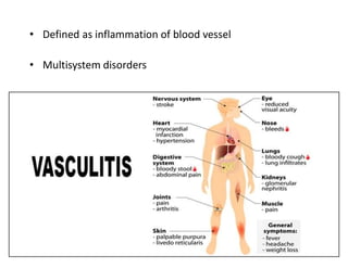
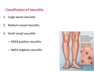
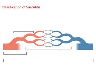
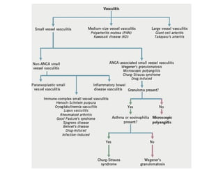
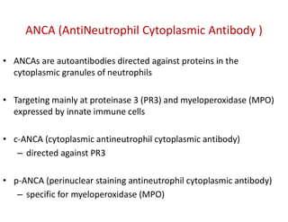
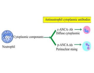
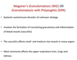
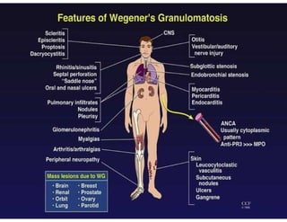
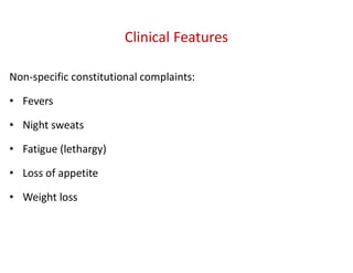
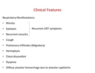
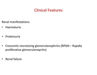
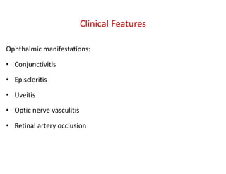
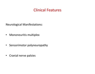
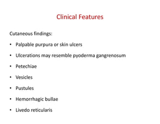
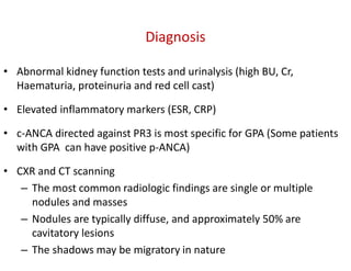
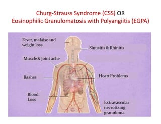
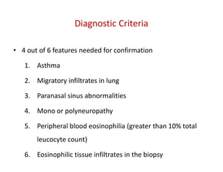
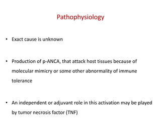
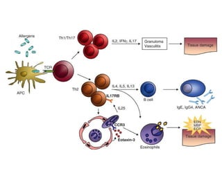
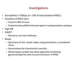
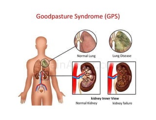
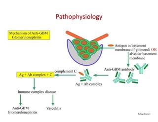
Vasculitis and Lungs
Dr P Mayurathan
MBBS (Jaffna), MD in Medicine (Colombo), MRCP (UK), FRCP (London), FRCP (Edinburgh)
Senior Lecturer in Medicine, FHCS
Consultant Physician in Internal Medicine
• Defined as inflammation of blood vessel
• Multisystem disorders
1. Large vessel vasculitis
2. Medium vessel vasculitis
3. Small vessel vasculitis
– ANCA positive vasculitis
– ANCA negative vasculitis
Classification of Vasculitis
Small vessel vasculitis
ANCA (AntiNeutrophil Cytoplasmic Antibody )
• ANCAs are autoantibodies directed against proteins in the
cytoplasmic granules of neutrophils
• Targeting mainly at proteinase 3 (PR3) and myeloperoxidase (MPO)
expressed by innate immune cells
• c-ANCA (cytoplasmic antineutrophil cytoplasmic antibody)
– directed against PR3
• p-ANCA (perinuclear staining antineutrophil cytoplasmic antibody)
– specific for myeloperoxidase (MPO)
Wegener’s Granulomatosis (WG) OR
Granulomatosis with Polyangiitis (GPA)
• Systemic autoimmune disorder of unknown etiology
• Involves the formation of necrotizing granulomas and inflammation
of blood vessels (vasculitis)
• The vasculitis affects small- and medium-size vessels in many organs
• Most commonly affects the upper respiratory tract, lungs and
kidneys
Clinical Features
Non-specific constitutional complaints:
• Fevers
• Night sweats
• Fatigue (lethargy)
• Loss of appetite
• Weight loss
Respiratory Manifestations:
• Rhinitis
• Epistaxis
• Recurrent sinusitis
• Cough
• Pulmonary infiltrates (Migratory)
• Hemoptysis
• Chest discomfort
• Dyspnea
• Diffuse alveolar hemorrhage due to alveolar capillaritis
Clinical Features
Recurrent URT symptoms
Renal manifestations:
• Haematuria
• Proteinuria
• Crescentic necrotizing glomerulonephritis (RPGN – Rapidly
proliferative glomerulonepritis)
• Renal failure
Clinical Features
Clinical Features
Ophthalmic manifestations:
• Conjunctivitis
• Episcleritis
• Uveitis
• Optic nerve vasculitis
• Retinal artery occlusion
Neurological Manifestations:
• Mononeuritis multiplex
• Sensorimotor polyneuropathy
• Cranial nerve palsies
Clinical Features
Musculoskeletal:
• Myalgia
• Arthralgia
• Arthritis
Clinical Features
Cutaneous findings:
• Palpable purpura or skin ulcers
• Ulcerations may resemble pyoderma gangrenosum
• Petechiae
• Vesicles
• Pustules
• Hemorrhagic bullae
• Livedo reticularis
Clinical Features
Diagnosis
• Abnormal kidney function tests and urinalysis (high BU, Cr,
Haematuria, proteinuria and red cell cast)
• Elevated inflammatory markers (ESR, CRP)
• c-ANCA directed against PR3 is most specific for GPA (Some patients
with GPA can have positive p-ANCA)
• CXR and CT scanning
– The most common radiologic findings are single or multiple
nodules and masses
– Nodules are typically diffuse, and approximately 50% are
cavitatory lesions
– The shadows may be migratory in nature
• Sinus CT scanning: To evaluate sinus disease
• Spirometry – Restrictive lung disease with high DLCO commonly due
to pulmonary haemorrhage
• Bronchoscopy: Helpful in the evaluation of alveolar hemorrhage,
infection, airway disease, and endobronchial lesions
• Biopsy: The diagnosis of GPA is generally confirmed with tissue
biopsy from a site of active disease; renal and lung biopsies are most
specific for GPA
Diagnosis
Management
• Steroids – Prednisolone
• Cyclophosphamide
• Rituximab
• Methotrexate
• Plasma exchange may be considered in patients with RPGN
Churg-Strauss Syndrome (CSS) OR
Eosinophilic Granulomatosis with Polyangiitis (EGPA)
Churg-Strauss Syndrome (CSS) OR
Eosinophilic Granulomatosis with Polyangiitis (EGPA)
• Characterized by necrotizing vasculitis of small and medium-sized
systemic blood vessels
• Same type of vasculitis can be seen in granulomatosis with polyangiitis
(GPA), microscopic polyangiitis (MPA) and polyarteritis nodosa (PAN)
• The classic distinction of CSS/EGPA from the other diseases in the
category is the coexistence of asthma, rhinosinusitis and presence of
peripheral eosinophilia
Diagnostic Criteria
• 4 out of 6 features needed for confirmation
1. Asthma
2. Migratory infiltrates in lung
3. Paranasal sinus abnormalities
4. Mono or polyneuropathy
5. Peripheral blood eosinophilia (greater than 10% total
leucocyte count)
6. Eosinophilic tissue infiltrates in the biopsy
Pathophysiology
• Exact cause is unknown
• Production of p-ANCA, that attack host tissues because of
molecular mimicry or some other abnormality of immune
tolerance
• An independent or adjuvant role in this activation may be played
by tumor necrosis factor (TNF)
Investigations
• Eosinophilia (> 1500/µL or > 10% of total peripheral WBCs)
• Elevations of ANCA titers
– Found in 40% of cases
– Predominantly pANCAs directed against myeloperoxidase epitopes
• High ESR
• CXR/CT
– Normal or can have infiltrates
• Biopsy
– Specimens of skin, lymph nodes, lung parenchyma, or peripheral
nerve
– Demonstrate the characteristic vasculitis
– Kidney biopsy sample may show segmental necrotizing
glomerulonephritis with crescent formation in RPGN
Treatment
• Immunosuppression
• Plasma exchange
Anti-glomerular basement membrane (anti-GBM) disease
• Anti-GBM disease is an autoimmune disorder
• It is a rare disease associated with small vessel vasculitis that typically
affects the capillaries of kidneys
• Patients can develop rapidly progressive glomerulonephritis (RPGN)
• It is caused by the deposition in glomerular basement membrane of
circulating autoantibodies directed against antigens intrinsic to the
basement membrane
• The exact mechanism that induces the formation of autoantibodies is
unknown, but probably environmental factors, infections or direct damage
to kidneys and lungs
• ANCA is negative
• Goodpasture syndrome is one of the classical examples of anti-GBM
disease where kidneys and lungs are predominantly affected
Goodpasture Syndrome (GPS)
Goodpasture Syndrome
• Clinical entity of
– diffuse pulmonary hemorrhage and
– acute or rapidly progressive glomerulonephritis and
– the presence of circulating anti–glomerular basement membrane
(anti-GBM) antibodies
Pathophysiology
• Goodpasture syndrome is an autoimmune disorder
• Characterized by autoantibodies directed against the
glomerular/alveolar basement membrane
• The autoantibodies bind to their reactive epitopes in the basement
membranes and activate the complement cascade, resulting in tissue
injury
• The principal component of basement membrane is type IV collagen
Pathophysiology
alveolar basement
membrane
OR
Clinical Features
• Constitutional symptoms
– Malaise
– Chills
– Fever
– Arthralgias
• May precede or develop concurrently with pulmonary or renal
manifestations
Clinical Features
Respiratory manifestations:
• Hemoptysis
– Presenting symptom when the disease affects the lungs
– The level of hemoptysis may vary and may be absent
• Cough
• Shortness of breath
• Massive pulmonary hemorrhage leading to respiratory failure may
occur
• Pleuritic chest pain is present in less than half of the patients
Clinical Features
Renal manifestations:
• Hematuria
• Proteinuria
• Oedema
• High blood pressure
• Uremia
Investigations
• Urinalysis - low-grade proteinuria, gross or microscopic hematuria, and red
blood cell casts
• FBC
– Anemia (iron deficiency caused by intrapulmonary bleeding)
– Leukocytosis is common
• Elevated blood urea and serum creatinine levels
• Elevation of ESR
• Anti–GBM Antibody
– Useful to confirm the diagnosis and monitor the efficacy of treatment
– ELISA for anti-GBM antibodies are highly sensitive (>95%) and specific (>97%)
• CXR
– Patchy parenchymal consolidations, which are usually bilateral,
symmetric perihilar and bibasal
• Spirometry
– May reveal evidence of restriction
– DLCO is elevated secondary to pulmonary hemorrhage
• Biopsy
– Renal biopsy
Investigations
Management
• Plasmapheresis (therapeutic plasma exchange – TPE)
– Rapidly remove circulating antibody
• Stop further production of antibodies using immunosuppression
with medications
Non-Vasculitic Lung conditions
Pulmonary Eosinophilia
• Pulmonary diseases associated with tissue and/or blood eosinophilia
• Extrinsic or intrinsic in origin (some syndromes may overlap)
• Inhaled or ingested extrinsic factors
– Medications
– Infectious agents (eg, parasites, fungi, mycobacteria)
• Intrinsic pulmonary eosinophilic syndromes are generally idiopathic in nature
– Trophical Pulmonary Eosinophilia (TPE)
– Churg-Strauss Syndrome (CSS)
– Chronic Eosinophilic Pneumonia (CEP)
– Idiopathic Hypereosinophilic Syndrome (IHES)
– Eosinophilic granuloma (eg, pulmonary histiocytosis X or Langerhans cell
granulomatosis)
• Eosinophilia and pulmonary infiltrates have been reported in patients with AIDS,
lymphoma, a variety of inflammatory lung diseases and collagen vascular diseases
• Asthma may manifest with eosinophilia without infiltrates
Tropical Pulmonary Eosinophilia (TPE)
• Characterized by coughing, asthmatic attacks, and an enlarged spleen
• It occurs most frequently in tropical and subtropical areas like India and Southeast
Asia
• Tropical pulmonary eosinophilia is a rare, but well recognized
• Syndrome characterized by pulmonary interstitial infiltrates and marked peripheral
eosinophilia
• In non-endemic countries, patients are commonly thought to have bronchial
asthma
• The syndrome is caused by a distinct hypersensitive immunological reaction to
microfilariae of Wuchereria bancrofti and Brugia malayi
Microfilariae
Wuchereria bancrofti Brugia malayi
Clinical Presentation
• Persistent or recurrent cough that gets aggravated at night
• Rarely haemoptysis
• Wheeze
• Weakness
• Weight loss
• Low fever
• Enlarged lymph nodes in the neck and elsewhere
• Splenomegaly
Diagnosis
• The diagnostic criteria
– a history supportive of exposure to lymphatic filariasis
– a peripheral eosinophilia count greater than 3 × 10⁹/L)
– an elevated serum IgE levels (> 1000 kU/L);
– increased titers of antifilarial antibodies
– peripheral blood negative for microfilariae
– a clinical response to diethylcarbamazine (DEC)
• High antifilarial IgG titers to microfilariae often result in cross
reactivity with other non-filarial helminth antigens (strongyloides and
schistosoma antigens)
• It is important to exclude other parasitic infections before tropical
pulmonary eosinophilia is diagnosed by serological tests
• Radiological findings are nonspecific (normal CXR in up to 20%)
• Lung biopsy is not part of the routine diagnostic investigation
Diagnosis
Treatment
• The dramatic response to Diethylcarbamazine (6 mg/kg/day in 3
divided doses for 21 days)
• The eosinophil count often falls dramatically within 7–10 days of
starting treatment

Vasculitis and Lungs and its pathophysiology

  • 1.
    Vasculitis and Lungs DrP Mayurathan MBBS (Jaffna), MD in Medicine (Colombo), MRCP (UK), FRCP (London), FRCP (Edinburgh) Senior Lecturer in Medicine, FHCS Consultant Physician in Internal Medicine
  • 2.
    • Defined asinflammation of blood vessel • Multisystem disorders
  • 3.
    1. Large vesselvasculitis 2. Medium vessel vasculitis 3. Small vessel vasculitis – ANCA positive vasculitis – ANCA negative vasculitis Classification of Vasculitis
  • 4.
  • 6.
    ANCA (AntiNeutrophil CytoplasmicAntibody ) • ANCAs are autoantibodies directed against proteins in the cytoplasmic granules of neutrophils • Targeting mainly at proteinase 3 (PR3) and myeloperoxidase (MPO) expressed by innate immune cells • c-ANCA (cytoplasmic antineutrophil cytoplasmic antibody) – directed against PR3 • p-ANCA (perinuclear staining antineutrophil cytoplasmic antibody) – specific for myeloperoxidase (MPO)
  • 8.
    Wegener’s Granulomatosis (WG)OR Granulomatosis with Polyangiitis (GPA) • Systemic autoimmune disorder of unknown etiology • Involves the formation of necrotizing granulomas and inflammation of blood vessels (vasculitis) • The vasculitis affects small- and medium-size vessels in many organs • Most commonly affects the upper respiratory tract, lungs and kidneys
  • 10.
    Clinical Features Non-specific constitutionalcomplaints: • Fevers • Night sweats • Fatigue (lethargy) • Loss of appetite • Weight loss
  • 11.
    Respiratory Manifestations: • Rhinitis •Epistaxis • Recurrent sinusitis • Cough • Pulmonary infiltrates (Migratory) • Hemoptysis • Chest discomfort • Dyspnea • Diffuse alveolar hemorrhage due to alveolar capillaritis Clinical Features Recurrent URT symptoms
  • 12.
    Renal manifestations: • Haematuria •Proteinuria • Crescentic necrotizing glomerulonephritis (RPGN – Rapidly proliferative glomerulonepritis) • Renal failure Clinical Features
  • 13.
    Clinical Features Ophthalmic manifestations: •Conjunctivitis • Episcleritis • Uveitis • Optic nerve vasculitis • Retinal artery occlusion
  • 14.
    Neurological Manifestations: • Mononeuritismultiplex • Sensorimotor polyneuropathy • Cranial nerve palsies Clinical Features
  • 15.
  • 16.
    Cutaneous findings: • Palpablepurpura or skin ulcers • Ulcerations may resemble pyoderma gangrenosum • Petechiae • Vesicles • Pustules • Hemorrhagic bullae • Livedo reticularis Clinical Features
  • 17.
    Diagnosis • Abnormal kidneyfunction tests and urinalysis (high BU, Cr, Haematuria, proteinuria and red cell cast) • Elevated inflammatory markers (ESR, CRP) • c-ANCA directed against PR3 is most specific for GPA (Some patients with GPA can have positive p-ANCA) • CXR and CT scanning – The most common radiologic findings are single or multiple nodules and masses – Nodules are typically diffuse, and approximately 50% are cavitatory lesions – The shadows may be migratory in nature
  • 18.
    • Sinus CTscanning: To evaluate sinus disease • Spirometry – Restrictive lung disease with high DLCO commonly due to pulmonary haemorrhage • Bronchoscopy: Helpful in the evaluation of alveolar hemorrhage, infection, airway disease, and endobronchial lesions • Biopsy: The diagnosis of GPA is generally confirmed with tissue biopsy from a site of active disease; renal and lung biopsies are most specific for GPA Diagnosis
  • 19.
    Management • Steroids –Prednisolone • Cyclophosphamide • Rituximab • Methotrexate • Plasma exchange may be considered in patients with RPGN
  • 20.
    Churg-Strauss Syndrome (CSS)OR Eosinophilic Granulomatosis with Polyangiitis (EGPA)
  • 21.
    Churg-Strauss Syndrome (CSS)OR Eosinophilic Granulomatosis with Polyangiitis (EGPA) • Characterized by necrotizing vasculitis of small and medium-sized systemic blood vessels • Same type of vasculitis can be seen in granulomatosis with polyangiitis (GPA), microscopic polyangiitis (MPA) and polyarteritis nodosa (PAN) • The classic distinction of CSS/EGPA from the other diseases in the category is the coexistence of asthma, rhinosinusitis and presence of peripheral eosinophilia
  • 22.
    Diagnostic Criteria • 4out of 6 features needed for confirmation 1. Asthma 2. Migratory infiltrates in lung 3. Paranasal sinus abnormalities 4. Mono or polyneuropathy 5. Peripheral blood eosinophilia (greater than 10% total leucocyte count) 6. Eosinophilic tissue infiltrates in the biopsy
  • 23.
    Pathophysiology • Exact causeis unknown • Production of p-ANCA, that attack host tissues because of molecular mimicry or some other abnormality of immune tolerance • An independent or adjuvant role in this activation may be played by tumor necrosis factor (TNF)
  • 25.
    Investigations • Eosinophilia (>1500/µL or > 10% of total peripheral WBCs) • Elevations of ANCA titers – Found in 40% of cases – Predominantly pANCAs directed against myeloperoxidase epitopes • High ESR • CXR/CT – Normal or can have infiltrates • Biopsy – Specimens of skin, lymph nodes, lung parenchyma, or peripheral nerve – Demonstrate the characteristic vasculitis – Kidney biopsy sample may show segmental necrotizing glomerulonephritis with crescent formation in RPGN
  • 26.
  • 27.
    Anti-glomerular basement membrane(anti-GBM) disease • Anti-GBM disease is an autoimmune disorder • It is a rare disease associated with small vessel vasculitis that typically affects the capillaries of kidneys • Patients can develop rapidly progressive glomerulonephritis (RPGN) • It is caused by the deposition in glomerular basement membrane of circulating autoantibodies directed against antigens intrinsic to the basement membrane • The exact mechanism that induces the formation of autoantibodies is unknown, but probably environmental factors, infections or direct damage to kidneys and lungs • ANCA is negative • Goodpasture syndrome is one of the classical examples of anti-GBM disease where kidneys and lungs are predominantly affected
  • 28.
  • 29.
    Goodpasture Syndrome • Clinicalentity of – diffuse pulmonary hemorrhage and – acute or rapidly progressive glomerulonephritis and – the presence of circulating anti–glomerular basement membrane (anti-GBM) antibodies
  • 30.
    Pathophysiology • Goodpasture syndromeis an autoimmune disorder • Characterized by autoantibodies directed against the glomerular/alveolar basement membrane • The autoantibodies bind to their reactive epitopes in the basement membranes and activate the complement cascade, resulting in tissue injury • The principal component of basement membrane is type IV collagen
  • 31.
  • 32.
    Clinical Features • Constitutionalsymptoms – Malaise – Chills – Fever – Arthralgias • May precede or develop concurrently with pulmonary or renal manifestations
  • 33.
    Clinical Features Respiratory manifestations: •Hemoptysis – Presenting symptom when the disease affects the lungs – The level of hemoptysis may vary and may be absent • Cough • Shortness of breath • Massive pulmonary hemorrhage leading to respiratory failure may occur • Pleuritic chest pain is present in less than half of the patients
  • 34.
    Clinical Features Renal manifestations: •Hematuria • Proteinuria • Oedema • High blood pressure • Uremia
  • 35.
    Investigations • Urinalysis -low-grade proteinuria, gross or microscopic hematuria, and red blood cell casts • FBC – Anemia (iron deficiency caused by intrapulmonary bleeding) – Leukocytosis is common • Elevated blood urea and serum creatinine levels • Elevation of ESR • Anti–GBM Antibody – Useful to confirm the diagnosis and monitor the efficacy of treatment – ELISA for anti-GBM antibodies are highly sensitive (>95%) and specific (>97%)
  • 36.
    • CXR – Patchyparenchymal consolidations, which are usually bilateral, symmetric perihilar and bibasal • Spirometry – May reveal evidence of restriction – DLCO is elevated secondary to pulmonary hemorrhage • Biopsy – Renal biopsy Investigations
  • 37.
    Management • Plasmapheresis (therapeuticplasma exchange – TPE) – Rapidly remove circulating antibody • Stop further production of antibodies using immunosuppression with medications
  • 38.
  • 39.
    Pulmonary Eosinophilia • Pulmonarydiseases associated with tissue and/or blood eosinophilia • Extrinsic or intrinsic in origin (some syndromes may overlap) • Inhaled or ingested extrinsic factors – Medications – Infectious agents (eg, parasites, fungi, mycobacteria) • Intrinsic pulmonary eosinophilic syndromes are generally idiopathic in nature – Trophical Pulmonary Eosinophilia (TPE) – Churg-Strauss Syndrome (CSS) – Chronic Eosinophilic Pneumonia (CEP) – Idiopathic Hypereosinophilic Syndrome (IHES) – Eosinophilic granuloma (eg, pulmonary histiocytosis X or Langerhans cell granulomatosis) • Eosinophilia and pulmonary infiltrates have been reported in patients with AIDS, lymphoma, a variety of inflammatory lung diseases and collagen vascular diseases • Asthma may manifest with eosinophilia without infiltrates
  • 40.
    Tropical Pulmonary Eosinophilia(TPE) • Characterized by coughing, asthmatic attacks, and an enlarged spleen • It occurs most frequently in tropical and subtropical areas like India and Southeast Asia • Tropical pulmonary eosinophilia is a rare, but well recognized • Syndrome characterized by pulmonary interstitial infiltrates and marked peripheral eosinophilia • In non-endemic countries, patients are commonly thought to have bronchial asthma • The syndrome is caused by a distinct hypersensitive immunological reaction to microfilariae of Wuchereria bancrofti and Brugia malayi
  • 41.
  • 42.
    Clinical Presentation • Persistentor recurrent cough that gets aggravated at night • Rarely haemoptysis • Wheeze • Weakness • Weight loss • Low fever • Enlarged lymph nodes in the neck and elsewhere • Splenomegaly
  • 43.
    Diagnosis • The diagnosticcriteria – a history supportive of exposure to lymphatic filariasis – a peripheral eosinophilia count greater than 3 × 10⁹/L) – an elevated serum IgE levels (> 1000 kU/L); – increased titers of antifilarial antibodies – peripheral blood negative for microfilariae – a clinical response to diethylcarbamazine (DEC)
  • 44.
    • High antifilarialIgG titers to microfilariae often result in cross reactivity with other non-filarial helminth antigens (strongyloides and schistosoma antigens) • It is important to exclude other parasitic infections before tropical pulmonary eosinophilia is diagnosed by serological tests • Radiological findings are nonspecific (normal CXR in up to 20%) • Lung biopsy is not part of the routine diagnostic investigation Diagnosis
  • 45.
    Treatment • The dramaticresponse to Diethylcarbamazine (6 mg/kg/day in 3 divided doses for 21 days) • The eosinophil count often falls dramatically within 7–10 days of starting treatment