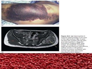
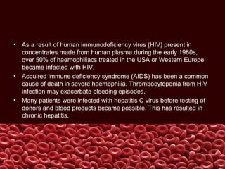
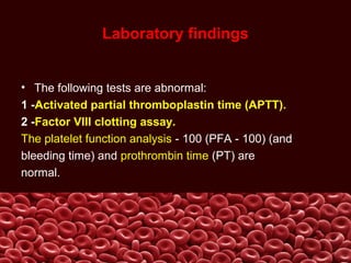
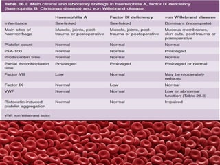
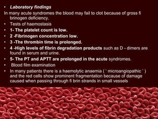
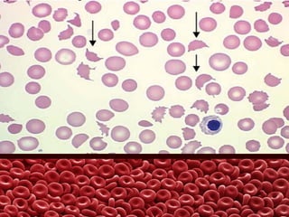
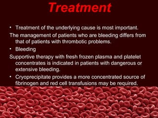
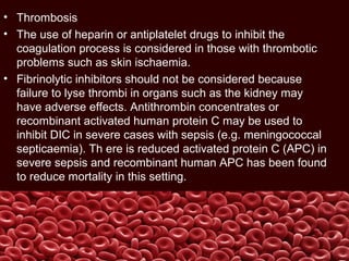
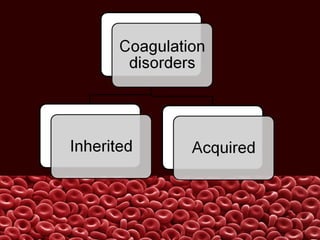
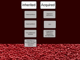
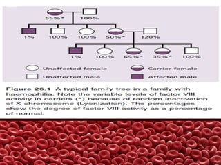
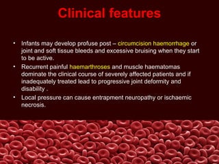
This document summarizes several coagulation disorders including haemophilia A, haemophilia B, von Willebrand disease, other hereditary coagulation factor deficiencies, and disseminated intravascular coagulation. It describes the clinical features, inheritance, laboratory findings, treatment, and management of each condition. Key points include that haemophilia A is the most common clotting factor deficiency, von Willebrand disease is the most common inherited bleeding disorder, and disseminated intravascular coagulation can be triggered by trauma, infection, cancer and other conditions.
Thrombocytopenia is most frequently encountered Hematological problem in hospitalized patients. The most common causes and differential diagnosis of In-patient and Outpatient presentations of Thrombocytopenia is discussed here. Useful for Internal Medicine Boards . Archer Internal Medicine Board review lectures will be released soon.
Investigation of bleeding disorder || bleeding disorderparveen singh
this is a topic on investigation of bleeding disorder.
This may result from:
1 Vascular disorders
a] Thrombocytopenia
2Platelet Disorder
b] Defective platelet function
3Defective coagulation
4Defective Fibrinolysis
it is due to
-Inherited bleeding disorders
-Acquired bleeding disorders
investigation include:
first line test {basic test daily done in coagulation lab}
second line test {some important test done whenever all first line investigation test are normal }
Main Credit Goes To__-----___--- nitin dudeja {senior}
Understanding of hemophilia increased over years, better understanding now lead us to better comprehensive care for such unfortunate patients. this presentation is derived from the text of world federation of hemophilia and indian academy of pediatrics.
Primary and Secondary Hemostasis is Discussed
This is a copy of a lecture provided as an overview of platelet disorders for board preparation to the MercyOne Des Moines Internal Medicine Residency
Thrombocytopenia is generally defined as platelet count <150 × 109/L. It can occur due to several reasons, like decreased platelet production (e.g., inherited bone marrow failure syndromes, acquired aplastic anemia, leukemia), ineffective platelet production (myelodysplastic syndrome, megaloblastic anemia), increased destruction (ITP, HLH), increased consumption (DIC, TTP, HUS), sequestration (hypersplenism), or may be due to combination of multiple mechanisms described above.
During evaluating a case of thrombocytopenia, the first step is getting a detailed history and doing a proper clinical examination. Then the next step would be checking the other parameters of complete blood count (CBC), especially hemoglobin (Hb) and the total WBC count, complemented by a peripheral smear (PS) examination, which will clear many doubts and will help us pinpointing our diagnostic approach.
Many a times pseudo-thrombocytopenia is encountered in a PS due to platelet clumping by EDTA and can be rectified by collecting blood samples in a citrate or heparin vials or by doing a direct finger prick smear. Any accompanying cytopenia will expand the differential diagnosis and an isolated thrombocytopenia will further narrow it down. Presence of any additional abnormalities of red cells (megaloblasts) or white cells (presence of hyper-segmented neutrophils, atypical lymphoid/myeloid cells) could be present in megaloblastic anemia/MDS, leukemia respectively, while in the presence of fragmented red cells microangiopathic hemolytic anemia should always be ruled out by doing PT and aPTT (DIC, TTP, HUS). In case of isolated thrombocytopenia, the platelet morphology is also important. In many patients in India, especially in eastern region many people have large platelets with their normal platelet count around 100 × 109/L with normal platelet function (Harris platelet syndrome). However, presence of any abnormal platelet morphology along with a low platelet count may indicate a platelet function disorder (large platelets in Bernard Soulier syndrome/ Glanzmann thrombasthenia or small platelets in Wiskott-Aldrich syndrome), especially if encountered in early part of life during evaluation for bleeding symptoms. In case of isolated thrombocytopenia, presence of additional congenital anomalies may point out towards an inherited marrow failure syndrome, e.g. amegakayocytic thrombocytopenia. Exposure to certain drugs may result in isolated low platelet count, e.g., ceftriaxone, piperacillin, heparin. Presence of toxic changes in neutrophils may indicate sepsis related thrombocytopenia. By excluding all these, immune thrombocytopenia (ITP) to be thought as no specific tests or markers are available for this entity and its diagnosis is largely clinical. A further work up complemented by bone marrow examination and in few cases a platelet function test will definitely help in reaching the final diagnosis.
So, summarizing, in the evaluation of a case of thrombocytopenia, all the
Von Willebrand Disease is the most common hereditary bleeding disorder; roughly 1 in every 100 people suffers from the disease. People who suffer from VWD have blood that does not clot properly.
Normally when a person is injured and starts to bleed, the von Willebrand factor in the blood attaches to small blood cells called platelets. This helps the platelets stick together to form a clot at the site of the injury and stop bleeding. When a person has VWD, the clot might take longer to form or not form the way it should and bleeding might take longer to stop. This can lead to heavy, hard-to-stop bleeding. Although rare, the bleeding can be severe enough to damage joints or internal organs, or even be life-threatening.
http://www.nlm.nih.gov/medlineplus/plateletdisorders.html#cat1
http://www.cdc.gov/ncbddd/vwd/facts.html
UAEU - CMHS - Hematology-Oncology Course - MMH 302 - HONC 320. Education material for medical students - It cover basic principles of hematology and oncology, including CAR-T and gene editing. It can be used for study and review. It illustrates main principles of hematology and oncology.
Hemophilia is perhaps the most well-known inherited bleeding disorder, although it is relatively rare. It affects mostly males. Many more people are affected by von Willebrand disease, the most common inherited bleeding disorder in America caused by clotting proteins.
Haemophilias: Medically Compromised Children in DentistryRajesh Bariker
Haemophilia is a group of hereditary genetic disorders that impair the body's ability to control blood clotting or coagulation, which is used to stop bleeding when a blood vessel is broken.
Factor v deficiency is rare
first described in a Norwegian patient in 1943, Identified by Dr. Paul Owren .
Fewer than 200 cases of congenital factor V deficiency have been reported worldwide since 1943.
inheritance of factor V deficiency is autosomal recessive.
usually only needed for severe bleeds or before surgery.
there is no concentrate containing only factor V.
fresh plasma or (FFP) infusions are used to correct the deficiency temporarily and should be given daily during a bleeding episode.
Operation “Blue Star” is the only event in the history of Independent India where the state went into war with its own people. Even after about 40 years it is not clear if it was culmination of states anger over people of the region, a political game of power or start of dictatorial chapter in the democratic setup.
The people of Punjab felt alienated from main stream due to denial of their just demands during a long democratic struggle since independence. As it happen all over the word, it led to militant struggle with great loss of lives of military, police and civilian personnel. Killing of Indira Gandhi and massacre of innocent Sikhs in Delhi and other India cities was also associated with this movement.
Unit 8 - Information and Communication Technology (Paper I).pdfThiyagu K
This slides describes the basic concepts of ICT, basics of Email, Emerging Technology and Digital Initiatives in Education. This presentations aligns with the UGC Paper I syllabus.
Acetabularia Information For Class 9 .docxvaibhavrinwa19
Acetabularia acetabulum is a single-celled green alga that in its vegetative state is morphologically differentiated into a basal rhizoid and an axially elongated stalk, which bears whorls of branching hairs. The single diploid nucleus resides in the rhizoid.
Biological screening of herbal drugs: Introduction and Need for
Phyto-Pharmacological Screening, New Strategies for evaluating
Natural Products, In vitro evaluation techniques for Antioxidants, Antimicrobial and Anticancer drugs. In vivo evaluation techniques
for Anti-inflammatory, Antiulcer, Anticancer, Wound healing, Antidiabetic, Hepatoprotective, Cardio protective, Diuretics and
Antifertility, Toxicity studies as per OECD guidelines
Model Attribute Check Company Auto PropertyCeline George
In Odoo, the multi-company feature allows you to manage multiple companies within a single Odoo database instance. Each company can have its own configurations while still sharing common resources such as products, customers, and suppliers.
Francesca Gottschalk - How can education support child empowerment.pptxEduSkills OECD
Francesca Gottschalk from the OECD’s Centre for Educational Research and Innovation presents at the Ask an Expert Webinar: How can education support child empowerment?
Introduction to AI for Nonprofits with Tapp NetworkTechSoup
Dive into the world of AI! Experts Jon Hill and Tareq Monaur will guide you through AI's role in enhancing nonprofit websites and basic marketing strategies, making it easy to understand and apply.
The Roman Empire A Historical Colossus.pdfkaushalkr1407
The Roman Empire, a vast and enduring power, stands as one of history's most remarkable civilizations, leaving an indelible imprint on the world. It emerged from the Roman Republic, transitioning into an imperial powerhouse under the leadership of Augustus Caesar in 27 BCE. This transformation marked the beginning of an era defined by unprecedented territorial expansion, architectural marvels, and profound cultural influence.
The empire's roots lie in the city of Rome, founded, according to legend, by Romulus in 753 BCE. Over centuries, Rome evolved from a small settlement to a formidable republic, characterized by a complex political system with elected officials and checks on power. However, internal strife, class conflicts, and military ambitions paved the way for the end of the Republic. Julius Caesar’s dictatorship and subsequent assassination in 44 BCE created a power vacuum, leading to a civil war. Octavian, later Augustus, emerged victorious, heralding the Roman Empire’s birth.
Under Augustus, the empire experienced the Pax Romana, a 200-year period of relative peace and stability. Augustus reformed the military, established efficient administrative systems, and initiated grand construction projects. The empire's borders expanded, encompassing territories from Britain to Egypt and from Spain to the Euphrates. Roman legions, renowned for their discipline and engineering prowess, secured and maintained these vast territories, building roads, fortifications, and cities that facilitated control and integration.
The Roman Empire’s society was hierarchical, with a rigid class system. At the top were the patricians, wealthy elites who held significant political power. Below them were the plebeians, free citizens with limited political influence, and the vast numbers of slaves who formed the backbone of the economy. The family unit was central, governed by the paterfamilias, the male head who held absolute authority.
Culturally, the Romans were eclectic, absorbing and adapting elements from the civilizations they encountered, particularly the Greeks. Roman art, literature, and philosophy reflected this synthesis, creating a rich cultural tapestry. Latin, the Roman language, became the lingua franca of the Western world, influencing numerous modern languages.
Roman architecture and engineering achievements were monumental. They perfected the arch, vault, and dome, constructing enduring structures like the Colosseum, Pantheon, and aqueducts. These engineering marvels not only showcased Roman ingenuity but also served practical purposes, from public entertainment to water supply.
Honest Reviews of Tim Han LMA Course Program.pptxtimhan337
Personal development courses are widely available today, with each one promising life-changing outcomes. Tim Han’s Life Mastery Achievers (LMA) Course has drawn a lot of interest. In addition to offering my frank assessment of Success Insider’s LMA Course, this piece examines the course’s effects via a variety of Tim Han LMA course reviews and Success Insider comments.
June 3, 2024 Anti-Semitism Letter Sent to MIT President Kornbluth and MIT Cor...Levi Shapiro
Letter from the Congress of the United States regarding Anti-Semitism sent June 3rd to MIT President Sally Kornbluth, MIT Corp Chair, Mark Gorenberg
Dear Dr. Kornbluth and Mr. Gorenberg,
The US House of Representatives is deeply concerned by ongoing and pervasive acts of antisemitic
harassment and intimidation at the Massachusetts Institute of Technology (MIT). Failing to act decisively to ensure a safe learning environment for all students would be a grave dereliction of your responsibilities as President of MIT and Chair of the MIT Corporation.
This Congress will not stand idly by and allow an environment hostile to Jewish students to persist. The House believes that your institution is in violation of Title VI of the Civil Rights Act, and the inability or
unwillingness to rectify this violation through action requires accountability.
Postsecondary education is a unique opportunity for students to learn and have their ideas and beliefs challenged. However, universities receiving hundreds of millions of federal funds annually have denied
students that opportunity and have been hijacked to become venues for the promotion of terrorism, antisemitic harassment and intimidation, unlawful encampments, and in some cases, assaults and riots.
The House of Representatives will not countenance the use of federal funds to indoctrinate students into hateful, antisemitic, anti-American supporters of terrorism. Investigations into campus antisemitism by the Committee on Education and the Workforce and the Committee on Ways and Means have been expanded into a Congress-wide probe across all relevant jurisdictions to address this national crisis. The undersigned Committees will conduct oversight into the use of federal funds at MIT and its learning environment under authorities granted to each Committee.
• The Committee on Education and the Workforce has been investigating your institution since December 7, 2023. The Committee has broad jurisdiction over postsecondary education, including its compliance with Title VI of the Civil Rights Act, campus safety concerns over disruptions to the learning environment, and the awarding of federal student aid under the Higher Education Act.
• The Committee on Oversight and Accountability is investigating the sources of funding and other support flowing to groups espousing pro-Hamas propaganda and engaged in antisemitic harassment and intimidation of students. The Committee on Oversight and Accountability is the principal oversight committee of the US House of Representatives and has broad authority to investigate “any matter” at “any time” under House Rule X.
• The Committee on Ways and Means has been investigating several universities since November 15, 2023, when the Committee held a hearing entitled From Ivory Towers to Dark Corners: Investigating the Nexus Between Antisemitism, Tax-Exempt Universities, and Terror Financing. The Committee followed the hearing with letters to those institutions on January 10, 202
5. Haemophilia A
• Haemophilia A is the most common of the hereditary
clotting factor deficiencies.
• The prevalence is of the order of 30 – 100 per million
population.
• The inheritance is sex – linked recessive but up to one -
third of patients have no family history and result
from recent mutation.
6.
7. Clinical features
• Infants may develop profuse post – circumcision haemorrhage or
joint and soft tissue bleeds and excessive bruising when they start
to be active.
• Recurrent painful haemarthroses and muscle haematomas
dominate the clinical course of severely affected patients and if
inadequately treated lead to progressive joint deformity and
disability .
• Local pressure can cause entrapment neuropathy or ischaemic
necrosis.
8.
9.
10. • Prolonged bleeding occurs after dental extractions.
• Spontaneous haematuria and gastrointestinal
haemorrhage,
• sometimes with obstruction resulting from intramucosal
bleeding, can also occur.
• The clinical severity of the disease correlates inversely
with the factor VIII level
11.
12. • Operative and post -traumatic haemorrhage are life -
threatening both in severely and mildly affected patients.
Although not common, spontaneous intracerebral
haemorrhage occurs more frequently than in the general
population and is an important cause of death in patients
with severe disease.
13. • Haemophilic pseudotumours are large encapsulated haematomas
with progressive cystic swelling from repeated haemorrhage.
They are best visualized by magnetic resonance imaging (MRI) They
may occur in fascial and muscle planes, large muscle groups and in
the long bones, pelvis and cranium.
The latter result from repeated subperiosteal haemorrhages with bone
destruction and new bone formation.
14.
15. • As a result of human immunodeficiency virus (HIV) present in
concentrates made from human plasma during the early 1980s,
over 50% of haemophiliacs treated in the USA or Western Europe
became infected with HIV.
• Acquired immune deficiency syndrome (AIDS) has been a common
cause of death in severe haemophilia. Thrombocytopenia from HIV
infection may exacerbate bleeding episodes.
• Many patients were infected with hepatitis C virus before testing of
donors and blood products became possible. This has resulted in
chronic hepatitis,
.
16. Laboratory findings
• The following tests are abnormal:
1 -Activated partial thromboplastin time (APTT).
2 -Factor VIII clotting assay.
The platelet function analysis - 100 (PFA - 100) (and
bleeding time) and prothrombin time (PT) are
normal.
17.
18. Carrier detection and antenatal diagnosis
• Carriers are detected with DNA probes.
• Chorionic biopsies at 8 – 10 weeks ’ gestation provide
sufficient fetal DNA for analysis. Antenatal diagnosis also
possible followingthe demonstration of low levels of
factor VIII in fetal blood obtained at 16 – 20 weeks ’
gestation from the umbilical vein by ultrasound - guided
needle aspiration.
This method is now only used if
DNA analysis is uninformative (1% of carriers).
19. Treatment
• Bleeding episodes are treated with factor VIII replacement therapy and
spontaneous bleeding is usually controlled if the patient ’ s factor VIII level is
raised to30 – 50% of normal.
• Guidelines exist for the plasma level to be achieved for different types of
haemorrhage.For major surgery, serious post – traumatic bleeding or when
haemorrhage is occurring at a dangerous site, the factor VIII level should be
elevated to 100% and then maintained above 50% when acute bleeding
has stopped, until healing has occurred. On average, factor VIII infusion
produces a plasma increment of 2 U/dL per unit infused perkilogram body
weight. Roughly, the dose to be infused (units) = (weight (kg) × increment
needed
(U/dL))/2.
20. • Recombinant factor VIII and plasma - derived
purified factor VIII preparations, which are heat
and solvent - detergent treated, are available for
clinical use and have never transmitted viral
infections.
21. 1 -Diamino - 8 - D - arginine vasopressin (DDAVP; desmopressin) provides an
alternative means of increasing the plasma factor VIII level in milder
haemophiliacs.
Following the intravenous administrationof this drug, there is a two - to fourfold
rise maximum at 30 – 60 min in the patient ’ s own factor VIII by release
from endothelial cells and this rise is proportional to the resting level.
DDAVP may also be taken subcutaneously or nasally – this has been used
as immediate treatment for mild haemophilia after accidental trauma or
haemorrhage. DDAVP has an antidiuretic action and should be avoided in
the elderly; fluid restriction is advised after its use.
• Local supportive measures used in treating haemarthroses and
haematomas include resting the aff ected part, application of ice and the
preventionof further trauma.
22. Prophylaxis
• The increased availability of factor VIII concentrates that may be
stored in domestic refrigerators has dramatically altered
haemophilia treatment.
• At the earliest suggestion of bleeding, the haemophilic child may be
treated at home. This advance has reduced the occurrence of
crippling haemarthroses and the need for inpatient care. Severely
affected patients are now reaching adult life with little or no arthritis.
23. . After the first spontaneous joint bleed,most boys with severe
haemophilia are started on prophylactic factor VIII three times a
week, aiming to keep their factor VIII trough levels above 1%.
• This may require the placement of a vascular access device such as
Port - a - Cath if venous access is difficult. A controlled trial has
proven that regular prophylaxis is far superior to on - demand
treatment as judged by progression of joint damage, which was
virtually absent in children on prophylaxis but always seen in boys
treated on - demand.
24. • Gene therapy
• Because it is only necessary to maintain factor levels
> 1% to prevent most of the mortality and morbidity
of factor VIII or IX deficiency, there is great
interest in gene - based therapy. Various viral vectors
(retroviral, adeno - associated) as well as non - viral
vectors are being explored. Phase 1 trials are being
carried out for both haemophilia A and B.
25. Inhibitors
• One of the most serious complications of haemophilia is the
development of antibodies (inhibitors) to infused factor VIII which
occurs in 30 – 40% of severely effected patients, usually within the
first 50 days of exposure.
• This renders the patient refractory to further replacement therapy.
Immunosuppression and immune tolerance regimens have been
used in an attempt to eradicate the antibody with success (at great
cost) in about two - thirds of cases.
• Recombinant activated factor VII (VIIa) and activated prothrombin
complex concentrates (FEIBA
• – factor VIII inhibitor bypassing activity) can be useful in the
treatment of bleeding episodes.
26. Factor IX deficiency
(Haemophilia B)
• The inheritance and clinical features of factor IX deficiency
(Christmas disease, haemophilia B) are identical to those of
haemophilia A.
• Indeed, the two disorders can only be distinguished by specific
coagulation factor assays. The incidence is one - fifth that
of haemophilia A. Factor IX is coded by a gene close to the gene for
factor VIII near the tip of the long arm of the X chromosome at
Xq2.6.
27. • Its synthesis, like that of prothrombin, factor VII, factor X and protein C, is
vitamin K - dependent.
• Carrier detection and antenatal diagnosis is performed as for
haemophilia A.
• The principles of replacement therapy are similar to those of haemophilia A.
• Bleeding episodes are treated with high - purity factor IX concentrates.
Because of its longer biological half - life, infusions do not have to be given
as frequently as do factor VIII concentrates in haemophilia A.
• Recombinant factor IX is preferred, but higher doses are needed than with
plasma - derived factor IX to attain the same response. Also the distribution
and kinetics of clearance differ from the natural product, but it is certainly
safe and effective.
28. Laboratory findings
• Th e following tests are abnormal:
• 1 APTT;
• 2 Factor IX clotting assay.
• As in haemophilia A, the PFA - 100 (and
bleeding time) and PT tests are normal.
29. Von Willebrand disease
• In this disorder there is either a reduced level or abnormal function
of von Willebrand factor (VWF) resulting from a missense mutation
or null mutation.
• VWF is produced in endothelial cells and megakaryocytes.
• It has two roles :It promotes platelet adhesion to subendothelium at
high shear rates and it is the carrier molecule for factor VIII,
protecting it from premature destruction. The latter property explains
the reduced factor VIII levels found in VWD.
30. • A Chronic elevation of VWF is part of the acute phase response to
injury, inflammation, neoplasia or pregnancy.
• VWF is synthesized as a large 600 -kDa dimeric protein which then
forms multimers up to 20 × 10 6 Da in weight which are the largest
molecules in blood. Three types of VWD have beend escribed .
Type 2 is divided into four subtypes depending on the type of
functional defect.
• Type 1 accounts for 75% of cases.
31.
32. • VWD is the most common inherited bleeding disorder. Usually, the
inheritance is autosomal dominant.
• The severity of the bleeding is highly variable depending on mutation type
and epistatic genetic effects such as ABO blood group.
• Women are worse affected than men at a given VWF level.
• Typically, there is mucous membrane bleeding (e.g. epistaxes,
menorrhagia), excessive blood loss from superficial cuts and abrasions, and
operative and post – traumatic haemorrhage. Th e severity is variable in the
different types. Haemarthroses and muscle haematomas are rare, except in
type 3 disease.
33. Laboratory findings
• 1-The PFA - 100 test is abnormal. This
has largely replaced the bleeding time test.
• 2- Factor VIII levels are often low. If low, a factor
VIII/VWF binding assay is performed.
• 3 -The APTT may be prolonged.
• 4 -VWF levels are usually low.
34. • 5- There is defective platelet aggregation by patient
plasma in the presence of ristocetin (VWF: Rco).
Aggregation to other agents (adenosine diphosphate
(ADP), thrombin or adrenaline) is usually normal.
• 6-Collagen - binding function (VWF: CB) is usually
reduced.
• 7 Multimer analysis is useful for diagnosing diff erent
subtypes (Table 26.3 ).
• 8 Th e platelet count is normal except for type 2B
disease (where it is low).
35.
36. Treatment
• Options are as follows:
• 1- Local measures and antifibrinolytic agent (e.g. tranexamic acid for
mild bleeding).
• 2 -DDAVP infusion for those with type 1 VWD.
This releases VWF from endothelial cells 30 min after intravenous infusion.
• 3- High - purity VWF concentrates for patients with very low VWF levels.
Plasma - derived factor VIII/ VWF concentrates are used. Recombinant
VWF is now in phase II clinical trials.
37. Hereditary disorders of other coagulation factors
• All these disorders (deficiency of fibrinogen, prothrombin, factors V, VII,
combined V and VIII, factors X, XI, XIII) are rare.
• In all the inheritance is autosomal recessive except for factor XI deficiency
where there is variable penetrance. Factor XI deficiency is seen mainly in
Ashkenazi Jews and occurs in either sex.
• The bleeding risk shows incomplete correlation to severity of the deficiency,
and bleeding only occurs after trauma such as surgery.
• Treatment is with fibrinolytic inhibitor, factor XI concentrate or fresh frozen
plasma.
• Factor XIII deficiency produces a severe bleeding tendency,
characteristically with umbilical stump bleeding.
38.
39. • Fat - soluble vitamin K is obtained from green
vegetables and bacterial synthesis in the gut.
Deficiency may present in the newborn
(haemorrhagic disease of the newborn) or in
later life.
40. • Deficiency of vitamin K is caused by an inadequate diet,
malabsorption or inhibition of vitamin K by drugs such as warfarin
which act as vitamin K antagonists.
• Warfarin is associated with a decrease in the functional activity of
factors II, VII, IX and X and proteins C and S, but immunological
methods show normal levels of these factors. The non - functional
proteins are called PIVKA (proteins formed in vitamin K absence).
41. • Vitamin K - dependent factors are low at birth and
fall further in breast - fed infants in the first few
days of life. Liver cell immaturity, lack of gut bacterial
synthesis of the vitamin and low quantities in
breast milk may all contribute to a deficiency which
causes haemorrhage, usually on the second to fourth
day of life, but occasionally during the first 2
months.
42. Diagnosis
• The PT and APTT are both abnormal. The
platelet count and fibrinogen are normal
with absent fibrin degradation products.
43. Treatment
• Prophylaxis. For many years vitamin K has been given to all
newborn babies as a single intramuscular injection of 1 mg.
• This remains the most appropriate and safest treatment.
• Following epidemiological evidence suggesting a possible link
between intramuscular vitamin K and an increased risk of childhood
tumours (which has not been substantiated), some centres
recommended an oral regimen but this has never been subjected to
randomized controlled trial.
44. Vitamin K deficiency in children or adults
• Deficiency resulting from obstructive jaundice, pancreatic
or small bowel disease occasionally causes a bleeding
diathesis in children or adults.
• Diagnosis
Both PT and APTT are prolonged. There are low plasma
levels of factors II, VII, IX and X.
45. Treatment
• 1 Prophylaxis: vitamin K 5 mg/day orally.
• 2 Active bleeding or prior to liver biopsy: vitamin K 10 mg slowly
intravenously. Some correction of PT is usual within 6 hours. Th e
dose should be repeated on the next 2 days after which optimal
correction is usual.
• 3 Rapid correction may be achieved by infusion of prothrombin
complex concentrate.
46. Disseminated intravascular coagulation
• Widespread inappropriate intravascular deposition of fibrin with
consumption of coagulation factors and platelets occurs as a
consequence of many disorders that release procoagulant material
into the circulation or cause widespread endothelial damag eor
platelet aggregation (Table 26.5 ). It may be associated with a
fulminant haemorrhagic or thrombotic syndrome with organ
dysfunction or run a less severe and more chronic course. The main
clinical presentation is with bleeding but 5 – 10% of patients
manifest thrombotic lesions (e.g. with gangrene of limbs).
47.
48. PATHOGENESIS
• The key event underlying DIC is increased activity of thrombin in the
circulation that overwhelms its normal rate of removal by natural
anticoagulants.
• This can come from tissue factor (TF) release into the circulation
from damaged tissues present on tumour cells or from up -
regulation of TF on circulating monocytes or endothelial cells in
response to pro - infl ammatory cytokines (e.g. interleukin - 1,tumour
necrosis factor, endotoxin).
49. • DIC may be triggered by the entry of procoagulant
material into the circulation in the following situations: severe trauma
amniotic fluid embolism, premature separation of the placenta,
widespread mucin - secreting adenocarcinomas, acute
promyelocytic leukaemia (t(15; 17)), liver disease, severe falciparum
malaria, haemolytic transfusion reaction and some snake bites.
• DIC may also be initiated by widespread endothelial damage
and collagen exposure (e.g. endotoxaemia, Gram - negative and
meningococcal septicaemia, septic abortion), certain virus infections
and severe burns or hypothermia
50.
51. Clinical features
• These are usually dominated by bleeding, particularly
from venepuncture sites or wounds (Fig. 26.10 a). There
may be generalized bleeding in the gastrointestinal tract,
the oropharynx, into the lungs, urogenital tract and in
obstetric cases, vaginal bleeding may be particularly
severe. Less frequently, microthrombi may cause skin
lesions, renal failure, gangrene of the fingers or toes
(Fig. 26.10 b) or cerebral ischaemia. Some patients may
develop subacute or chronic DIC, especially with mucin -
secreting adenocarcinoma.Compensation by the liver
may render some of the coagulation tests normal.
52.
53. • Laboratory findings
In many acute syndromes the blood may fail to clot because of gross fi
brinogen deficiency.
• Tests of haemostasis
• 1- The platelet count is low.
• 2 -Fibrinogen concentration low.
• 3 -The thrombin time is prolonged.
• 4 -High levels of fibrin degradation products such as D - dimers are
found in serum and urine.
• 5- The PT and APTT are prolonged in the acute syndromes.
• Blood film examination
• In many patients there is a haemolytic anaemia ( ‘ microangiopathic ’ )
and the red cells show prominent fragmentation because of damage
caused when passing through fi brin strands in small vessels
54.
55. Treatment
• Treatment of the underlying cause is most important.
The management of patients who are bleeding differs from
that of patients with thrombotic problems.
• Bleeding
Supportive therapy with fresh frozen plasma and platelet
concentrates is indicated in patients with dangerous or
extensive bleeding.
• Cryoprecipitate provides a more concentrated source of
fibrinogen and red cell transfusions may be required.
56. • Thrombosis
• The use of heparin or antiplatelet drugs to inhibit the
coagulation process is considered in those with thrombotic
problems such as skin ischaemia.
• Fibrinolytic inhibitors should not be considered because
failure to lyse thrombi in organs such as the kidney may
have adverse effects. Antithrombin concentrates or
recombinant activated human protein C may be used to
inhibit DIC in severe cases with sepsis (e.g. meningococcal
septicaemia). Th ere is reduced activated protein C (APC) in
severe sepsis and recombinant human APC has been found
to reduce mortality in this setting.
57. • Circulating antibodies to coagulation factors are
occasionally seen with an incidence of approximately 1
per million per year rising markedly with age.
Alloantibodies to factor VIII occur in 5 – 10% of
haemophiliacs. Factor VIII autoantibodies may also
result in a bleeding syndrome. These immunoglobulin G
(IgG) antibodies occur rarely post - partum, in certain
immunological disorders (e.g. rheumatoid arthritis), in
cancer and in old age.
58. Coagulation deficiency caused by
antibodies
• Treatment usually consists of a combination of
immunosuppression and treatment with factor
replacement, usually as human factor VIII, recombinant
VIIa or activated prothrombin complex
concentrate (FEIBA).