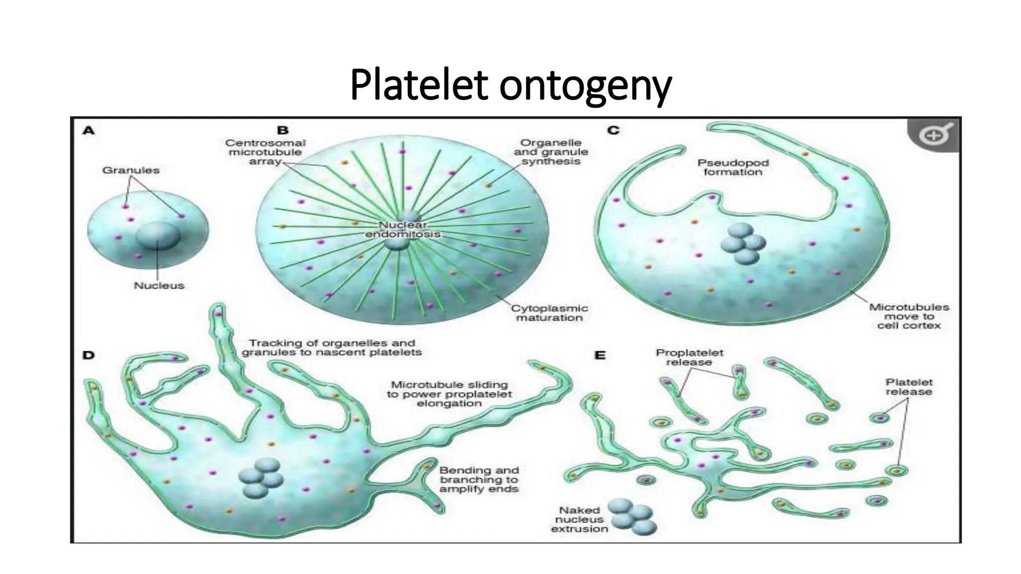
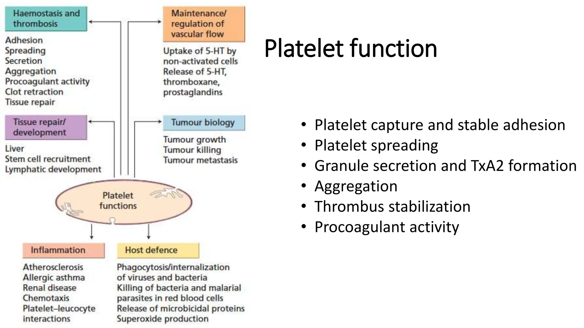
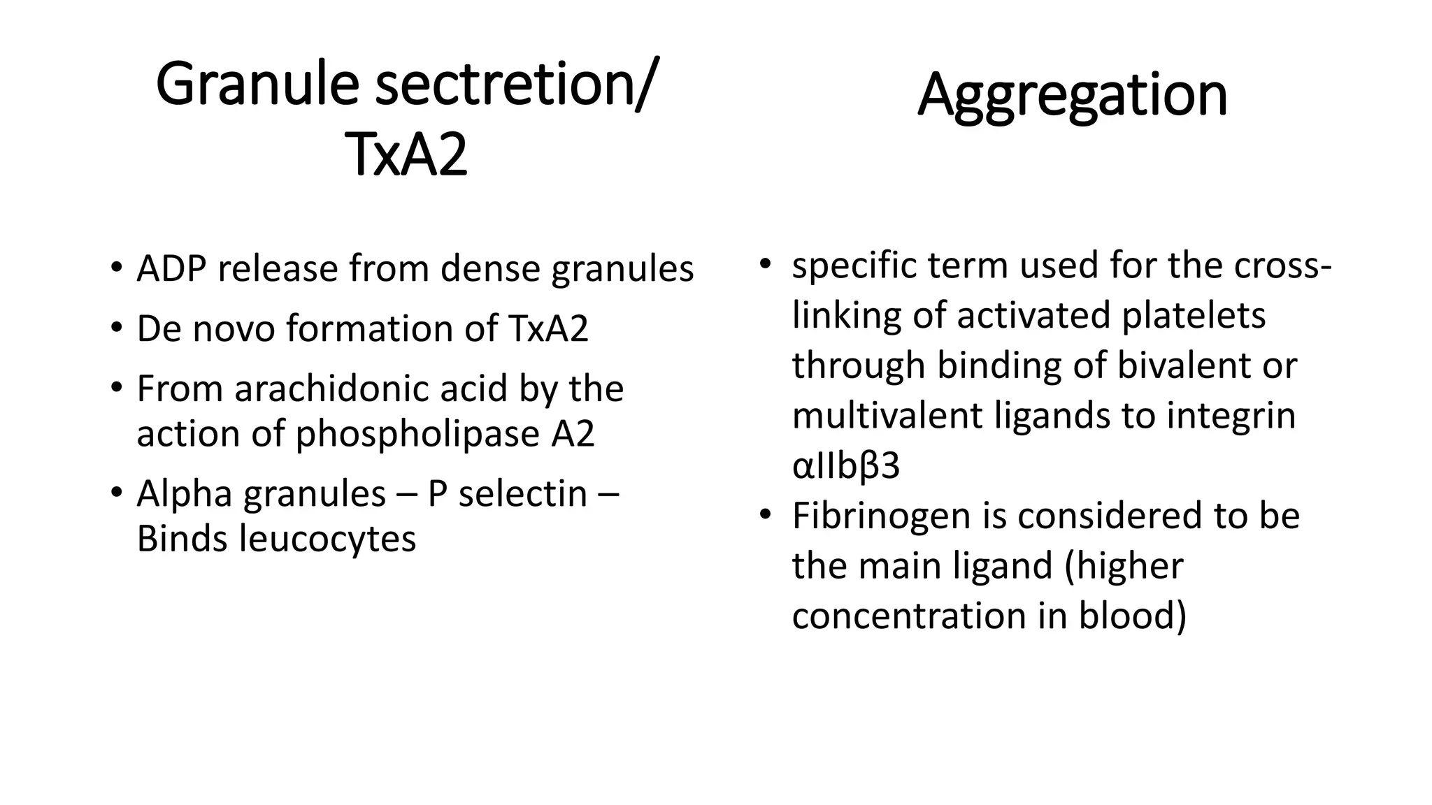
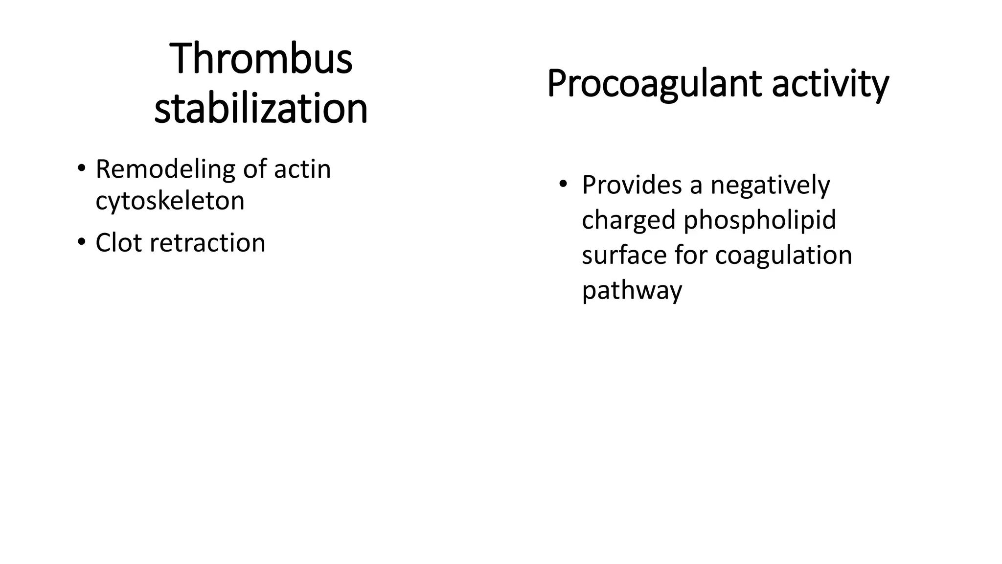
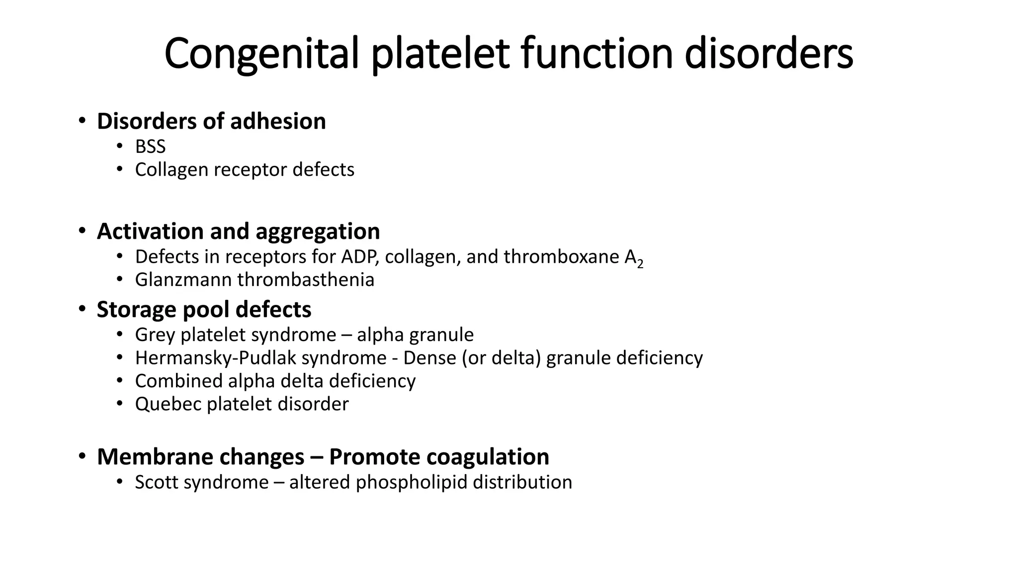
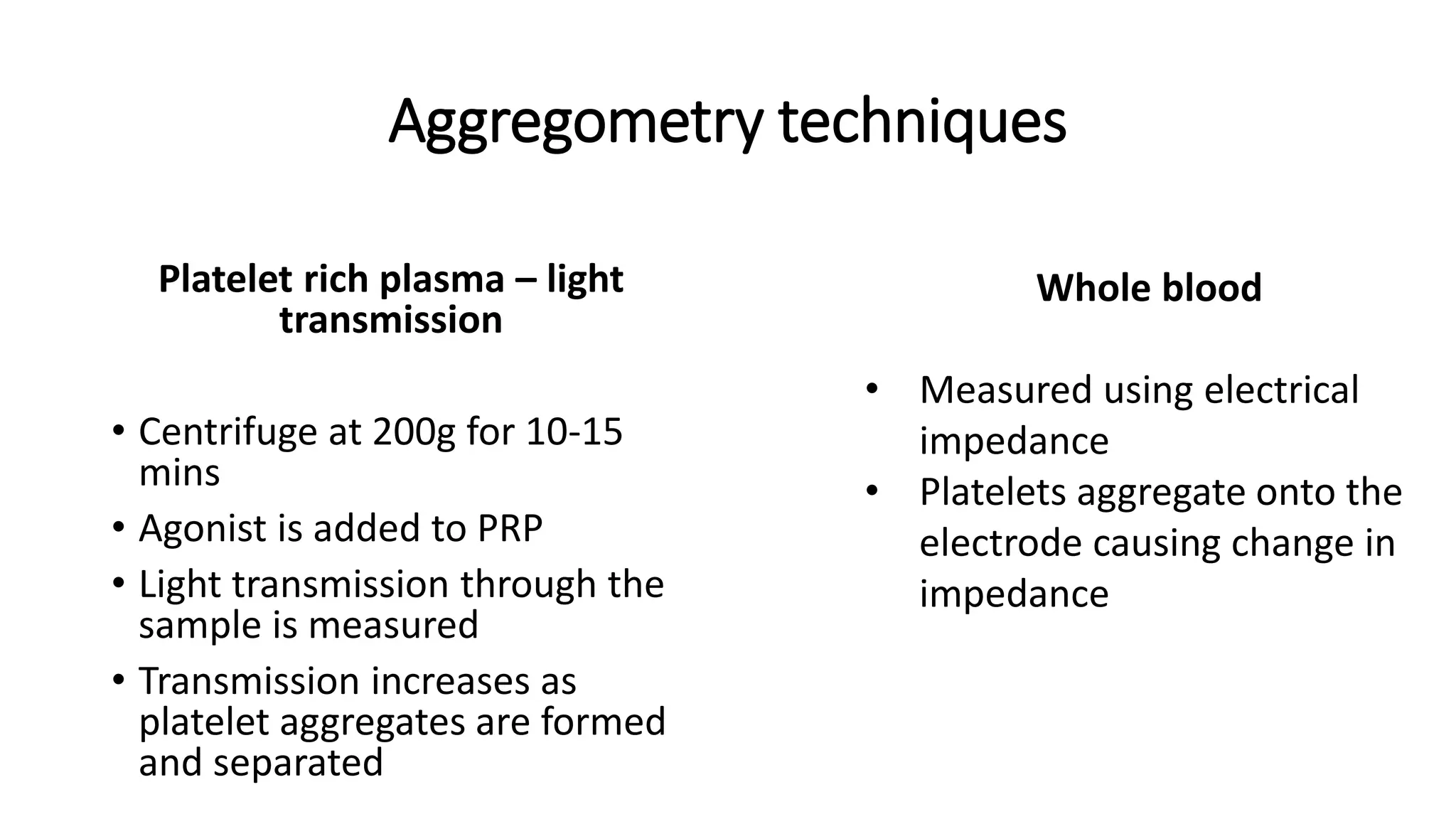
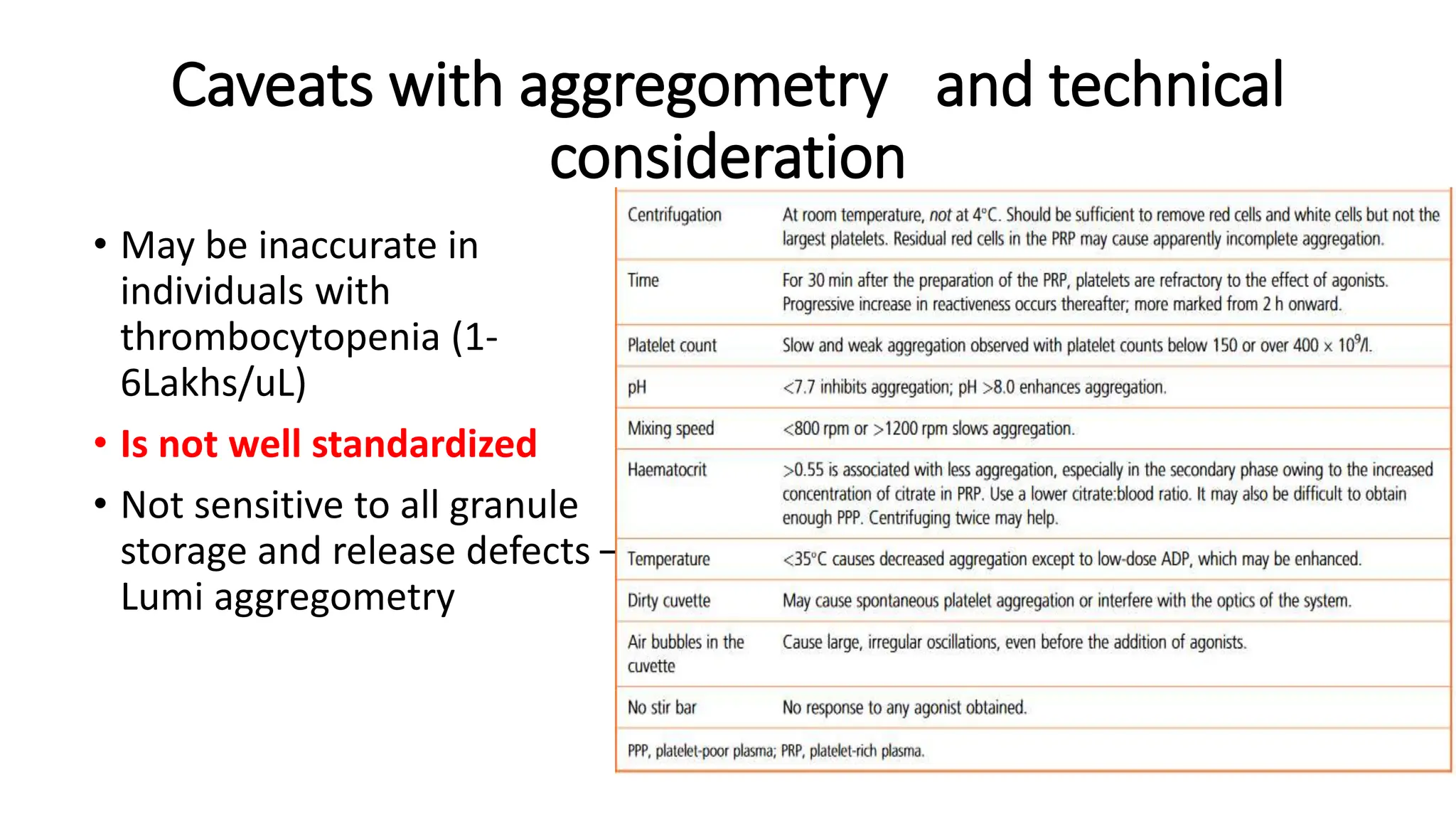
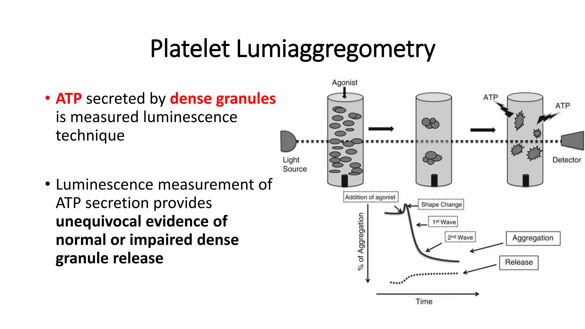
Platelet function disorders,
assessment and testing
Platelet ontogeny
• Originates from the
common
megakaryocyte
erythrocyte precursor
• Promegakaryocyte,
megakaryocyte,
proplatelet, platelet
Platelet ontogeny
Normal platelet function
• Megakaryopoietic agents
• stem cell factor, GM-CSF, IL3, IL6
• Thrombopoietin, c-mpl – hepatocytes, bone marrow stromal cells
• Endomitosis – upto 128n
• Produce 2000-3000 platelet per megakaryocyte
• Maturation - surface GPIb, GPII, von Willebrand factor
• Reside in bone marrow adjacent to sinusoids with pseudopods
projecting into the sinuses
Megakaryocyte showing emperipolesis
Endomitosis
Platelet physiology
• Discoid shape – normal volume 7-11fL – margination
• Rigid structure – Microtubules, actin filaments linked to GPIb
• Lifespan – 10 days
• 4 types of granules –
• Dense granules – ADP, ATP, phosphates,serotonis, Ca++
• Alpha granules – fibrinogen, VWF, fV, protein S
• Lysosomes
• Peroxisomes
Platelet function
• Platelet capture and stable adhesion
• Platelet spreading
• Granule secretion and TxA2 formation
• Aggregation
• Thrombus stabilization
• Procoagulant activity
Platelet capture and
stable adhesion
• Low flow/ shear rates
– directly adhere to
exposed
subendothelial
connective tissue
• High flow/ shear
rates – mediated by
the action of VWF
with GPIb-IX-V
Spreading
• Fillopodia formation
• Fried egg appearance
Granule sectretion/
TxA2
• ADP release from dense granules
• De novo formation of TxA2
• From arachidonic acid by the
action of phospholipase A2
• Alpha granules – P selectin –
Binds leucocytes
Aggregation
• specific term used for the cross-
linking of activated platelets
through binding of bivalent or
multivalent ligands to integrin
αIIbβ3
• Fibrinogen is considered to be
the main ligand (higher
concentration in blood)
Thrombus
stabilization
• Remodeling of actin
cytoskeleton
• Clot retraction
Procoagulant activity
• Provides a negatively
charged phospholipid
surface for coagulation
pathway
VWF – what, where and how
• Multimer varying in size from 500 – 20,000kDa
• Ultra-large VWF is stored in endothelial cells and released (Weibel-
palade bodies)
• Broken down into smaller multimers by ADAMTS-13
• VWF stabilizes factor VIII
• Globular form and extended forms (activated)
• Binds to GPIb-V-IX
Important molecules summarized
• GPIb–IX–V
• GPVI and integrin 𝛂2𝛃1 – Collagen receptor
• Integrin 𝛂IIb𝛃3 (GPIIb/IIIa)
• P2Y1 and P2Y12 ADP receptors – Adenosine receptor
• TxA2 receptor
• PAR-1 and PAR-4 – Thrombin receptors
• Ca++
Inhibitory agents
Cyclic
nucleotides
(cAMP, cGMP)
NO
Endothelial
cells
Congenital platelet function disorders
• Disorders of adhesion
• BSS
• Collagen receptor defects
• Activation and aggregation
• Defects in receptors for ADP, collagen, and thromboxane A2
• Glanzmann thrombasthenia
• Storage pool defects
• Grey platelet syndrome – alpha granule
• Hermansky-Pudlak syndrome - Dense (or delta) granule deficiency
• Combined alpha delta deficiency
• Quebec platelet disorder
• Membrane changes – Promote coagulation
• Scott syndrome – altered phospholipid distribution
Indication of platelet function testing
• Bleeding manifestations with unrevealing initial evaluation for
common bleeding disorders
• Positive family history for bleeding disorders
• Genetic test that point to platelet function disorder
• Usually not done for abnormal platelet morphology
Not useful for
• Monitoring antiplatelet therapy
• Predicting bleeding risk in people with thrombocytopenia
Adult with bleeding disorder- Actively bleeding
individual
• Search for anatomic and surgical lesions
• PT, aPTT, platelet count, fibrinogen levels, peripheral smear
• PFA-100, vWf testing
• Intervention –
• Platelet transfusion if thrombocytopenia
• Fibrinogen or Vit K in prolonged clotting time
• Clotting factors
• Antifibrinolytic drugs
Adult with bleeding disorder- Not actively
bleeding
• History
• Underlying conditions
• Cancer
• Excess alcohol use
• Liver disease
• Kidney disease
• Connective tissue disorders
• Hypothyroidism
• Response to bleeding
challenges
• Prior bleeding history
• During infancy/ stump bleeding
• Adolescence/ menstrual bleeding
• Severe bleeds requiring intervention
• History of iron deficiency
• Pregnancy history
• Bruising history
• Family history
Interpretation of history
BAT
• Low BAT score predicts absence of a bleeding disorder and can limit
excess testing
• Elevated BAT score predicts risk of subsequent bleeding
• Validated for
• Inherited platelet disorders
• VWD
• Heavy menstrual bleeding
• General bleeding evaluation
Bleeding assessment tool (BAT)
Pre testing evaluation
• Testing required prior to platelet function testing:-
• Thorough history, Bleeding assessment tool (BAT)
• Complete blood counts and peripheral smear study
• PT, aPTT
• Screening for von Willebrand disease (optional)
What are the available tests for
assessing platelet function
• Platelet aggregometry is gold standard
• PFA-100 – screening test
• Genetic testing
• Flow cytometry
• Bleeding time is not recommended
Pretesting considerations
• Medications: antiplatelet agents, NSAIDs, SSRIs
• Discontinue for at least 10 days prior to testing
• Thrombocytopenia: usually not recommended below a count of
80,000. Some cases require flow cytometric testing
• Preferably overnight fasting before collection of sample -
chylomicrons
Collection and transportation
• 20ml blood to be collected
• Minimal venous occlusion
• 1/10 volume of trisodium citrate
• Should not be chilled
• Testing done 30mins – 3hrs post centrifuging
Aggregometry techniques
Platelet rich plasma – light
transmission
• Centrifuge at 200g for 10-15
mins
• Agonist is added to PRP
• Light transmission through the
sample is measured
• Transmission increases as
platelet aggregates are formed
and separated
Whole blood
• Measured using electrical
impedance
• Platelets aggregate onto the
electrode causing change in
impedance
Platelet aggregometry – First panel
• Aggregometry measures platelet response (aggregation) to a panel of
agonists:
1. Collagen---------------------------------- Physiologic platelet activator
2. Adenosine diphosphate (ADP)------ Physiologic platelet activator
3. Epinephrine------------------------------ Physiologic platelet activator
4. Ristocetin -------------------------------- Antibiotic with platelet activation
5. Arachidonic acid------------------------ Physiologic platelet activator
• Agonists added at a ratio of 1:10 by volume
Second line of agonists that may be used
• 46619 (a thromboxane receptor agonist)
• Gamma thrombin
• TRAP (thrombin receptor activating peptides) that stimulate PAR-1
(peptide sequence SFLRRN) or PAR-4 (peptide sequence AYPGKF)
• Collagen Related Peptide (CRP) and Convulxin (a rattlesnake toxin),
which stimulate platelet GpVI
• Calcium ionophore A23187
• Phorbol 12-myristate-13-acetate
Interpretation of aggregometry
• A- shape
change
• B- primary wave
aggregation
• X- angle of initial
aggregation
• Y – height of
aggregation
trace
• D – lag phase
Caveats with aggregometry and technical
consideration
• May be inaccurate in
individuals with
thrombocytopenia (1-
6Lakhs/uL)
• Is not well standardized
• Not sensitive to all granule
storage and release defects –
Lumi aggregometry
Expected aggregometry panel for
thrombasthenias
PFA - 100
Attempts to reproduce
under high shear rates VWF
binding, platelet
attachment,
activation and aggregation,
which slowly build a stable
platelet plug at the
aperture
Can be used to exclude a diagnosis of function defect
Platelet Lumiaggregometry
• ATP secreted by dense granules
is measured luminescence
technique
• Luminescence measurement of
ATP secretion provides
unequivocal evidence of
normal or impaired dense
granule release
Flow cytometry
• Assay platelet surface glycoprotein deficiencies seen in heritable
platelet disorders
• Assay for activation markers after exposure to platelet agonists.
• Assay for dense granule deficiency and storage pool disease using a
granule binding dye
References
References
• ISTH/SSC bleeding assessment tool: a standardized questionnaire and
a proposal for a new bleeding score for inherited bleeding disorders.
Rodeghiero F, Tosetto A, Abshire T, Arnold DM. 2010;8(9):2063.
• https://www.uptodate.com/contents/approach-to-the-adult-with-a-
suspected-bleeding-
disorder?search=Platelet%20disorders&source=search_result&select
edTitle=2~150&usage_type=default&display_rank=2#H2100446941
• https://www.uptodate.com/contents/inherited-platelet-function-
disorders-
ipfds?search=Platelet%20disorders&source=search_result&selectedTi
tle=1~150&usage_type=default&display_rank=1

Platelet function disorders, assessment and testing.pptx

  • 1.
  • 2.
    Platelet ontogeny • Originatesfrom the common megakaryocyte erythrocyte precursor • Promegakaryocyte, megakaryocyte, proplatelet, platelet
  • 3.
  • 4.
    Normal platelet function •Megakaryopoietic agents • stem cell factor, GM-CSF, IL3, IL6 • Thrombopoietin, c-mpl – hepatocytes, bone marrow stromal cells • Endomitosis – upto 128n • Produce 2000-3000 platelet per megakaryocyte • Maturation - surface GPIb, GPII, von Willebrand factor • Reside in bone marrow adjacent to sinusoids with pseudopods projecting into the sinuses
  • 6.
  • 7.
  • 8.
    Platelet physiology • Discoidshape – normal volume 7-11fL – margination • Rigid structure – Microtubules, actin filaments linked to GPIb • Lifespan – 10 days • 4 types of granules – • Dense granules – ADP, ATP, phosphates,serotonis, Ca++ • Alpha granules – fibrinogen, VWF, fV, protein S • Lysosomes • Peroxisomes
  • 9.
    Platelet function • Plateletcapture and stable adhesion • Platelet spreading • Granule secretion and TxA2 formation • Aggregation • Thrombus stabilization • Procoagulant activity
  • 10.
    Platelet capture and stableadhesion • Low flow/ shear rates – directly adhere to exposed subendothelial connective tissue • High flow/ shear rates – mediated by the action of VWF with GPIb-IX-V Spreading • Fillopodia formation • Fried egg appearance
  • 11.
    Granule sectretion/ TxA2 • ADPrelease from dense granules • De novo formation of TxA2 • From arachidonic acid by the action of phospholipase A2 • Alpha granules – P selectin – Binds leucocytes Aggregation • specific term used for the cross- linking of activated platelets through binding of bivalent or multivalent ligands to integrin αIIbβ3 • Fibrinogen is considered to be the main ligand (higher concentration in blood)
  • 12.
    Thrombus stabilization • Remodeling ofactin cytoskeleton • Clot retraction Procoagulant activity • Provides a negatively charged phospholipid surface for coagulation pathway
  • 13.
    VWF – what,where and how • Multimer varying in size from 500 – 20,000kDa • Ultra-large VWF is stored in endothelial cells and released (Weibel- palade bodies) • Broken down into smaller multimers by ADAMTS-13 • VWF stabilizes factor VIII • Globular form and extended forms (activated) • Binds to GPIb-V-IX
  • 14.
    Important molecules summarized •GPIb–IX–V • GPVI and integrin 𝛂2𝛃1 – Collagen receptor • Integrin 𝛂IIb𝛃3 (GPIIb/IIIa) • P2Y1 and P2Y12 ADP receptors – Adenosine receptor • TxA2 receptor • PAR-1 and PAR-4 – Thrombin receptors • Ca++
  • 15.
  • 16.
    Congenital platelet functiondisorders • Disorders of adhesion • BSS • Collagen receptor defects • Activation and aggregation • Defects in receptors for ADP, collagen, and thromboxane A2 • Glanzmann thrombasthenia • Storage pool defects • Grey platelet syndrome – alpha granule • Hermansky-Pudlak syndrome - Dense (or delta) granule deficiency • Combined alpha delta deficiency • Quebec platelet disorder • Membrane changes – Promote coagulation • Scott syndrome – altered phospholipid distribution
  • 17.
    Indication of plateletfunction testing • Bleeding manifestations with unrevealing initial evaluation for common bleeding disorders • Positive family history for bleeding disorders • Genetic test that point to platelet function disorder • Usually not done for abnormal platelet morphology
  • 18.
    Not useful for •Monitoring antiplatelet therapy • Predicting bleeding risk in people with thrombocytopenia
  • 19.
    Adult with bleedingdisorder- Actively bleeding individual • Search for anatomic and surgical lesions • PT, aPTT, platelet count, fibrinogen levels, peripheral smear • PFA-100, vWf testing • Intervention – • Platelet transfusion if thrombocytopenia • Fibrinogen or Vit K in prolonged clotting time • Clotting factors • Antifibrinolytic drugs
  • 20.
    Adult with bleedingdisorder- Not actively bleeding • History • Underlying conditions • Cancer • Excess alcohol use • Liver disease • Kidney disease • Connective tissue disorders • Hypothyroidism • Response to bleeding challenges • Prior bleeding history • During infancy/ stump bleeding • Adolescence/ menstrual bleeding • Severe bleeds requiring intervention • History of iron deficiency • Pregnancy history • Bruising history • Family history
  • 21.
  • 22.
    BAT • Low BATscore predicts absence of a bleeding disorder and can limit excess testing • Elevated BAT score predicts risk of subsequent bleeding • Validated for • Inherited platelet disorders • VWD • Heavy menstrual bleeding • General bleeding evaluation
  • 23.
  • 24.
    Pre testing evaluation •Testing required prior to platelet function testing:- • Thorough history, Bleeding assessment tool (BAT) • Complete blood counts and peripheral smear study • PT, aPTT • Screening for von Willebrand disease (optional)
  • 25.
    What are theavailable tests for assessing platelet function • Platelet aggregometry is gold standard • PFA-100 – screening test • Genetic testing • Flow cytometry • Bleeding time is not recommended
  • 26.
    Pretesting considerations • Medications:antiplatelet agents, NSAIDs, SSRIs • Discontinue for at least 10 days prior to testing • Thrombocytopenia: usually not recommended below a count of 80,000. Some cases require flow cytometric testing • Preferably overnight fasting before collection of sample - chylomicrons
  • 27.
    Collection and transportation •20ml blood to be collected • Minimal venous occlusion • 1/10 volume of trisodium citrate • Should not be chilled • Testing done 30mins – 3hrs post centrifuging
  • 28.
    Aggregometry techniques Platelet richplasma – light transmission • Centrifuge at 200g for 10-15 mins • Agonist is added to PRP • Light transmission through the sample is measured • Transmission increases as platelet aggregates are formed and separated Whole blood • Measured using electrical impedance • Platelets aggregate onto the electrode causing change in impedance
  • 29.
    Platelet aggregometry –First panel • Aggregometry measures platelet response (aggregation) to a panel of agonists: 1. Collagen---------------------------------- Physiologic platelet activator 2. Adenosine diphosphate (ADP)------ Physiologic platelet activator 3. Epinephrine------------------------------ Physiologic platelet activator 4. Ristocetin -------------------------------- Antibiotic with platelet activation 5. Arachidonic acid------------------------ Physiologic platelet activator • Agonists added at a ratio of 1:10 by volume
  • 30.
    Second line ofagonists that may be used • 46619 (a thromboxane receptor agonist) • Gamma thrombin • TRAP (thrombin receptor activating peptides) that stimulate PAR-1 (peptide sequence SFLRRN) or PAR-4 (peptide sequence AYPGKF) • Collagen Related Peptide (CRP) and Convulxin (a rattlesnake toxin), which stimulate platelet GpVI • Calcium ionophore A23187 • Phorbol 12-myristate-13-acetate
  • 31.
    Interpretation of aggregometry •A- shape change • B- primary wave aggregation • X- angle of initial aggregation • Y – height of aggregation trace • D – lag phase
  • 32.
    Caveats with aggregometryand technical consideration • May be inaccurate in individuals with thrombocytopenia (1- 6Lakhs/uL) • Is not well standardized • Not sensitive to all granule storage and release defects – Lumi aggregometry
  • 34.
    Expected aggregometry panelfor thrombasthenias
  • 36.
    PFA - 100 Attemptsto reproduce under high shear rates VWF binding, platelet attachment, activation and aggregation, which slowly build a stable platelet plug at the aperture Can be used to exclude a diagnosis of function defect
  • 37.
    Platelet Lumiaggregometry • ATPsecreted by dense granules is measured luminescence technique • Luminescence measurement of ATP secretion provides unequivocal evidence of normal or impaired dense granule release
  • 38.
    Flow cytometry • Assayplatelet surface glycoprotein deficiencies seen in heritable platelet disorders • Assay for activation markers after exposure to platelet agonists. • Assay for dense granule deficiency and storage pool disease using a granule binding dye
  • 39.
  • 40.
    References • ISTH/SSC bleedingassessment tool: a standardized questionnaire and a proposal for a new bleeding score for inherited bleeding disorders. Rodeghiero F, Tosetto A, Abshire T, Arnold DM. 2010;8(9):2063. • https://www.uptodate.com/contents/approach-to-the-adult-with-a- suspected-bleeding- disorder?search=Platelet%20disorders&source=search_result&select edTitle=2~150&usage_type=default&display_rank=2#H2100446941 • https://www.uptodate.com/contents/inherited-platelet-function- disorders- ipfds?search=Platelet%20disorders&source=search_result&selectedTi tle=1~150&usage_type=default&display_rank=1

Editor's Notes

  • #3 Proplatelets are released into circulation
  • #6 Identifying megakaryocyte Localization of megakaryocyte Sinus vs fat spaces
  • #7 Multinucleated???
  • #8 Endomitosis – normal finding
  • #10 Questionable function
  • #11 FG- fibrinogen CRP – collagen related peptide THR - thrombin
  • #12 P selectin binds ??microvesicles
  • #13 A complex of FIXa and FVIIIa on the negatively charged lipid surface converts FX to FXa (tenase complex), which in turn forms a complex with FVa on the same surface to efficiently convert prothrombin to thrombin (prothrombinase complex). Scott syndrome
  • #14 TTP – lack of ADAMTS13 – large multimers – thrombosis Mutations in GPIbα, GPIbβ and GPIX give rise to the rare bleeding disorder Bernard–Soulier syndrome, which is characterized by macrothrombocytopenia
  • #15 Clopidogrel, prasugrel,,,, ticargrelor Aspirin Abciximab
  • #21 History of easy bruising, which is commonly reported, lacks sensitivity and specificity and may be a sign of trauma or skin fragility rather than a true bleeding disorder. Excessive menstrual bleeding has many causes (eg, an anatomic lesion, hormonal factors, or impaired hemostasis) Platelet and vascular disorders are generally characterized by mucosal bleeding Coagulation factor disorders are generally characterized by bleeding into muscles and joints and delayed bleeding after a bleeding challenge Inherited disorders tend to present in childhood if severe but frequently present in adulthood or only after a bleeding challenge if mild. Some inherited disorders have additional syndromic features Some acquired disorders are associated with an underlying condition such as pregnancy, cancer, or connective tissue diseases.
  • #22 Difficulty - Vague symptomatology Different presentation for identical disease processes Environmental effect – different exposure to bleeding challenges – trauma, surgeries, physical work
  • #23 Vwd - cutoff score of ≥4 points had a sensitivity of 100 percent, specificity of 87 percent, positive predictive value (PPV) of 0.20, and negative predictive value (NPV) of 1 for the diagnosis of VWD IPFD – median score approx. 9 Heavy menstrual bleeding – In a 2012 study involving 30 females with heavy menstrual bleeding for whom anatomic, hormonal, and medication-related causes had been excluded, use of the score with a cutoff of 3.5 points had a sensitivity of 85 percent, specificity of 90 percent, PPV of 0.89, and NPV of 0.86
  • #24 Distinction between 0 and 1 is of critical importance Normal range is <4 in adult males, <6 in adult females and <3 in children (3). https://bleedingscore.certe.nl
  • #30 Control sample will be platelet poor plasma 2000g for 20 mins
  • #31 ADP - aggregation testing, prepare 100, 50, 25, 10 and 5 mmol/l Collagen- 1 and 4 mg/ml Ristocetin sulphate – 12.5mg/ml Epinephrine – 20-200umol/L Arachidonic acid – 5, 10 mmol/l solution