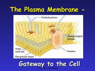
The Plasma Membrane  - Gateway to the Cell copyright cmassengale
Photograph of a Cell Membrane copyright cmassengale
Cell Membrane The cell membrane is  flexible  and allows a  unicellular  organism to  move copyright cmassengale
Homeostasis Balanced  internal condition of cells Also called  equilibrium Maintained by plasma membrane  controlling what enters & leaves the cell copyright cmassengale
Functions of Plasma Membrane Protective barrier Regulate transport in & out of cell  (selectively permeable) Allow cell recognition Provide anchoring sites for filaments  of cytoskeleton copyright cmassengale
Functions of Plasma Membrane Provide a binding site for enzymes Interlocking surfaces bind cells  together (junctions) Contains the cytoplasm (fluid in cell)  copyright cmassengale
Structure of the Cell Membrane copyright cmassengale
Phospholipids Cholesterol Proteins ( peripheral and integral) Carbohydrates (glucose) Membrane Components copyright cmassengale
copyright cmassengale
Phospholipids Make up the cell membrane Contains 2 fatty acid chains that are  nonpolar Head is  polar  & contains a  –PO 4  group & glycerol copyright cmassengale
FLUID - because individual phospholipids and proteins can move side-to-side within the layer, like it’s a liquid. MOSAIC - because of the pattern produced by the scattered protein molecules when the membrane is viewed from above . FLUID MOSAIC MODEL copyright cmassengale
Cell Membrane Polar heads are  hydrophilic  “water loving ” Nonpolar tails are  hydrophobic  “water fearing” Makes membrane  “Selective”  in what crosses copyright cmassengale
copyright cmassengale
Cell Membrane Hydrophobic  molecules pass  easily ; hydrophilic  DO NOT The cell membrane is made of  2 layers of  phospholipid s  called the lipid  bilayer copyright cmassengale
Solubility Materials that are soluble in  lipids  can pass through the cell membrane  easily copyright cmassengale
Semipermeable Membrane Small  molecules and  larger hydrophobic  molecules move through easily. e.g. O 2 , CO 2 , H 2 O copyright cmassengale
Ions ,  hydrophilic  molecules larger than water, and large molecules such as  proteins   do not move  through the membrane on their own.  Semipermeable Membrane copyright cmassengale
Types of Transport Across Cell Membranes copyright cmassengale
Simple Diffusion Requires  NO  energy Molecules move from area of  HIGH to LOW  concentration copyright cmassengale
DIFFUSION Diffusion is a  PASSIVE  process which means no energy is used to make the molecules move, they have a natural  KINETIC ENERGY copyright cmassengale
Diffusion of Liquids copyright cmassengale
Diffusion through a Membrane Cell membrane Solute moves  DOWN  concentration gradient  (HIGH to LOW) copyright cmassengale
Osmosis Diffusion of water   across a membrane Moves from   HIGH water potential   (low solute) to   LOW water potential   (high solute) Diffusion across a membrane Semipermeable membrane copyright cmassengale
Diffusion of H 2 O Across A Membrane High H 2 O potential Low solute concentration Low H 2 O potential High solute concentration copyright cmassengale
Aquaporins Water Channels Protein pores used during OSMOSIS WATER MOLECULES copyright cmassengale
Cell in Isotonic Solution CELL 10% NaCL 90% H 2 O 10% NaCL 90% H 2 O What is the direction of water movement? The cell is at _______________. equilibrium ENVIRONMENT NO NET MOVEMENT copyright cmassengale
Cell in Hypotonic Solution CELL 10% NaCL 90% H 2 O 20% NaCL 80% H 2 O What is the direction of water movement? copyright cmassengale
Cell in Hypertonic Solution CELL 15% NaCL 85% H 2 O 5% NaCL 95% H 2 O What is the direction of water movement? ENVIRONMENT copyright cmassengale
Cells in Solutions copyright cmassengale
Isotonic Solution NO NET MOVEMENT OF H 2 O (equal amounts entering & leaving) Hypotonic Solution CYTOLYSIS Hypertonic Solution PLASMOLYSIS copyright cmassengale
Cytolysis & Plasmolysis Cytolysis Plasmolysis copyright cmassengale
Osmosis in Red Blood Cells Isotonic Hypotonic Hypertonic copyright cmassengale
What Happens to Blood Cells? copyright cmassengale
hypotonic hypertonic isotonic hypertonic isotonic hypotonic copyright cmassengale
Three Forms of Transport Across the Membrane   copyright cmassengale
Passive Transport Simple Diffusion Doesn’t  require energy Moves  high to low  concentration Example:  Oxygen  or  water  diffusing into a cell and  carbon dioxide  diffusing out . copyright cmassengale
Passive Transport Facilitated diffusion Doesn’t  require energy Uses  transport proteins  to move  high to low  concentration Examples:  Glucose  or  amino acids  moving from blood into a cell. copyright cmassengale
Proteins Are Critical to Membrane Function copyright cmassengale
Types of Transport Proteins Channel proteins  are embedded in the cell membrane & have a  pore  for materials to cross Carrier proteins  can  change shape  to move material from one side of the membrane to the other copyright cmassengale
Facilitated Diffusion   Molecules will randomly move through the  pores  in  Channel Proteins . copyright cmassengale
Facilitated Diffusion Some  Carrier proteins  do not extend through the membrane. They  bond and drag molecules  through the lipid bilayer and release them on the opposite side.   copyright cmassengale
Carrier Proteins Other carrier proteins  change shape  to move materials across the cell membrane copyright cmassengale
Active Transport Requires energy or  ATP Moves materials from  LOW to HIGH  concentration AGAINST  concentration gradient copyright cmassengale
Active transport Examples: Pumping  Na +  (sodium ions) out and  K +  (potassium ions) in  against  strong concentration gradients . Called  Na+-K+ Pump copyright cmassengale
Sodium-Potassium Pump 3 Na+ pumped in for every 2 K+ pumped out; creates a membrane potential copyright cmassengale
Moving the “Big Stuff” Molecules are  moved out  of the cell by  vesicles  that  fuse  with the plasma membrane. Exocytosis -  moving things out. This is how many  hormones  are secreted and how  nerve cells  communicate with one another . copyright cmassengale
Exocytosis copyright cmassengale Exocytic vesicle immediately after fusion with plasma membrane.
Moving the “Big Stuff” Large molecules move materials into the cell by one of  three forms of endocytosis .  copyright cmassengale
Pinocytosis Most  common  form of endocytosis .   Takes in  dissolved  molecules as a vesicle .  copyright cmassengale
Pinocytosis Cell forms an  invagination Materials  dissolve in water  to be brought into cell Called  “Cell Drinking” copyright cmassengale
Example of Pinocytosis pinocytic vesicles forming mature transport vesicle Transport across a  capillary cell  (blue). copyright cmassengale
Receptor-Mediated Endocytosis Some  integral proteins  have  receptors  on their surface to recognize & take in  hormones, cholesterol , etc. copyright cmassengale
Receptor-Mediated Endocytosis copyright cmassengale
copyright cmassengale
Endocytosis – Phagocytosis  Used to  engulf large particles  such as food,  bacteria , etc. into vesicles Called  “Cell Eating” copyright cmassengale
copyright cmassengale
Phagocytosis About to Occur copyright cmassengale
Phagocytosis  - Capture of a  Yeast  Cell (yellow) by Membrane Extensions of an  Immune System Cell  (blue) copyright cmassengale
Exocytosis   The opposite of endocytosis is exocytosis.  Large molecules  that are manufactured in the cell are  released  through the cell membrane . Inside Cell Cell environment copyright cmassengale
copyright cmassengale

Plasma Membrane

  • 1.
    The Plasma Membrane - Gateway to the Cell copyright cmassengale
  • 2.
    Photograph of aCell Membrane copyright cmassengale
  • 3.
    Cell Membrane Thecell membrane is flexible and allows a unicellular organism to move copyright cmassengale
  • 4.
    Homeostasis Balanced internal condition of cells Also called equilibrium Maintained by plasma membrane controlling what enters & leaves the cell copyright cmassengale
  • 5.
    Functions of PlasmaMembrane Protective barrier Regulate transport in & out of cell (selectively permeable) Allow cell recognition Provide anchoring sites for filaments of cytoskeleton copyright cmassengale
  • 6.
    Functions of PlasmaMembrane Provide a binding site for enzymes Interlocking surfaces bind cells together (junctions) Contains the cytoplasm (fluid in cell) copyright cmassengale
  • 7.
    Structure of theCell Membrane copyright cmassengale
  • 8.
    Phospholipids Cholesterol Proteins( peripheral and integral) Carbohydrates (glucose) Membrane Components copyright cmassengale
  • 9.
  • 10.
    Phospholipids Make upthe cell membrane Contains 2 fatty acid chains that are nonpolar Head is polar & contains a –PO 4 group & glycerol copyright cmassengale
  • 11.
    FLUID - becauseindividual phospholipids and proteins can move side-to-side within the layer, like it’s a liquid. MOSAIC - because of the pattern produced by the scattered protein molecules when the membrane is viewed from above . FLUID MOSAIC MODEL copyright cmassengale
  • 12.
    Cell Membrane Polarheads are hydrophilic “water loving ” Nonpolar tails are hydrophobic “water fearing” Makes membrane “Selective” in what crosses copyright cmassengale
  • 13.
  • 14.
    Cell Membrane Hydrophobic molecules pass easily ; hydrophilic DO NOT The cell membrane is made of 2 layers of phospholipid s called the lipid bilayer copyright cmassengale
  • 15.
    Solubility Materials thatare soluble in lipids can pass through the cell membrane easily copyright cmassengale
  • 16.
    Semipermeable Membrane Small molecules and larger hydrophobic molecules move through easily. e.g. O 2 , CO 2 , H 2 O copyright cmassengale
  • 17.
    Ions , hydrophilic molecules larger than water, and large molecules such as proteins do not move through the membrane on their own. Semipermeable Membrane copyright cmassengale
  • 18.
    Types of TransportAcross Cell Membranes copyright cmassengale
  • 19.
    Simple Diffusion Requires NO energy Molecules move from area of HIGH to LOW concentration copyright cmassengale
  • 20.
    DIFFUSION Diffusion isa PASSIVE process which means no energy is used to make the molecules move, they have a natural KINETIC ENERGY copyright cmassengale
  • 21.
    Diffusion of Liquidscopyright cmassengale
  • 22.
    Diffusion through aMembrane Cell membrane Solute moves DOWN concentration gradient (HIGH to LOW) copyright cmassengale
  • 23.
    Osmosis Diffusion ofwater across a membrane Moves from HIGH water potential (low solute) to LOW water potential (high solute) Diffusion across a membrane Semipermeable membrane copyright cmassengale
  • 24.
    Diffusion of H2 O Across A Membrane High H 2 O potential Low solute concentration Low H 2 O potential High solute concentration copyright cmassengale
  • 25.
    Aquaporins Water ChannelsProtein pores used during OSMOSIS WATER MOLECULES copyright cmassengale
  • 26.
    Cell in IsotonicSolution CELL 10% NaCL 90% H 2 O 10% NaCL 90% H 2 O What is the direction of water movement? The cell is at _______________. equilibrium ENVIRONMENT NO NET MOVEMENT copyright cmassengale
  • 27.
    Cell in HypotonicSolution CELL 10% NaCL 90% H 2 O 20% NaCL 80% H 2 O What is the direction of water movement? copyright cmassengale
  • 28.
    Cell in HypertonicSolution CELL 15% NaCL 85% H 2 O 5% NaCL 95% H 2 O What is the direction of water movement? ENVIRONMENT copyright cmassengale
  • 29.
    Cells in Solutionscopyright cmassengale
  • 30.
    Isotonic Solution NONET MOVEMENT OF H 2 O (equal amounts entering & leaving) Hypotonic Solution CYTOLYSIS Hypertonic Solution PLASMOLYSIS copyright cmassengale
  • 31.
    Cytolysis & PlasmolysisCytolysis Plasmolysis copyright cmassengale
  • 32.
    Osmosis in RedBlood Cells Isotonic Hypotonic Hypertonic copyright cmassengale
  • 33.
    What Happens toBlood Cells? copyright cmassengale
  • 34.
    hypotonic hypertonic isotonichypertonic isotonic hypotonic copyright cmassengale
  • 35.
    Three Forms ofTransport Across the Membrane copyright cmassengale
  • 36.
    Passive Transport SimpleDiffusion Doesn’t require energy Moves high to low concentration Example: Oxygen or water diffusing into a cell and carbon dioxide diffusing out . copyright cmassengale
  • 37.
    Passive Transport Facilitateddiffusion Doesn’t require energy Uses transport proteins to move high to low concentration Examples: Glucose or amino acids moving from blood into a cell. copyright cmassengale
  • 38.
    Proteins Are Criticalto Membrane Function copyright cmassengale
  • 39.
    Types of TransportProteins Channel proteins are embedded in the cell membrane & have a pore for materials to cross Carrier proteins can change shape to move material from one side of the membrane to the other copyright cmassengale
  • 40.
    Facilitated Diffusion Molecules will randomly move through the pores in Channel Proteins . copyright cmassengale
  • 41.
    Facilitated Diffusion Some Carrier proteins do not extend through the membrane. They bond and drag molecules through the lipid bilayer and release them on the opposite side. copyright cmassengale
  • 42.
    Carrier Proteins Othercarrier proteins change shape to move materials across the cell membrane copyright cmassengale
  • 43.
    Active Transport Requiresenergy or ATP Moves materials from LOW to HIGH concentration AGAINST concentration gradient copyright cmassengale
  • 44.
    Active transport Examples:Pumping Na + (sodium ions) out and K + (potassium ions) in against strong concentration gradients . Called Na+-K+ Pump copyright cmassengale
  • 45.
    Sodium-Potassium Pump 3Na+ pumped in for every 2 K+ pumped out; creates a membrane potential copyright cmassengale
  • 46.
    Moving the “BigStuff” Molecules are moved out of the cell by vesicles that fuse with the plasma membrane. Exocytosis - moving things out. This is how many hormones are secreted and how nerve cells communicate with one another . copyright cmassengale
  • 47.
    Exocytosis copyright cmassengaleExocytic vesicle immediately after fusion with plasma membrane.
  • 48.
    Moving the “BigStuff” Large molecules move materials into the cell by one of three forms of endocytosis . copyright cmassengale
  • 49.
    Pinocytosis Most common form of endocytosis . Takes in dissolved molecules as a vesicle . copyright cmassengale
  • 50.
    Pinocytosis Cell formsan invagination Materials dissolve in water to be brought into cell Called “Cell Drinking” copyright cmassengale
  • 51.
    Example of Pinocytosispinocytic vesicles forming mature transport vesicle Transport across a capillary cell (blue). copyright cmassengale
  • 52.
    Receptor-Mediated Endocytosis Some integral proteins have receptors on their surface to recognize & take in hormones, cholesterol , etc. copyright cmassengale
  • 53.
  • 54.
  • 55.
    Endocytosis – Phagocytosis Used to engulf large particles such as food, bacteria , etc. into vesicles Called “Cell Eating” copyright cmassengale
  • 56.
  • 57.
    Phagocytosis About toOccur copyright cmassengale
  • 58.
    Phagocytosis -Capture of a Yeast Cell (yellow) by Membrane Extensions of an Immune System Cell (blue) copyright cmassengale
  • 59.
    Exocytosis The opposite of endocytosis is exocytosis. Large molecules that are manufactured in the cell are released through the cell membrane . Inside Cell Cell environment copyright cmassengale
  • 60.

Editor's Notes

  • #2 The Plasma Membrane 01/11/10 G. Podgorski, Biol. 1010
  • #3 The Plasma Membrane 01/11/10 G. Podgorski, Biol. 1010
  • #4 The Plasma Membrane 01/11/10 G. Podgorski, Biol. 1010
  • #5 The Plasma Membrane 01/11/10 G. Podgorski, Biol. 1010
  • #6 The Plasma Membrane 01/11/10 G. Podgorski, Biol. 1010
  • #7 The Plasma Membrane 01/11/10 G. Podgorski, Biol. 1010
  • #8 The Plasma Membrane 01/11/10 G. Podgorski, Biol. 1010
  • #9 The Plasma Membrane 01/11/10 G. Podgorski, Biol. 1010
  • #11 The Plasma Membrane 01/11/10 G. Podgorski, Biol. 1010
  • #12 The Plasma Membrane 01/11/10 G. Podgorski, Biol. 1010
  • #13 The Plasma Membrane 01/11/10 G. Podgorski, Biol. 1010
  • #14 The Plasma Membrane 01/11/10 G. Podgorski, Biol. 1010
  • #15 The Plasma Membrane 01/11/10 G. Podgorski, Biol. 1010
  • #16 The Plasma Membrane 01/11/10 G. Podgorski, Biol. 1010
  • #17 The Plasma Membrane 01/11/10 G. Podgorski, Biol. 1010
  • #18 The Plasma Membrane 01/11/10 G. Podgorski, Biol. 1010
  • #19 The Plasma Membrane 01/11/10 G. Podgorski, Biol. 1010
  • #20 The Plasma Membrane 01/11/10 G. Podgorski, Biol. 1010
  • #21 The Plasma Membrane 01/11/10 G. Podgorski, Biol. 1010
  • #22 The Plasma Membrane 01/11/10 G. Podgorski, Biol. 1010
  • #23 The Plasma Membrane 01/11/10 G. Podgorski, Biol. 1010
  • #24 The Plasma Membrane 01/11/10 G. Podgorski, Biol. 1010
  • #25 The Plasma Membrane 01/11/10 G. Podgorski, Biol. 1010
  • #26 The Plasma Membrane 01/11/10 G. Podgorski, Biol. 1010
  • #27 The Plasma Membrane 01/11/10 G. Podgorski, Biol. 1010
  • #28 The Plasma Membrane 01/11/10 G. Podgorski, Biol. 1010
  • #29 The Plasma Membrane 01/11/10 G. Podgorski, Biol. 1010
  • #30 The Plasma Membrane 01/11/10 G. Podgorski, Biol. 1010
  • #31 The Plasma Membrane 01/11/10 G. Podgorski, Biol. 1010
  • #32 The Plasma Membrane 01/11/10 G. Podgorski, Biol. 1010
  • #33 The Plasma Membrane 01/11/10 G. Podgorski, Biol. 1010
  • #35 The Plasma Membrane 01/11/10 G. Podgorski, Biol. 1010
  • #36 The Plasma Membrane 01/11/10 G. Podgorski, Biol. 1010
  • #37 The Plasma Membrane 01/11/10 G. Podgorski, Biol. 1010
  • #38 The Plasma Membrane 01/11/10 G. Podgorski, Biol. 1010
  • #39 The Plasma Membrane 01/11/10 G. Podgorski, Biol. 1010
  • #40 The Plasma Membrane 01/11/10 G. Podgorski, Biol. 1010
  • #41 The Plasma Membrane 01/11/10 G. Podgorski, Biol. 1010
  • #42 The Plasma Membrane 01/11/10 G. Podgorski, Biol. 1010
  • #43 The Plasma Membrane 01/11/10 G. Podgorski, Biol. 1010
  • #44 The Plasma Membrane 01/11/10 G. Podgorski, Biol. 1010
  • #45 The Plasma Membrane 01/11/10 G. Podgorski, Biol. 1010
  • #46 The Plasma Membrane 01/11/10 G. Podgorski, Biol. 1010
  • #47 The Plasma Membrane 01/11/10 G. Podgorski, Biol. 1010
  • #48 The Plasma Membrane 01/11/10 G. Podgorski, Biol. 1010
  • #49 The Plasma Membrane 01/11/10 G. Podgorski, Biol. 1010
  • #50 The Plasma Membrane 01/11/10 G. Podgorski, Biol. 1010
  • #51 The Plasma Membrane 01/11/10 G. Podgorski, Biol. 1010
  • #52 The Plasma Membrane 01/11/10 G. Podgorski, Biol. 1010
  • #53 The Plasma Membrane 01/11/10 G. Podgorski, Biol. 1010
  • #54 The Plasma Membrane 01/11/10 G. Podgorski, Biol. 1010
  • #56 The Plasma Membrane 01/11/10 G. Podgorski, Biol. 1010
  • #58 The Plasma Membrane 01/11/10 G. Podgorski, Biol. 1010
  • #59 The Plasma Membrane 01/11/10 G. Podgorski, Biol. 1010
  • #60 The Plasma Membrane 01/11/10 G. Podgorski, Biol. 1010
  • #61 The Plasma Membrane 01/11/10 G. Podgorski, Biol. 1010