Download to read offline

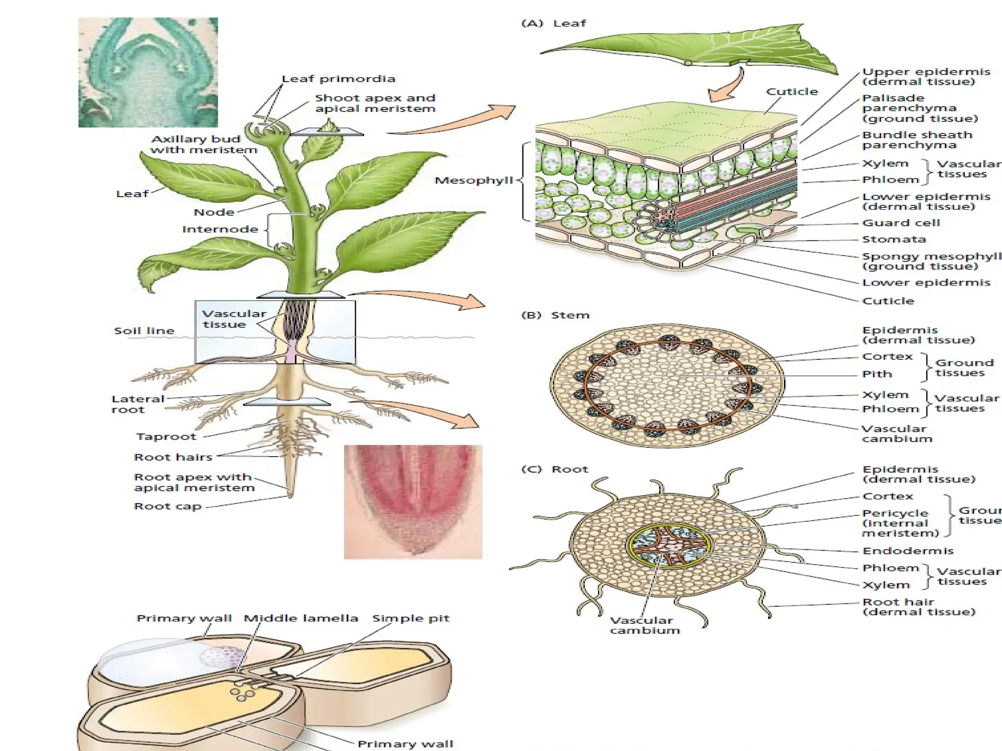
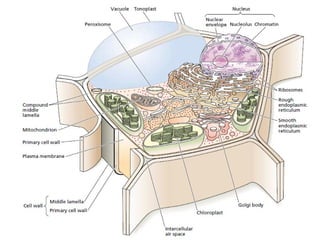
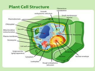
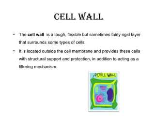
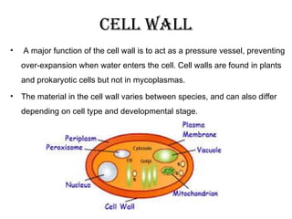
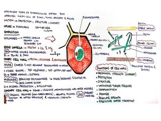
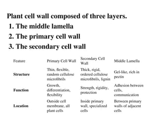
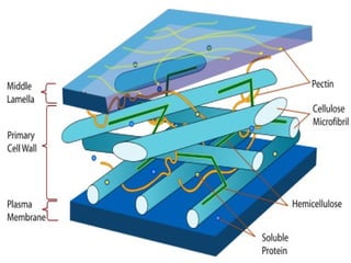
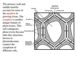
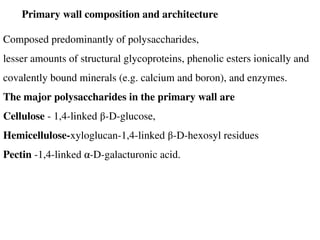
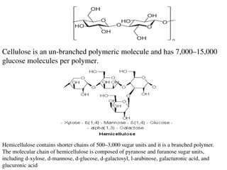
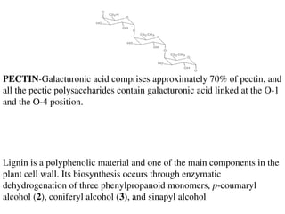
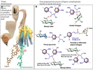
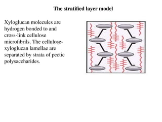
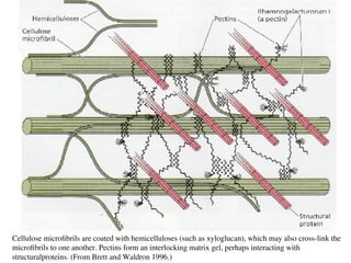
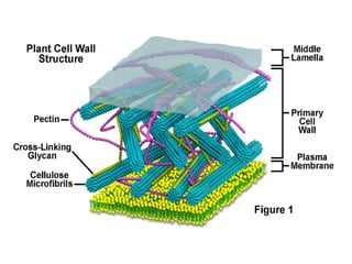
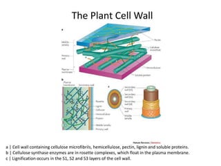
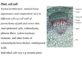
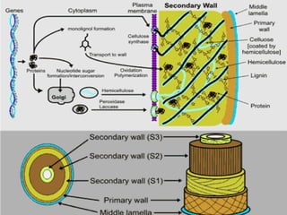
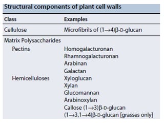
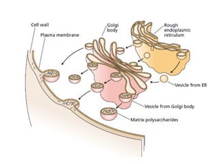

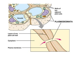

The document discusses the structure and functions of plant cell walls, highlighting their roles in providing support, protection, and regulating the flow of nutrients. It details the composition of the cell wall, including cellulose, hemicellulose, lignin, and pectin, and describes the three distinct layers: middle lamella, primary wall, and secondary wall. Furthermore, it addresses the implications of cell wall properties for plant growth and commercial uses of cell wall components.