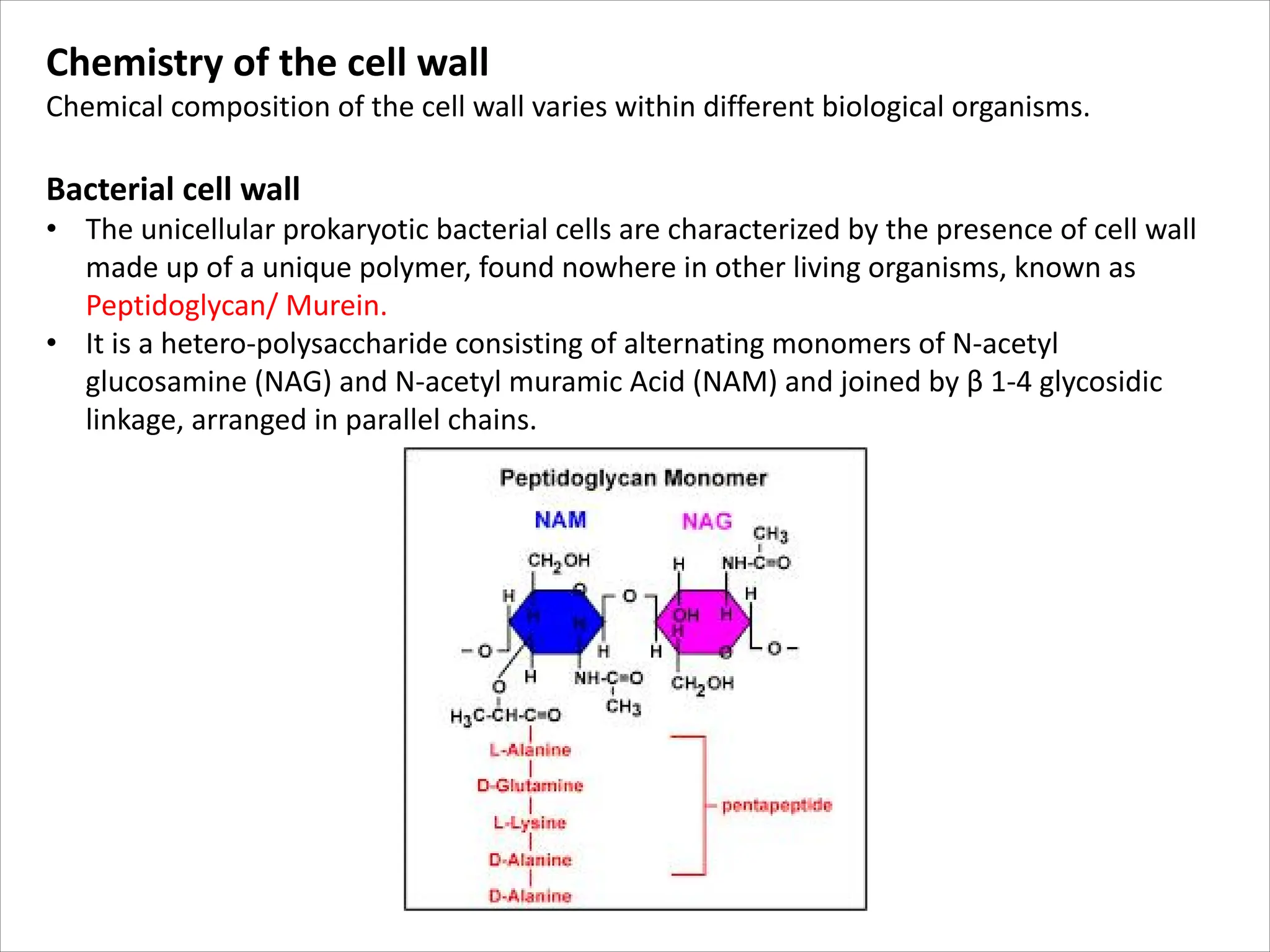
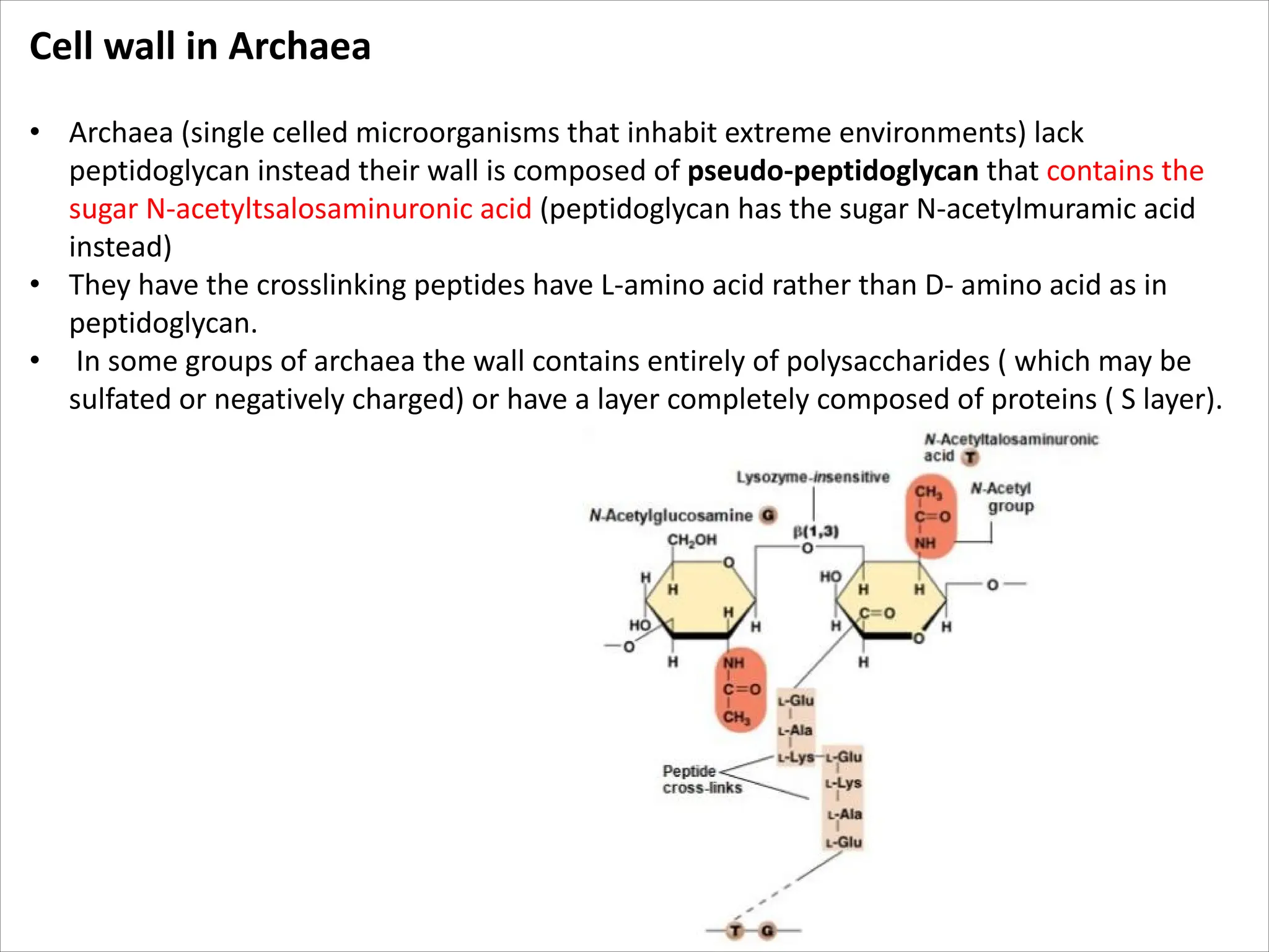
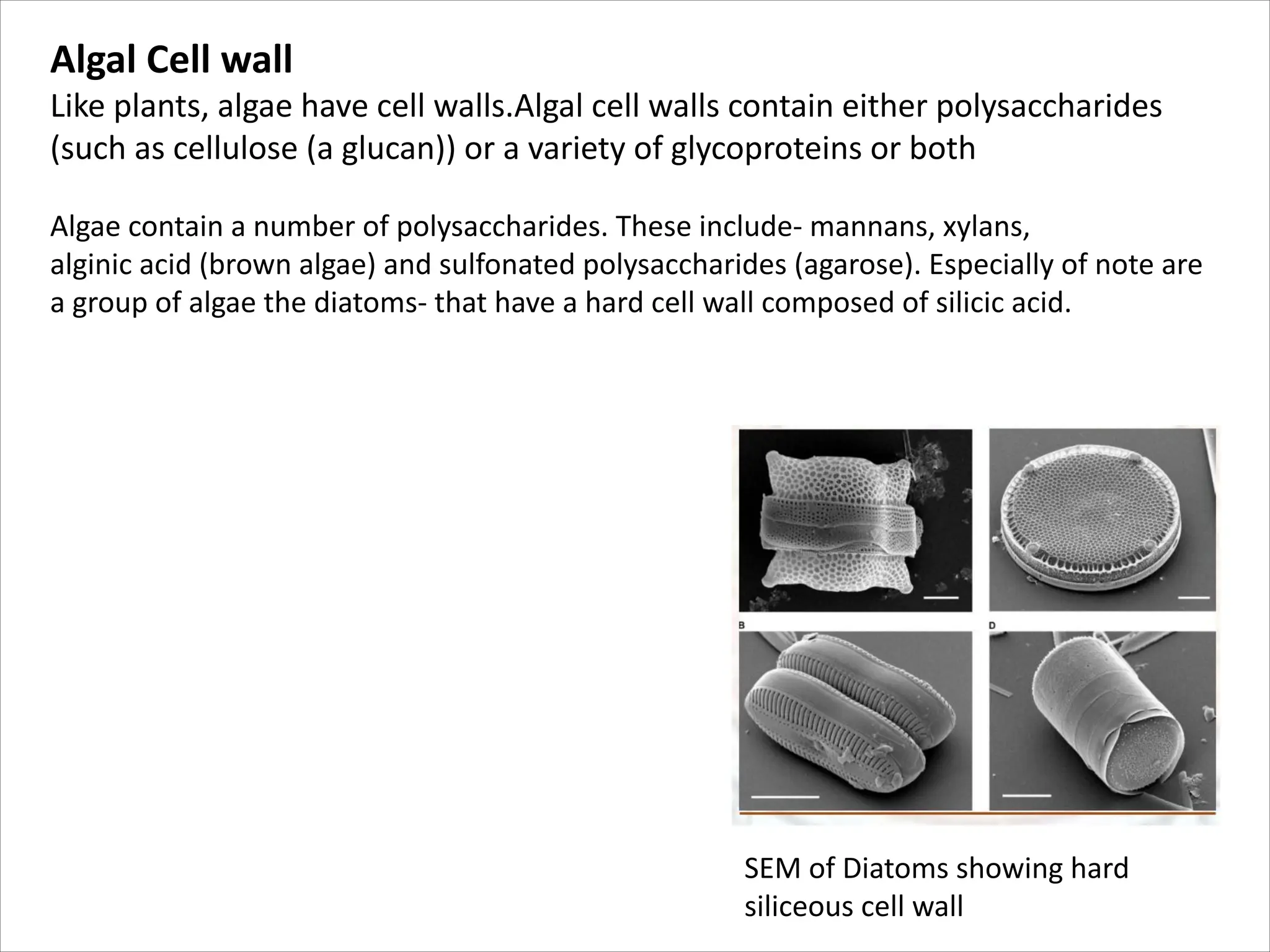
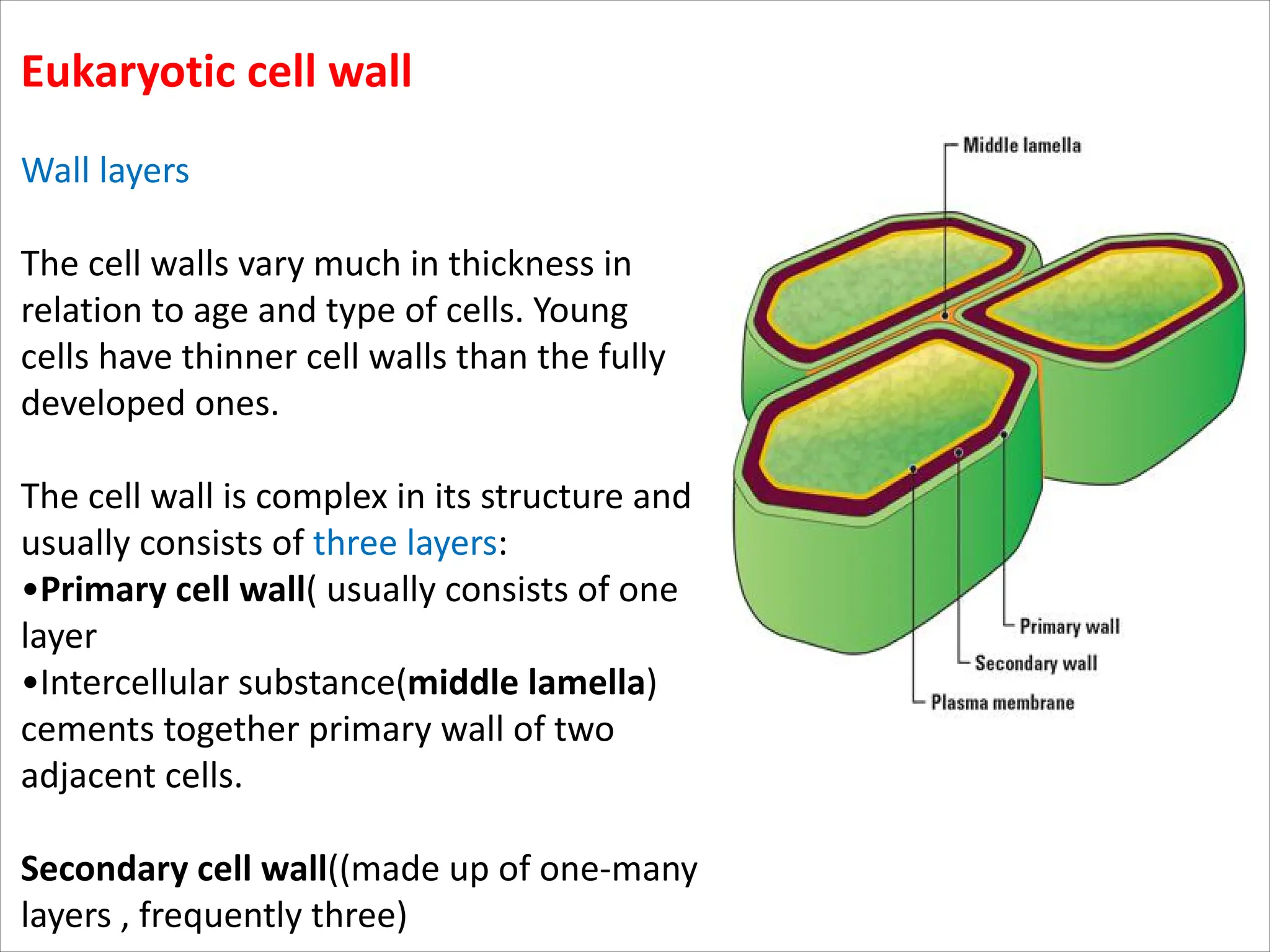
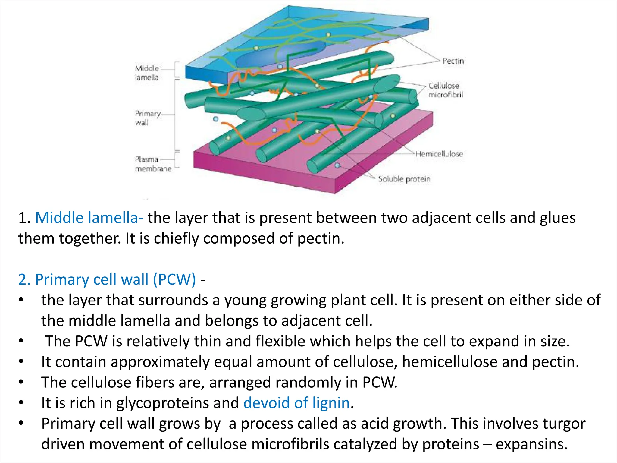
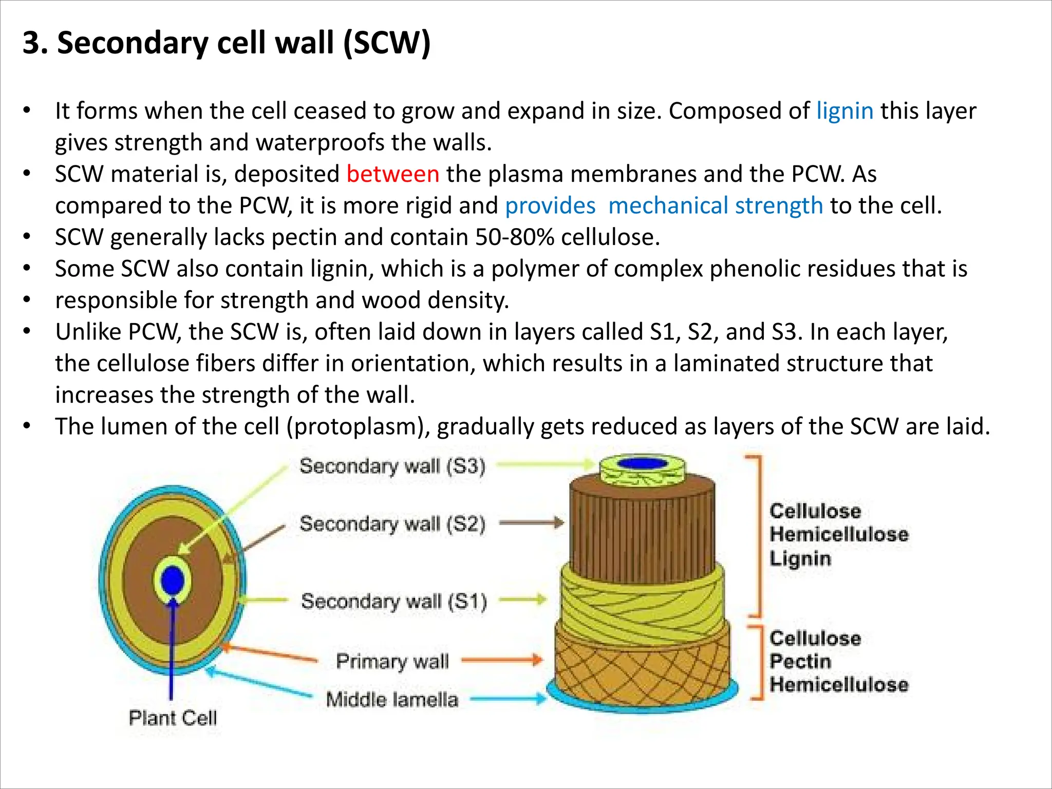
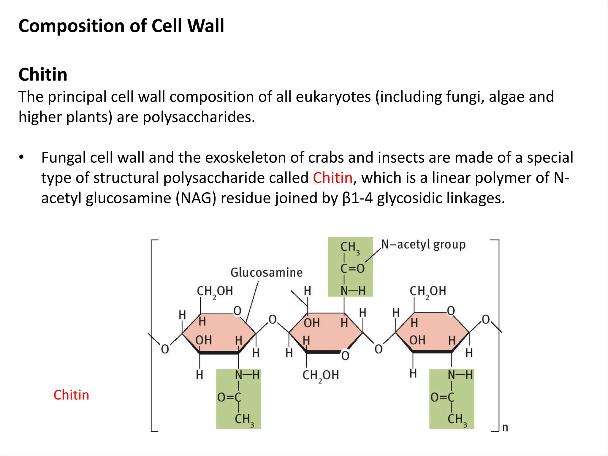
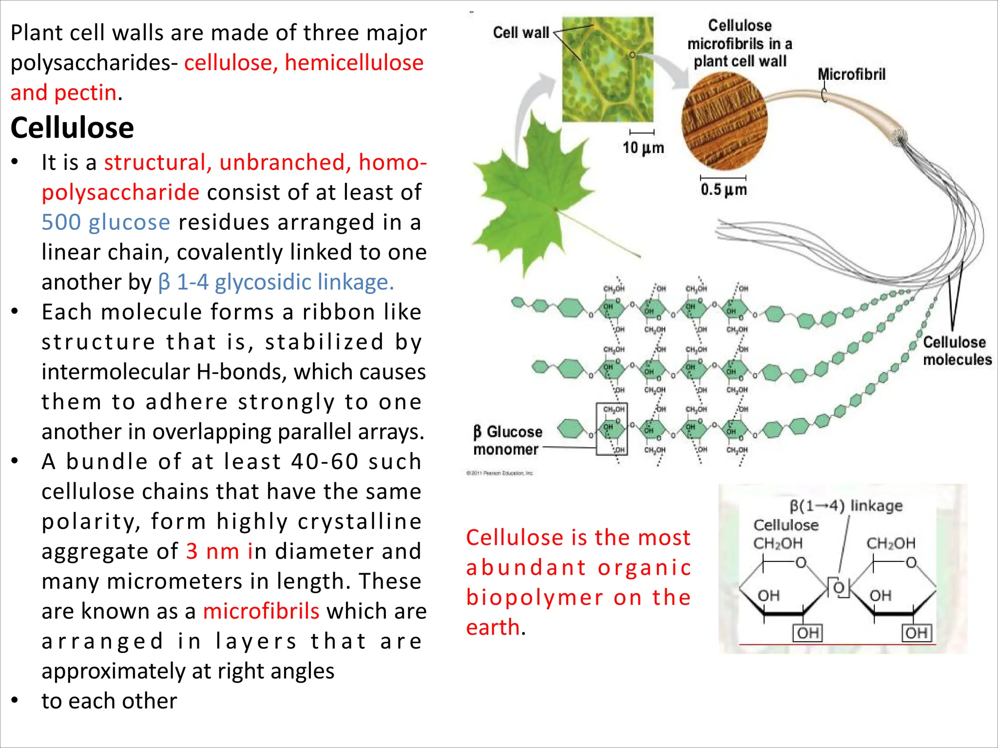
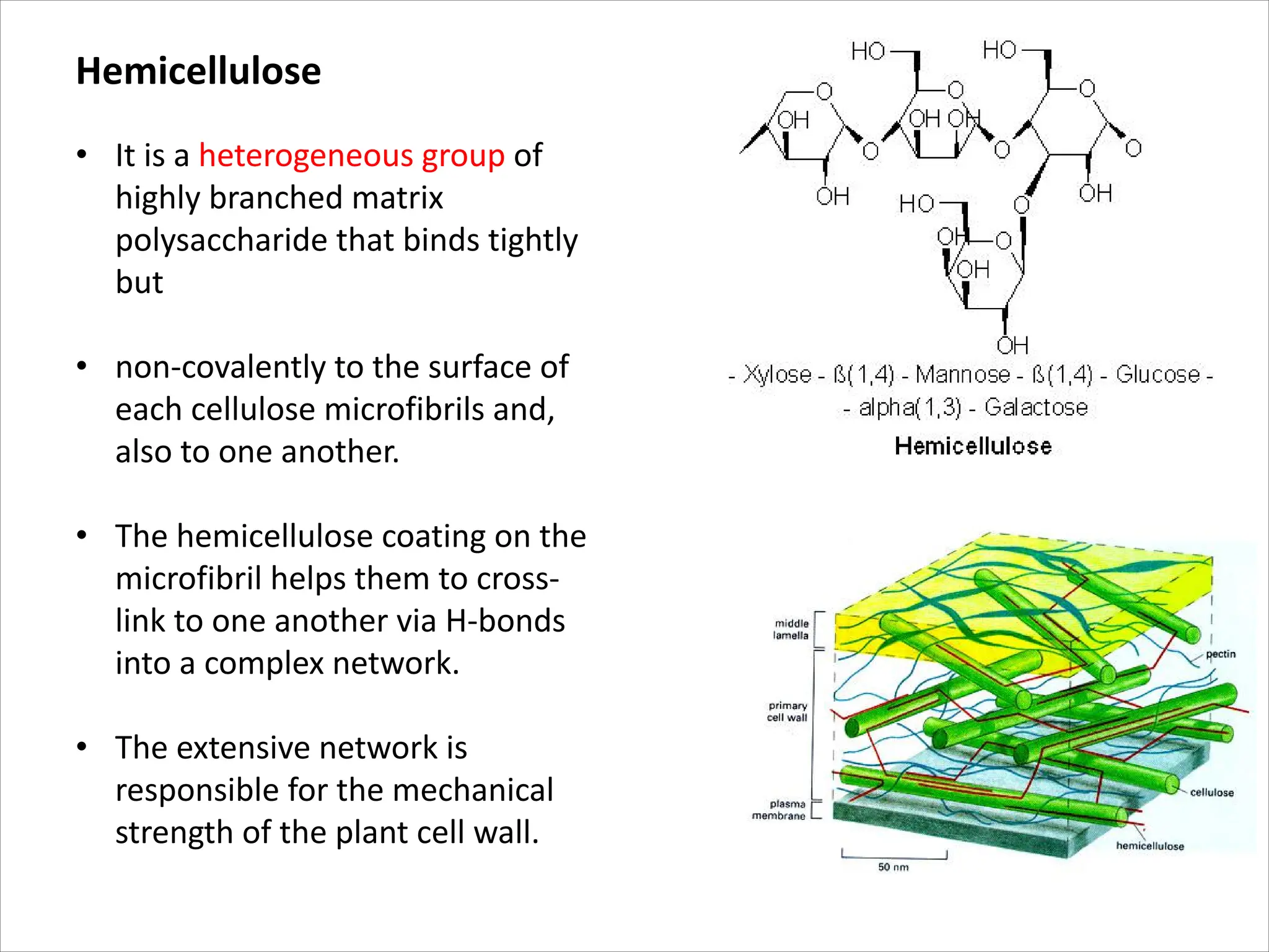
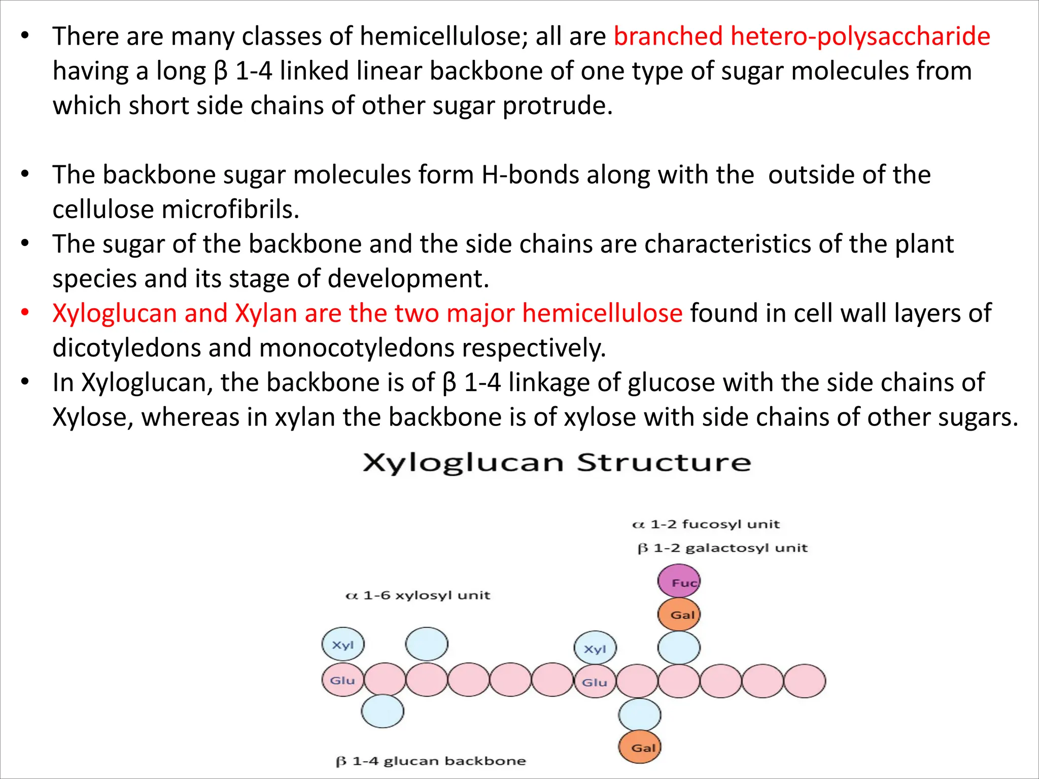
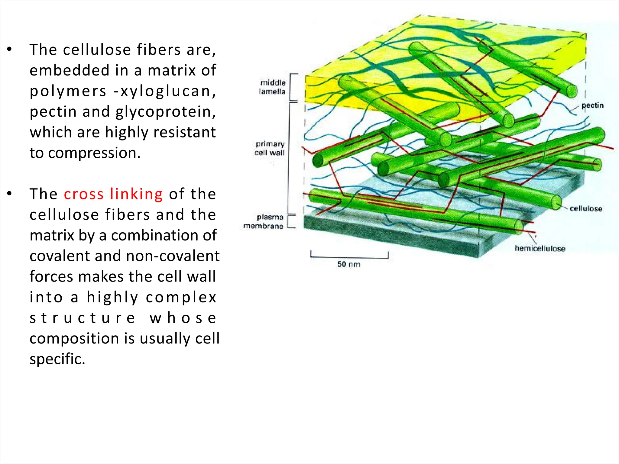
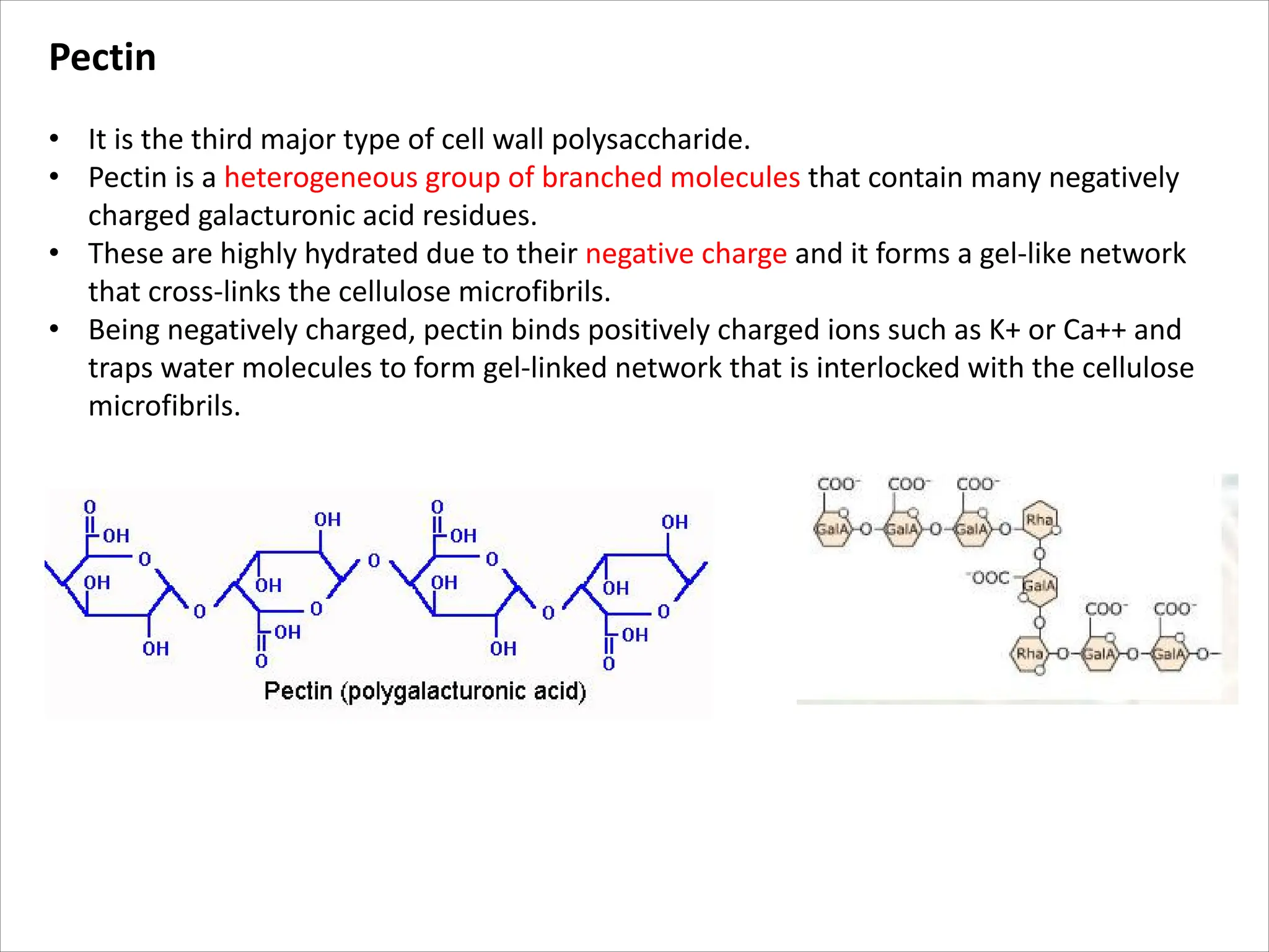
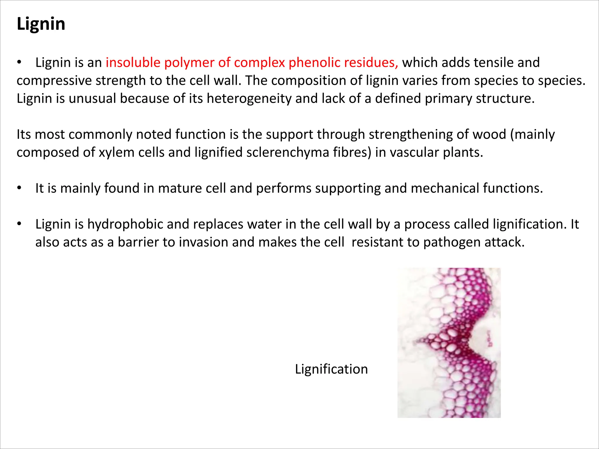
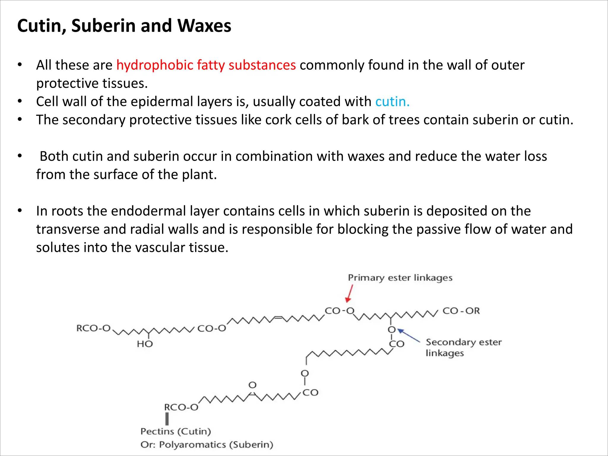
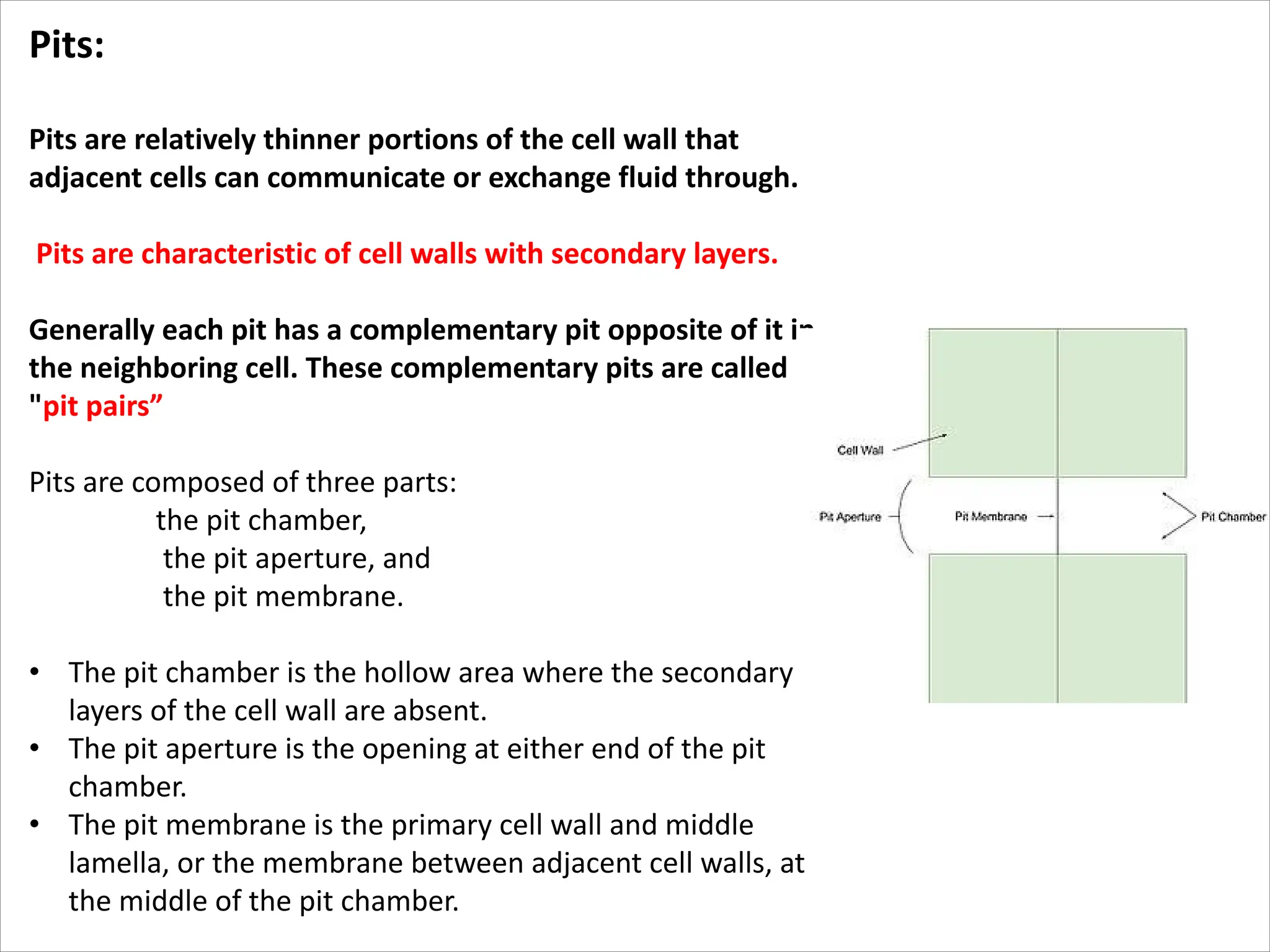
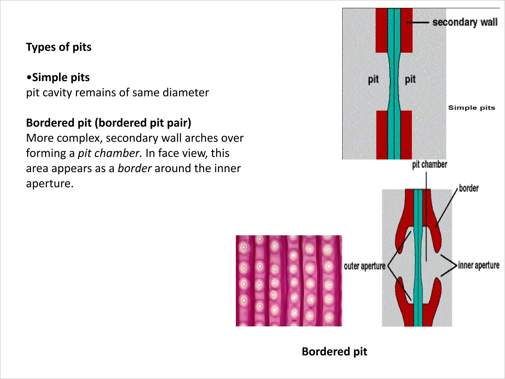
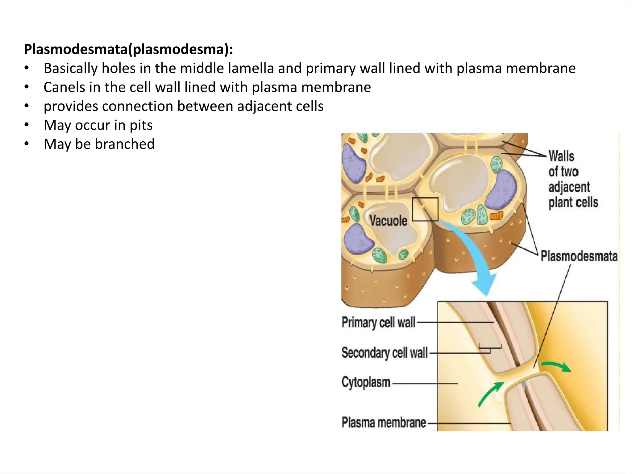
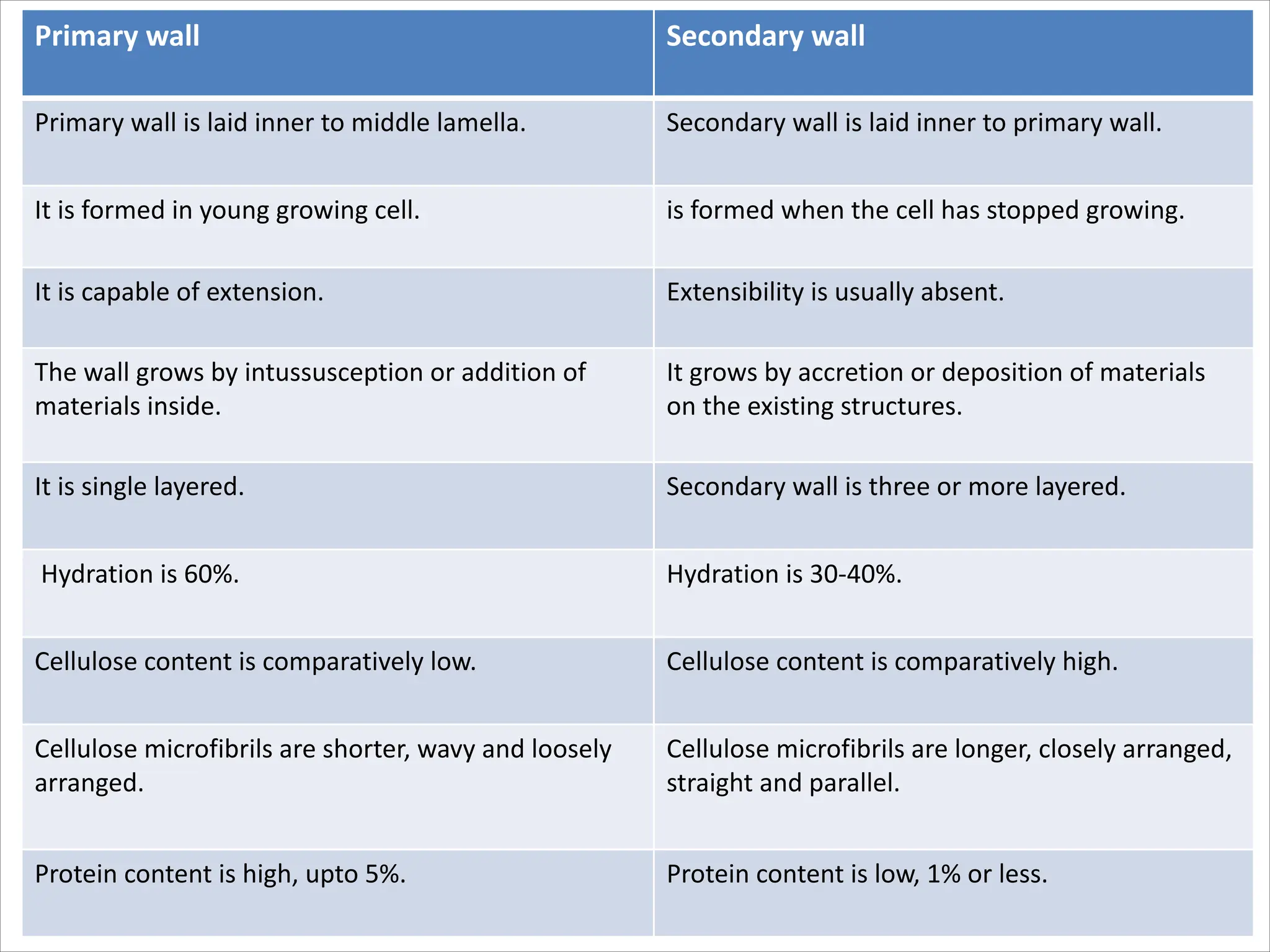
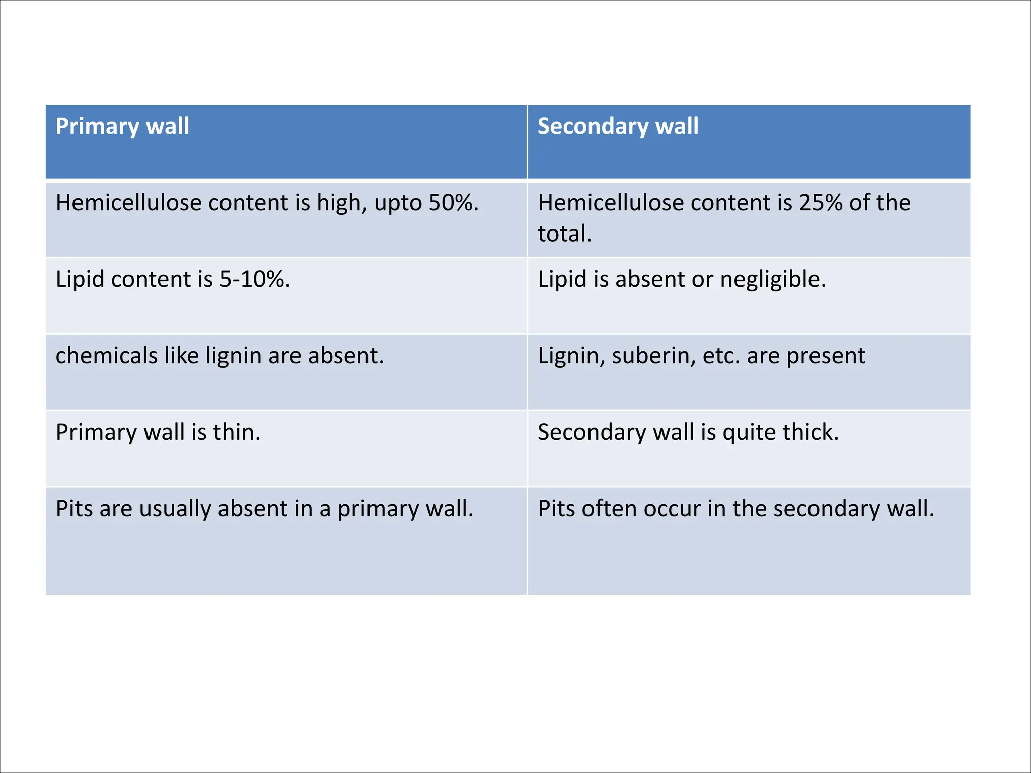
Cell wall
• Introduction
• Functions
• Chemistry
Fungal cell walls
Bacterial cell walls &Archaeal cell walls
Algal cell wall
Plant cell wall
Structure & composition of Primary & secondary cell wall
Pits, Plasmodesmata
Growth
• Differences between Primary & secondary cell wall
• Cell wall is one of the unique features in the plant cell that
distinguishes it from the animal cell.
• The boundary of each cell in plants is, defined by the cell wall.
• It is made of insoluble structural polysaccharide secreted by the cells.
• The plasma membrane forms the outermost layer in animal cells
where as in plants the plasma membrane lies next to the cell wall,
which provides an elaborate extracellular matrix.
• Plant cell wall can be modified in response to changing
environmental conditions.
Some of the important functions of cell wall are:
• Gives structural and mechanical support thus determining the
overall form and architecture of the plant
• Gives definite shape to the cell
• Prevents cell from bursting by resisting the turgor pressure
from inside the cell.
• Regulates exchange of material through the apoplast
• Determines the growth direction and rate
• Protection for the cellular constituents.
• Cell – cell interaction
• Storage in the form of wall carbohydrates
• Source of biological signaling molecules
Chemistry of the cell wall
Chemical composition of the cell wall varies within different biological organisms.
Bacterial cell wall
• The unicellular prokaryotic bacterial cells are characterized by the presence of cell wall
made up of a unique polymer, found nowhere in other living organisms, known as
Peptidoglycan/ Murein.
• It is a hetero-polysaccharide consisting of alternating monomers of N-acetyl
glucosamine (NAG) and N-acetyl muramic Acid (NAM) and joined by β 1-4 glycosidic
linkage, arranged in parallel chains.
These parallel chains are, cross-linked by tetra-peptide attached to the NAM residues.
The amino acids forming the tetra-peptides vary in different species of bacteria.
Gram positive and Gram negative bacteria
Cell wall in Archaea
• Archaea (single celled microorganisms that inhabit extreme environments) lack
peptidoglycan instead their wall is composed of pseudo-peptidoglycan that contains the
sugar N-acetyltsalosaminuronic acid (peptidoglycan has the sugar N-acetylmuramic acid
instead)
• They have the crosslinking peptides have L-amino acid rather than D- amino acid as in
peptidoglycan.
• In some groups of archaea the wall contains entirely of polysaccharides ( which may be
sulfated or negatively charged) or have a layer completely composed of proteins ( S layer).
Algal Cell wall
Like plants, algae have cell walls.Algal cell walls contain either polysaccharides
(such as cellulose (a glucan)) or a variety of glycoproteins or both
Algae contain a number of polysaccharides. These include- mannans, xylans,
alginic acid (brown algae) and sulfonated polysaccharides (agarose). Especially of note are
a group of algae the diatoms- that have a hard cell wall composed of silicic acid.
SEM of Diatoms showing hard
siliceous cell wall
Eukaryotic cell wall
Wall layers
The cell walls vary much in thickness in
relation to age and type of cells. Young
cells have thinner cell walls than the fully
developed ones.
The cell wall is complex in its structure and
usually consists of three layers:
•Primary cell wall( usually consists of one
layer
•Intercellular substance(middle lamella)
cements together primary wall of two
adjacent cells.
Secondary cell wall((made up of one-many
layers , frequently three)
1. Middle lamella- the layer that is present between two adjacent cells and glues
them together. It is chiefly composed of pectin.
2. Primary cell wall (PCW) -
• the layer that surrounds a young growing plant cell. It is present on either side of
the middle lamella and belongs to adjacent cell.
• The PCW is relatively thin and flexible which helps the cell to expand in size.
• It contain approximately equal amount of cellulose, hemicellulose and pectin.
• The cellulose fibers are, arranged randomly in PCW.
• It is rich in glycoproteins and devoid of lignin.
• Primary cell wall grows by a process called as acid growth. This involves turgor
driven movement of cellulose microfibrils catalyzed by proteins – expansins.
3. Secondary cell wall (SCW)
• It forms when the cell ceased to grow and expand in size. Composed of lignin this layer
gives strength and waterproofs the walls.
• SCW material is, deposited between the plasma membranes and the PCW. As
compared to the PCW, it is more rigid and provides mechanical strength to the cell.
• SCW generally lacks pectin and contain 50-80% cellulose.
• Some SCW also contain lignin, which is a polymer of complex phenolic residues that is
• responsible for strength and wood density.
• Unlike PCW, the SCW is, often laid down in layers called S1, S2, and S3. In each layer,
the cellulose fibers differ in orientation, which results in a laminated structure that
increases the strength of the wall.
• The lumen of the cell (protoplasm), gradually gets reduced as layers of the SCW are laid.
Composition of Cell Wall
Chitin
The principal cell wall composition of all eukaryotes (including fungi, algae and
higher plants) are polysaccharides.
• Fungal cell wall and the exoskeleton of crabs and insects are made of a special
type of structural polysaccharide called Chitin, which is a linear polymer of N-
acetyl glucosamine (NAG) residue joined by β1-4 glycosidic linkages.
Chitin
Plant cell walls are made of three major
polysaccharides- cellulose, hemicellulose
and pectin.
Cellulose
• It is a structural, unbranched, homo-
polysaccharide consist of at least of
500 glucose residues arranged in a
linear chain, covalently linked to one
another by β 1-4 glycosidic linkage.
• Each molecule forms a ribbon like
structure that is, stabilized by
intermolecular H-bonds, which causes
them to adhere strongly to one
another in overlapping parallel arrays.
• A bundle of at least 40-60 such
cellulose chains that have the same
polarity, form highly crystalline
aggregate of 3 nm in diameter and
many micrometers in length. These
are known as a microfibrils which are
a r r a n g e d i n l a y e r s t h a t a r e
approximately at right angles
• to each other
Cellulose is the most
abundant organic
biopolymer on the
earth.
Hemicellulose
• It is a heterogeneous group of
highly branched matrix
polysaccharide that binds tightly
but
• non-covalently to the surface of
each cellulose microfibrils and,
also to one another.
• The hemicellulose coating on the
microfibril helps them to cross-
link to one another via H-bonds
into a complex network.
• The extensive network is
responsible for the mechanical
strength of the plant cell wall.
• There are many classes of hemicellulose; all are branched hetero-polysaccharide
having a long β 1-4 linked linear backbone of one type of sugar molecules from
which short side chains of other sugar protrude.
• The backbone sugar molecules form H-bonds along with the outside of the
cellulose microfibrils.
• The sugar of the backbone and the side chains are characteristics of the plant
species and its stage of development.
• Xyloglucan and Xylan are the two major hemicellulose found in cell wall layers of
dicotyledons and monocotyledons respectively.
• In Xyloglucan, the backbone is of β 1-4 linkage of glucose with the side chains of
Xylose, whereas in xylan the backbone is of xylose with side chains of other sugars.
• The cellulose fibers are,
embedded in a matrix of
polymers -xyloglucan,
pectin and glycoprotein,
which are highly resistant
to compression.
• The cross linking of the
cellulose fibers and the
matrix by a combination of
covalent and non-covalent
forces makes the cell wall
into a highly complex
s t r u c t u r e w h o s e
composition is usually cell
specific.
Pectin
• It is the third major type of cell wall polysaccharide.
• Pectin is a heterogeneous group of branched molecules that contain many negatively
charged galacturonic acid residues.
• These are highly hydrated due to their negative charge and it forms a gel-like network
that cross-links the cellulose microfibrils.
• Being negatively charged, pectin binds positively charged ions such as K+ or Ca++ and
traps water molecules to form gel-linked network that is interlocked with the cellulose
microfibrils.
Glycoprotein
• In addition to the three major polymers, the matrix of the primary cell wall also
contains proteins associated with short chains of carbohydrates called glycoproteins.
• These comprise almost 10% of the dry weight of many primary cell walls.
Glycoproteins are both structural and enzymes.
• Extensins are the best-characterized glycoproteins, which are rich in hydroxy-proline.
The deposition of extensin makes the cell wall more rigid and less extensible.
• A large number of enzymes such as peroxidase, phosphatase, cellulase, pectinase etc.
have been reported to be present in the first formed cell wall
Lignin
• Lignin is an insoluble polymer of complex phenolic residues, which adds tensile and
compressive strength to the cell wall. The composition of lignin varies from species to species.
Lignin is unusual because of its heterogeneity and lack of a defined primary structure.
Its most commonly noted function is the support through strengthening of wood (mainly
composed of xylem cells and lignified sclerenchyma fibres) in vascular plants.
• It is mainly found in mature cell and performs supporting and mechanical functions.
• Lignin is hydrophobic and replaces water in the cell wall by a process called lignification. It
also acts as a barrier to invasion and makes the cell resistant to pathogen attack.
Lignification
Cutin, Suberin and Waxes
• All these are hydrophobic fatty substances commonly found in the wall of outer
protective tissues.
• Cell wall of the epidermal layers is, usually coated with cutin.
• The secondary protective tissues like cork cells of bark of trees contain suberin or cutin.
• Both cutin and suberin occur in combination with waxes and reduce the water loss
from the surface of the plant.
• In roots the endodermal layer contains cells in which suberin is deposited on the
transverse and radial walls and is responsible for blocking the passive flow of water and
solutes into the vascular tissue.
Pits:
Pits are relatively thinner portions of the cell wall that
adjacent cells can communicate or exchange fluid through.
Pits are characteristic of cell walls with secondary layers.
Generally each pit has a complementary pit opposite of it in
the neighboring cell. These complementary pits are called
"pit pairs”
Pits are composed of three parts:
the pit chamber,
the pit aperture, and
the pit membrane.
• The pit chamber is the hollow area where the secondary
layers of the cell wall are absent.
• The pit aperture is the opening at either end of the pit
chamber.
• The pit membrane is the primary cell wall and middle
lamella, or the membrane between adjacent cell walls, at
the middle of the pit chamber.
Types of pits
•Simple pits
pit cavity remains of same diameter
Bordered pit (bordered pit pair)
More complex, secondary wall arches over
forming a pit chamber. In face view, this
area appears as a border around the inner
aperture.
Bordered pit
Plasmodesmata(plasmodesma):
• Basically holes in the middle lamella and primary wall lined with plasma membrane
• Canels in the cell wall lined with plasma membrane
• provides connection between adjacent cells
• May occur in pits
• May be branched
Growth of cell wall
•Laid down during cytokinesis when cell plate is formed.
•Phragmoplast–an assemblage of microtubules extending between two
daughter nuclei (spindle microtubules)
-guide Golgi vesicles to cell plate where they fuse
-Begins in middle and works outwardly
•Vesicles fuse releasing pectins(middle lamella and forming plasmalemma
in both cells)
Plasmodesmata formed when fusion of vesicles into cell plate leaving gaps,
gaps may trapped ER tubules
Growth in thickness of walls is evident in secondary wall as well as primary
walls.
•Occurs by deposition of wall material layer after layer, it occurs from
outside to inside.
Primary wall Secondary wall
Primary wall is laid inner to middle lamella. Secondary wall is laid inner to primary wall.
It is formed in young growing cell. is formed when the cell has stopped growing.
It is capable of extension. Extensibility is usually absent.
The wall grows by intussusception or addition of
materials inside.
It grows by accretion or deposition of materials
on the existing structures.
It is single layered. Secondary wall is three or more layered.
Hydration is 60%. Hydration is 30-40%.
Cellulose content is comparatively low. Cellulose content is comparatively high.
Cellulose microfibrils are shorter, wavy and loosely
arranged.
Cellulose microfibrils are longer, closely arranged,
straight and parallel.
Protein content is high, upto 5%. Protein content is low, 1% or less.
Primary wall Secondary wall
Hemicellulose content is high, upto 50%. Hemicellulose content is 25% of the
total.
Lipid content is 5-10%. Lipid is absent or negligible.
chemicals like lignin are absent. Lignin, suberin, etc. are present
Primary wall is thin. Secondary wall is quite thick.
Pits are usually absent in a primary wall. Pits often occur in the secondary wall.

Cell wall is a part of cell structure that covers it from outside

  • 1.
    Cell wall • Introduction •Functions • Chemistry Fungal cell walls Bacterial cell walls &Archaeal cell walls Algal cell wall Plant cell wall Structure & composition of Primary & secondary cell wall Pits, Plasmodesmata Growth • Differences between Primary & secondary cell wall
  • 2.
    • Cell wallis one of the unique features in the plant cell that distinguishes it from the animal cell. • The boundary of each cell in plants is, defined by the cell wall. • It is made of insoluble structural polysaccharide secreted by the cells. • The plasma membrane forms the outermost layer in animal cells where as in plants the plasma membrane lies next to the cell wall, which provides an elaborate extracellular matrix. • Plant cell wall can be modified in response to changing environmental conditions.
  • 3.
    Some of theimportant functions of cell wall are: • Gives structural and mechanical support thus determining the overall form and architecture of the plant • Gives definite shape to the cell • Prevents cell from bursting by resisting the turgor pressure from inside the cell. • Regulates exchange of material through the apoplast • Determines the growth direction and rate • Protection for the cellular constituents. • Cell – cell interaction • Storage in the form of wall carbohydrates • Source of biological signaling molecules
  • 4.
    Chemistry of thecell wall Chemical composition of the cell wall varies within different biological organisms. Bacterial cell wall • The unicellular prokaryotic bacterial cells are characterized by the presence of cell wall made up of a unique polymer, found nowhere in other living organisms, known as Peptidoglycan/ Murein. • It is a hetero-polysaccharide consisting of alternating monomers of N-acetyl glucosamine (NAG) and N-acetyl muramic Acid (NAM) and joined by β 1-4 glycosidic linkage, arranged in parallel chains.
  • 5.
    These parallel chainsare, cross-linked by tetra-peptide attached to the NAM residues. The amino acids forming the tetra-peptides vary in different species of bacteria.
  • 6.
    Gram positive andGram negative bacteria
  • 9.
    Cell wall inArchaea • Archaea (single celled microorganisms that inhabit extreme environments) lack peptidoglycan instead their wall is composed of pseudo-peptidoglycan that contains the sugar N-acetyltsalosaminuronic acid (peptidoglycan has the sugar N-acetylmuramic acid instead) • They have the crosslinking peptides have L-amino acid rather than D- amino acid as in peptidoglycan. • In some groups of archaea the wall contains entirely of polysaccharides ( which may be sulfated or negatively charged) or have a layer completely composed of proteins ( S layer).
  • 10.
    Algal Cell wall Likeplants, algae have cell walls.Algal cell walls contain either polysaccharides (such as cellulose (a glucan)) or a variety of glycoproteins or both Algae contain a number of polysaccharides. These include- mannans, xylans, alginic acid (brown algae) and sulfonated polysaccharides (agarose). Especially of note are a group of algae the diatoms- that have a hard cell wall composed of silicic acid. SEM of Diatoms showing hard siliceous cell wall
  • 11.
    Eukaryotic cell wall Walllayers The cell walls vary much in thickness in relation to age and type of cells. Young cells have thinner cell walls than the fully developed ones. The cell wall is complex in its structure and usually consists of three layers: •Primary cell wall( usually consists of one layer •Intercellular substance(middle lamella) cements together primary wall of two adjacent cells. Secondary cell wall((made up of one-many layers , frequently three)
  • 12.
    1. Middle lamella-the layer that is present between two adjacent cells and glues them together. It is chiefly composed of pectin. 2. Primary cell wall (PCW) - • the layer that surrounds a young growing plant cell. It is present on either side of the middle lamella and belongs to adjacent cell. • The PCW is relatively thin and flexible which helps the cell to expand in size. • It contain approximately equal amount of cellulose, hemicellulose and pectin. • The cellulose fibers are, arranged randomly in PCW. • It is rich in glycoproteins and devoid of lignin. • Primary cell wall grows by a process called as acid growth. This involves turgor driven movement of cellulose microfibrils catalyzed by proteins – expansins.
  • 13.
    3. Secondary cellwall (SCW) • It forms when the cell ceased to grow and expand in size. Composed of lignin this layer gives strength and waterproofs the walls. • SCW material is, deposited between the plasma membranes and the PCW. As compared to the PCW, it is more rigid and provides mechanical strength to the cell. • SCW generally lacks pectin and contain 50-80% cellulose. • Some SCW also contain lignin, which is a polymer of complex phenolic residues that is • responsible for strength and wood density. • Unlike PCW, the SCW is, often laid down in layers called S1, S2, and S3. In each layer, the cellulose fibers differ in orientation, which results in a laminated structure that increases the strength of the wall. • The lumen of the cell (protoplasm), gradually gets reduced as layers of the SCW are laid.
  • 14.
    Composition of CellWall Chitin The principal cell wall composition of all eukaryotes (including fungi, algae and higher plants) are polysaccharides. • Fungal cell wall and the exoskeleton of crabs and insects are made of a special type of structural polysaccharide called Chitin, which is a linear polymer of N- acetyl glucosamine (NAG) residue joined by β1-4 glycosidic linkages. Chitin
  • 15.
    Plant cell wallsare made of three major polysaccharides- cellulose, hemicellulose and pectin. Cellulose • It is a structural, unbranched, homo- polysaccharide consist of at least of 500 glucose residues arranged in a linear chain, covalently linked to one another by β 1-4 glycosidic linkage. • Each molecule forms a ribbon like structure that is, stabilized by intermolecular H-bonds, which causes them to adhere strongly to one another in overlapping parallel arrays. • A bundle of at least 40-60 such cellulose chains that have the same polarity, form highly crystalline aggregate of 3 nm in diameter and many micrometers in length. These are known as a microfibrils which are a r r a n g e d i n l a y e r s t h a t a r e approximately at right angles • to each other Cellulose is the most abundant organic biopolymer on the earth.
  • 16.
    Hemicellulose • It isa heterogeneous group of highly branched matrix polysaccharide that binds tightly but • non-covalently to the surface of each cellulose microfibrils and, also to one another. • The hemicellulose coating on the microfibril helps them to cross- link to one another via H-bonds into a complex network. • The extensive network is responsible for the mechanical strength of the plant cell wall.
  • 17.
    • There aremany classes of hemicellulose; all are branched hetero-polysaccharide having a long β 1-4 linked linear backbone of one type of sugar molecules from which short side chains of other sugar protrude. • The backbone sugar molecules form H-bonds along with the outside of the cellulose microfibrils. • The sugar of the backbone and the side chains are characteristics of the plant species and its stage of development. • Xyloglucan and Xylan are the two major hemicellulose found in cell wall layers of dicotyledons and monocotyledons respectively. • In Xyloglucan, the backbone is of β 1-4 linkage of glucose with the side chains of Xylose, whereas in xylan the backbone is of xylose with side chains of other sugars.
  • 18.
    • The cellulosefibers are, embedded in a matrix of polymers -xyloglucan, pectin and glycoprotein, which are highly resistant to compression. • The cross linking of the cellulose fibers and the matrix by a combination of covalent and non-covalent forces makes the cell wall into a highly complex s t r u c t u r e w h o s e composition is usually cell specific.
  • 19.
    Pectin • It isthe third major type of cell wall polysaccharide. • Pectin is a heterogeneous group of branched molecules that contain many negatively charged galacturonic acid residues. • These are highly hydrated due to their negative charge and it forms a gel-like network that cross-links the cellulose microfibrils. • Being negatively charged, pectin binds positively charged ions such as K+ or Ca++ and traps water molecules to form gel-linked network that is interlocked with the cellulose microfibrils.
  • 20.
    Glycoprotein • In additionto the three major polymers, the matrix of the primary cell wall also contains proteins associated with short chains of carbohydrates called glycoproteins. • These comprise almost 10% of the dry weight of many primary cell walls. Glycoproteins are both structural and enzymes. • Extensins are the best-characterized glycoproteins, which are rich in hydroxy-proline. The deposition of extensin makes the cell wall more rigid and less extensible. • A large number of enzymes such as peroxidase, phosphatase, cellulase, pectinase etc. have been reported to be present in the first formed cell wall
  • 21.
    Lignin • Lignin isan insoluble polymer of complex phenolic residues, which adds tensile and compressive strength to the cell wall. The composition of lignin varies from species to species. Lignin is unusual because of its heterogeneity and lack of a defined primary structure. Its most commonly noted function is the support through strengthening of wood (mainly composed of xylem cells and lignified sclerenchyma fibres) in vascular plants. • It is mainly found in mature cell and performs supporting and mechanical functions. • Lignin is hydrophobic and replaces water in the cell wall by a process called lignification. It also acts as a barrier to invasion and makes the cell resistant to pathogen attack. Lignification
  • 22.
    Cutin, Suberin andWaxes • All these are hydrophobic fatty substances commonly found in the wall of outer protective tissues. • Cell wall of the epidermal layers is, usually coated with cutin. • The secondary protective tissues like cork cells of bark of trees contain suberin or cutin. • Both cutin and suberin occur in combination with waxes and reduce the water loss from the surface of the plant. • In roots the endodermal layer contains cells in which suberin is deposited on the transverse and radial walls and is responsible for blocking the passive flow of water and solutes into the vascular tissue.
  • 23.
    Pits: Pits are relativelythinner portions of the cell wall that adjacent cells can communicate or exchange fluid through. Pits are characteristic of cell walls with secondary layers. Generally each pit has a complementary pit opposite of it in the neighboring cell. These complementary pits are called "pit pairs” Pits are composed of three parts: the pit chamber, the pit aperture, and the pit membrane. • The pit chamber is the hollow area where the secondary layers of the cell wall are absent. • The pit aperture is the opening at either end of the pit chamber. • The pit membrane is the primary cell wall and middle lamella, or the membrane between adjacent cell walls, at the middle of the pit chamber.
  • 24.
    Types of pits •Simplepits pit cavity remains of same diameter Bordered pit (bordered pit pair) More complex, secondary wall arches over forming a pit chamber. In face view, this area appears as a border around the inner aperture. Bordered pit
  • 25.
    Plasmodesmata(plasmodesma): • Basically holesin the middle lamella and primary wall lined with plasma membrane • Canels in the cell wall lined with plasma membrane • provides connection between adjacent cells • May occur in pits • May be branched
  • 26.
    Growth of cellwall •Laid down during cytokinesis when cell plate is formed. •Phragmoplast–an assemblage of microtubules extending between two daughter nuclei (spindle microtubules) -guide Golgi vesicles to cell plate where they fuse -Begins in middle and works outwardly •Vesicles fuse releasing pectins(middle lamella and forming plasmalemma in both cells) Plasmodesmata formed when fusion of vesicles into cell plate leaving gaps, gaps may trapped ER tubules Growth in thickness of walls is evident in secondary wall as well as primary walls. •Occurs by deposition of wall material layer after layer, it occurs from outside to inside.
  • 27.
    Primary wall Secondarywall Primary wall is laid inner to middle lamella. Secondary wall is laid inner to primary wall. It is formed in young growing cell. is formed when the cell has stopped growing. It is capable of extension. Extensibility is usually absent. The wall grows by intussusception or addition of materials inside. It grows by accretion or deposition of materials on the existing structures. It is single layered. Secondary wall is three or more layered. Hydration is 60%. Hydration is 30-40%. Cellulose content is comparatively low. Cellulose content is comparatively high. Cellulose microfibrils are shorter, wavy and loosely arranged. Cellulose microfibrils are longer, closely arranged, straight and parallel. Protein content is high, upto 5%. Protein content is low, 1% or less.
  • 28.
    Primary wall Secondarywall Hemicellulose content is high, upto 50%. Hemicellulose content is 25% of the total. Lipid content is 5-10%. Lipid is absent or negligible. chemicals like lignin are absent. Lignin, suberin, etc. are present Primary wall is thin. Secondary wall is quite thick. Pits are usually absent in a primary wall. Pits often occur in the secondary wall.