Downloaded 219 times

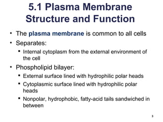
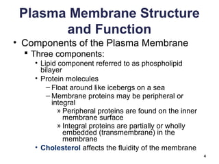


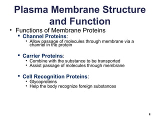
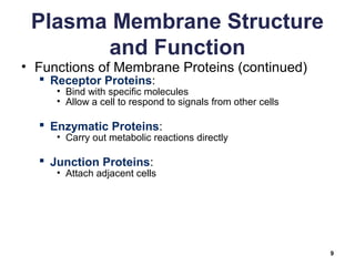


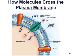


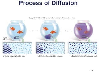


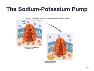


The document outlines chapter 5 of a biology textbook on membrane structure and function. It discusses: 1) The structure of the plasma membrane, including the phospholipid bilayer and embedded proteins. 2) Passive transport mechanisms like diffusion, osmosis, and facilitated transport that allow molecules to cross the membrane down a concentration gradient without cellular energy expenditure. 3) Active transport mechanisms that require cellular energy to move molecules across the membrane against a concentration gradient.


![FINAL CELLS ANATOMY (1) [Auip8uui;oy9'yiuy79y08ugtttosaved].pptx](https://cdn.slidesharecdn.com/ss_thumbnails/finalcellsanatomy1autosaved-250115165251-ffe2d15c-thumbnail.jpg?width=640&height=640&fit=bounds)


