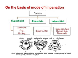
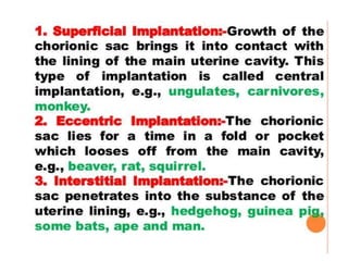
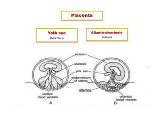
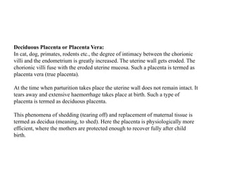
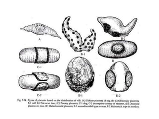
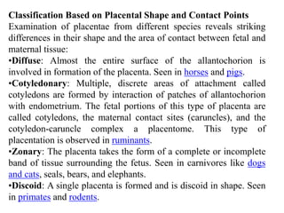
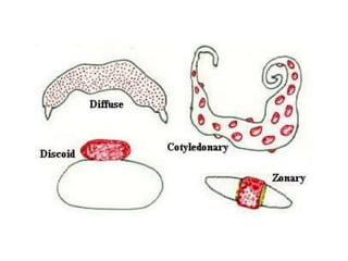
Placentation in Animals
Compiled by: Dr. Mehrajuddin Naikoo
Asst. Prof. ARGO-FVSc & AH
SKUAST-Kashmir
• It is an organ built up of maternal and foetal
tissues combination and serves for the
transportation of nutrients and exchange of
gases from mother to the foetus.
• It is a temporary organ that connects the
developing foetus via the umbilical cord to the
uterine wall.
• to allow nutrient uptake, thermo-regulation,
waste elimination, and gas exchange via the
mother's blood supply; to fight against
internal infection; and to produce hormones
which support pregnancy.
• Placentas are a defining characteristic of
placental mammals, but are also found in
marsupials and some non-mammals with
varying levels of development
Choriovitelline placenta
In higher mammals, the yolk sac is formed early in development, usually while the
blastocyst is still unattached in the uterine cavity.
In most mammals, the endoderm of the early yolk sac combines with the trophoblastic
layer of the blastocyst forming a bilaminar yolk sac.
When the vascular mesoderm becomes interposed between the chorion and the endoderm,
the bilaminar structure becomes a trilaminar yolk sac which functions as the embryonic
component of the choriovitelline placenta .
While the choriovitelline placenta persists as the definitive placenta in most marsupials,
among domestic mammals it exists only as an early temporary structure, losing its exchange
function when the extra‐embryonic coelom extends into the mesoderm of the trilaminar
yolk sac, separating the mesoderm into splanchnic and somatic layers.
As these changes take place rapidly in cattle, sheep and pigs, this yolk sac placenta
functions for only a short period of time.
In dogs and cats, the choriovitelline placenta functions up to the 21st day of pregnancy,
whereas in horses it functions up to the eighth week of pregnancy. The choriovitelline
placenta does not establish an extensive and intimate contact with the endometrium.
Chorioallantoic placenta
The embryonic component of a chorioallantoic placenta is formed by the
attachment and fusion of the outer wall of the expanding allantoic sac with the
adjacent chorion .
This is the definitive form of placentation which occurs in higher mammals and
it is characterised by an extensive area of contact between the embryonic
placental component and the endometrium.
Increased surface contact is achieved through folding of the chorioallantois and
the endometrial surface, formation of chorionic villi and the establishment of
chorionic labyrinths.
Non-Deciduous Placenta or Semi Placenta:
In non-deciduous placenta the implantation is superficial. This occurs in most
mammals where the blastocysts lie in the uterine cavity. At the point of
contact with the wall of the uterus, the blastocyst surface gives out finger like
projection called chorionic villi that penetrate into the depressions of the
uterine wall and are loosely united.
At the time of birth, when parturition takes place, the chorionic villi are
simply withdrawn from the cavities of the uterine wall without causing any
damage or bleeding. This type of placenta formation is seen in pig, cattle,
horse etc., where the less hazardous birth process allows the females to
resume full running speed soon after birth.
As the chorionic villi do not fuse with the endometrium, such a placenta is
also called semi placenta.
Deciduous Placenta or Placenta Vera:
In cat, dog, primates, rodents etc., the degree of intimacy between the chorionic
villi and the endometrium is greatly increased. The uterine wall gets eroded. The
chorionic villi fuse with the eroded uterine mucosa. Such a placenta is termed as
placenta vera (true placenta).
At the time when parturition takes place the uterine wall does not remain intact. It
tears away and extensive haemorrhage takes place at birth. Such a type of
placenta is termed as deciduous placenta.
This phenomena of shedding (tearing off) and replacement of maternal tissue is
termed as decidua (meaning, to shed). Here the placenta is physiologically more
efficient, where the mothers are protected enough to recover fully after child
birth.
iii. Contra-Deciduate Placenta:
A somewhat modified type of deciduate placenta is seen in Parameles and
Talpa (mole), where there is loss of both maternal tissue as well as foetal
portion of placenta. Such a placenta is called contra-deciduate placenta.
subtypes according to the manner of distribution of villi.
(a) Diffuse : Villi remain scattered all over the surface of
allantochorion. e.g., pig, horse, lemur.
(b) Cotyledonary : Villi are arranged in separate tufts or patches called
cotyledons. e.g., goat, sheep, cow, deer.
(iii) Intermediate : Villi are arranged in cotyledons as well as
scattered. e.g., camel, giraffe.
(ii) Deciduous placenta : Villi are complicated, branched and intimately
connected. At birth, a variable amount of maternal tissue is pulled out with the
shedding of blood. Deciduous or deciduate placenta is also differentiated in the
following subtypes
(a) Zonary : Villi form an incomplete (e.g., racoon) or complete girdle encircling
the blastocyst. e.g., cat, dog, seal, Lion, Tiger, Elephant etc.
(b) Discoidal : Villi are restricted to a circular disc or plate on the dorsal surface
of blastocyst. e.g., insectivores, bats, rodents (rat, mouse), rabbit, bear.
(c) Metadiscoidal : Villi are at first scattered but later become restricted to one or
two discs. It is monodiscoidal in man and bidiscoidal in monkeys and apes.
Classification Based on Placental Shape and Contact Points
Examination of placentae from different species reveals striking
differences in their shape and the area of contact between fetal and
maternal tissue:
•Diffuse: Almost the entire surface of the allantochorion is
involved in formation of the placenta. Seen in horses and pigs.
•Cotyledonary: Multiple, discrete areas of attachment called
cotyledons are formed by interaction of patches of allantochorion
with endometrium. The fetal portions of this type of placenta are
called cotyledons, the maternal contact sites (caruncles), and the
cotyledon-caruncle complex a placentome. This type of
placentation is observed in ruminants.
•Zonary: The placenta takes the form of a complete or incomplete
band of tissue surrounding the fetus. Seen in carnivores like dogs
and cats, seals, bears, and elephants.
•Discoid: A single placenta is formed and is discoid in shape. Seen
in primates and rodents.
Thank You !

placentation in animals ppt_014943.pptx

  • 1.
    Placentation in Animals Compiledby: Dr. Mehrajuddin Naikoo Asst. Prof. ARGO-FVSc & AH SKUAST-Kashmir
  • 2.
    • It isan organ built up of maternal and foetal tissues combination and serves for the transportation of nutrients and exchange of gases from mother to the foetus. • It is a temporary organ that connects the developing foetus via the umbilical cord to the uterine wall.
  • 3.
    • to allownutrient uptake, thermo-regulation, waste elimination, and gas exchange via the mother's blood supply; to fight against internal infection; and to produce hormones which support pregnancy. • Placentas are a defining characteristic of placental mammals, but are also found in marsupials and some non-mammals with varying levels of development
  • 8.
    Choriovitelline placenta In highermammals, the yolk sac is formed early in development, usually while the blastocyst is still unattached in the uterine cavity. In most mammals, the endoderm of the early yolk sac combines with the trophoblastic layer of the blastocyst forming a bilaminar yolk sac. When the vascular mesoderm becomes interposed between the chorion and the endoderm, the bilaminar structure becomes a trilaminar yolk sac which functions as the embryonic component of the choriovitelline placenta . While the choriovitelline placenta persists as the definitive placenta in most marsupials, among domestic mammals it exists only as an early temporary structure, losing its exchange function when the extra‐embryonic coelom extends into the mesoderm of the trilaminar yolk sac, separating the mesoderm into splanchnic and somatic layers. As these changes take place rapidly in cattle, sheep and pigs, this yolk sac placenta functions for only a short period of time. In dogs and cats, the choriovitelline placenta functions up to the 21st day of pregnancy, whereas in horses it functions up to the eighth week of pregnancy. The choriovitelline placenta does not establish an extensive and intimate contact with the endometrium.
  • 9.
    Chorioallantoic placenta The embryoniccomponent of a chorioallantoic placenta is formed by the attachment and fusion of the outer wall of the expanding allantoic sac with the adjacent chorion . This is the definitive form of placentation which occurs in higher mammals and it is characterised by an extensive area of contact between the embryonic placental component and the endometrium. Increased surface contact is achieved through folding of the chorioallantois and the endometrial surface, formation of chorionic villi and the establishment of chorionic labyrinths.
  • 10.
    Non-Deciduous Placenta orSemi Placenta: In non-deciduous placenta the implantation is superficial. This occurs in most mammals where the blastocysts lie in the uterine cavity. At the point of contact with the wall of the uterus, the blastocyst surface gives out finger like projection called chorionic villi that penetrate into the depressions of the uterine wall and are loosely united. At the time of birth, when parturition takes place, the chorionic villi are simply withdrawn from the cavities of the uterine wall without causing any damage or bleeding. This type of placenta formation is seen in pig, cattle, horse etc., where the less hazardous birth process allows the females to resume full running speed soon after birth. As the chorionic villi do not fuse with the endometrium, such a placenta is also called semi placenta.
  • 11.
    Deciduous Placenta orPlacenta Vera: In cat, dog, primates, rodents etc., the degree of intimacy between the chorionic villi and the endometrium is greatly increased. The uterine wall gets eroded. The chorionic villi fuse with the eroded uterine mucosa. Such a placenta is termed as placenta vera (true placenta). At the time when parturition takes place the uterine wall does not remain intact. It tears away and extensive haemorrhage takes place at birth. Such a type of placenta is termed as deciduous placenta. This phenomena of shedding (tearing off) and replacement of maternal tissue is termed as decidua (meaning, to shed). Here the placenta is physiologically more efficient, where the mothers are protected enough to recover fully after child birth.
  • 12.
    iii. Contra-Deciduate Placenta: Asomewhat modified type of deciduate placenta is seen in Parameles and Talpa (mole), where there is loss of both maternal tissue as well as foetal portion of placenta. Such a placenta is called contra-deciduate placenta.
  • 13.
    subtypes according tothe manner of distribution of villi. (a) Diffuse : Villi remain scattered all over the surface of allantochorion. e.g., pig, horse, lemur. (b) Cotyledonary : Villi are arranged in separate tufts or patches called cotyledons. e.g., goat, sheep, cow, deer. (iii) Intermediate : Villi are arranged in cotyledons as well as scattered. e.g., camel, giraffe. (ii) Deciduous placenta : Villi are complicated, branched and intimately connected. At birth, a variable amount of maternal tissue is pulled out with the shedding of blood. Deciduous or deciduate placenta is also differentiated in the following subtypes (a) Zonary : Villi form an incomplete (e.g., racoon) or complete girdle encircling the blastocyst. e.g., cat, dog, seal, Lion, Tiger, Elephant etc. (b) Discoidal : Villi are restricted to a circular disc or plate on the dorsal surface of blastocyst. e.g., insectivores, bats, rodents (rat, mouse), rabbit, bear. (c) Metadiscoidal : Villi are at first scattered but later become restricted to one or two discs. It is monodiscoidal in man and bidiscoidal in monkeys and apes.
  • 15.
    Classification Based onPlacental Shape and Contact Points Examination of placentae from different species reveals striking differences in their shape and the area of contact between fetal and maternal tissue: •Diffuse: Almost the entire surface of the allantochorion is involved in formation of the placenta. Seen in horses and pigs. •Cotyledonary: Multiple, discrete areas of attachment called cotyledons are formed by interaction of patches of allantochorion with endometrium. The fetal portions of this type of placenta are called cotyledons, the maternal contact sites (caruncles), and the cotyledon-caruncle complex a placentome. This type of placentation is observed in ruminants. •Zonary: The placenta takes the form of a complete or incomplete band of tissue surrounding the fetus. Seen in carnivores like dogs and cats, seals, bears, and elephants. •Discoid: A single placenta is formed and is discoid in shape. Seen in primates and rodents.
  • 17.