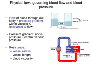
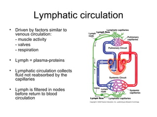
This document provides an outline and overview of the cardiovascular system, including blood flow, blood pressure, vasculature, arteries, capillaries, veins, and lymphatic circulation. It discusses the physical laws governing blood flow and pressure, factors that influence resistance and peripheral resistance. It also examines the regulation of blood flow and pressure through intrinsic controls like metabolic activity and stretch of arteries as well as extrinsic controls like neural and hormonal mechanisms. The goal is to explain how blood is circulated and pressure is maintained through the complex cardiovascular system.
The lymphatic system consists of lymph fluid, lymph vessels, and lymphatic organs that include lymph nodes and tonsils. Lymph is formed from interstitial fluid and contains water, proteins, cells, and other components. It flows from lymph capillaries through collecting vessels and trunks into the thoracic duct or right lymphatic duct and returns to the bloodstream. The lymphatic system helps remove excess fluid from tissues, transports fat and proteins, and plays a role in immune defense and absorption of nutrients. Disruptions in lymph flow can cause edema or other pathologies like lymphadenopathy or elephantiasis.
The document discusses capillary exchange and hemodynamics. It explains that capillaries allow exchange of substances between blood and tissues via diffusion, transcytosis, and bulk flow. Most exchange occurs via diffusion down concentration gradients. Bulk flow involves filtration of fluid from arteries into tissues and reabsorption into veins. Edema can occur if filtration exceeds reabsorption. Hemodynamics influence blood flow - it is highest where pressure differences are largest and resistance is lowest, such as from arteries to veins.
This document discusses leukopoiesis, the process by which white blood cells are formed. It explains that all blood cells, including white blood cells, are formed from pluripotent hematopoietic stem cells in the bone marrow. These stem cells can differentiate into either myeloid or lymphoid stem cells. Myeloid stem cells go on to produce red blood cells, platelets, monocytes, or granulocytes, while lymphoid stem cells produce lymphocytes. Leukopoiesis involves the maturation of these stem cells into the various types of white blood cells.
There are three main types of blood vessels: arteries, veins, and capillaries. Arteries carry oxygenated blood away from the heart to tissues throughout the body. They have thick muscular walls and elastic tissue to withstand high blood pressure. Veins carry deoxygenated blood back to the heart and have thinner walls and valves to prevent backflow. Capillaries are the smallest blood vessels and form a network between arteries and veins in tissues, allowing for the exchange of water, oxygen, nutrients and waste.
The heart has four chambers. The two superior receiving chambers are the atria (= entry halls or chambers), and the two inferior pumping chambers are the ventricles (= little bellies).
On the anterior surface of each atrium is a wrinkled pouchlike structure called an auricle
Tunica Interna – innermost endothelium of simple squamous epithelium + basement membrane
Arteries – have an “internal elastic lamina” of elastic CT to allow for expansion under pressure
Veins – may have “valves” (folds of endothelium + CT) to prevent backflow of blood due to low pressure Microscopic, very thin-walled vessels comprised of endothelium with basement membrane; allows for filtration and reabsorption Found in all tissues of the body except for those that are “avascular” Usually form branching networks (“capillary beds”) within tissues for increased surface area blood flow into capillaries may be regulated by “pre- capillary sphincters” may have a central or “thoroughfare” channel that provides direct connection between “metarteriole” (terminal end of arteriole) & venule
William Harvey was the first modern physiologist in the 16th century. He proved that blood circulates in a continuous loop from the heart to the arteries and back to the veins and heart, overturning the long-held Galenic view of two separate circulatory systems. The circulatory system consists of arteries, which carry blood away from the heart; capillaries, where gas and nutrient exchange occurs; and veins, which carry blood back to the heart. Arteries have thicker muscular walls than veins and carry oxygenated blood except in the pulmonary circulation.
The document summarizes a lecture on cell physiology given by Dr. Nilesh Kate on February 18, 2015. It covers the basic structure and components of cells, including the cell membrane, organelles like mitochondria and the endoplasmic reticulum, and intercellular junctions. It describes the key discoveries in cell biology from the 17th century onwards and the development of the cell theory. The lecture objectives are listed as cell structure, the cell membrane, and intercellular junctions.
The document discusses hemodynamics and blood flow through the circulatory system. It describes the structure of the heart including the atria and ventricles. Blood flows from the left ventricle through the aorta and arteries to tissues, then returns to the right atrium via veins. Heart valves like the mitral and tricuspid valves regulate blood flow. Blood pressure is highest in the arteries and driven by ventricular contraction. Common techniques to measure blood pressure include auscultation and sphygmomanometry.
1. The document discusses hemodynamic factors like pressure, blood flow, resistance, and compliance and their interrelationships.
2. It defines terms like blood pressure, blood flow, resistance, compliance, laminar and turbulent blood flow. It also discusses how changes in vessel diameter affect resistance and flow.
3. The document compares arterial and venous compliance, noting that veins are more compliant and act as reservoirs due to their thin walls, storing over 60% of the blood volume.
This document discusses renal physiology, including renal blood flow, oxygen consumption, regulation of blood flow, glomerular filtration, and factors affecting glomerular filtration rate (GFR). Some key points:
- Renal blood flow is approximately 1/4 of cardiac output, or 1200 ml/min. Blood flow to the cortex is higher than to the medulla.
- Glomerular filtration is determined by the net filtration pressure and filtration coefficient. Forces increasing filtration are glomerular hydrostatic pressure and oncotic pressure in Bowman's space. Forces decreasing filtration are plasma oncotic pressure and hydrostatic pressure in Bowman's space.
- GFR is regulated through autoregulation mechanisms like tub
Skeletal muscle circulation can increase blood flow up to 25-fold during exercise through vasodilation. The vascular supply begins with feed arteries that branch into arterioles and capillaries. Blood flow is regulated by vasoactive substances released from active muscle fibers and sympathetic nerve activity.
Cutaneous circulation regulates heat loss through the skin. The apical skin contains arteriovenous anastomoses that constrict to reduce heat loss. Non-apical skin uses sweat glands and vasoactive substances like bradykinin to dilate blood vessels. Cutaneous responses to stimuli include the triple response of redness, swelling, and wheal formation.
1. The document discusses the cardiovascular system, including circulation, pressure, flow, and resistance of blood.
2. It describes the functional parts of circulation including arteries, arterioles, capillaries, venules and veins. Arteries transport blood at high pressure while veins act as reservoirs and transport blood at low pressure back to the heart.
3. Blood flow through vessels is determined by pressure difference and vascular resistance according to Ohm's law. Laminar flow typically occurs at low velocities while turbulent flow can occur at higher velocities or where there are obstructions.
Renal blood flow (The Guyton and Hall physiology)Maryam Fida
In an average 70-kilogram man, the combined blood flow through both kidneys is about 1100 ml/min, or about 22 per cent of the cardiac output. Two kidneys makes about 0.4 % of total body weight but receive very high blood flow as compared with other body organ. The purpose of additional blood flow is to supply sufficient plasma for high rates of GF which is essential for regulating body fluid volumes & solute concentrations.
Characteristics of the renal blood flow:
1, High blood flow. 1100 ml/min, or 22 percent of the cardiac output. 94% to the cortex.
2, Two capillary beds
High hydrostatic pressure in glomerular capillary (about 60 mmHg) and low hydrostatic pressure in peritubular capillaries (about 13 mmHg)
Blood flow to renal medulla is supplied by vasa recta.
Blood flow in vasa recta of medulla is very low as compared to blood flow in cortex.
Blood flow in renal medulla is 1-2 % of total renal blood flow.
Vasa recta are important to form concentrated urine.
Stethography is a process that records respiratory movements in humans using a stethograph instrument. The stethograph is a corrugated rubber tube connected to a tambour, which is connected to a pen that writes on a moving drum. When the stethograph is applied to a person's chest, it can record their chest movements during respiration. The stethogram produced provides information about respiratory physiology by recording things like normal breathing, the effects of swallowing, hyperventilation, exercise, breath holding, and other respiratory actions.
The lymphatic system is a closed system of lymph vessels that drains lymph fluid from tissues and returns it to the bloodstream. It consists of lymph capillaries that drain into larger vessels and ultimately form the right lymphatic duct and thoracic duct, which empty into subclavian veins. The lymphatic system helps fight pathogens through innate immunity as the first line of defense and acquired immunity developed by T and B lymphocytes, which provide cellular and humoral immunity through memory and plasma cells. Cytokines are proteins secreted by immune cells that help coordinate immune responses.
1) The document discusses the three types of muscular tissue - skeletal, cardiac, and smooth muscle.
2) Skeletal muscle is striated, voluntary muscle attached to bones. It contains bundles of fibers surrounded by connective tissue. Microscopically, it contains myofibrils with repeating dark A and light I bands.
3) Cardiac muscle is striated and involuntary. Microscopically, its branching fibers are joined end to end at intercalated discs.
4) Smooth muscle is non-striated and present in organs like blood vessels. Microscopically, its spindle-shaped cells are arranged in bundles and layers connected by gap junctions.
The document summarizes the key structures and mechanisms involved in urine formation by the kidneys. The glomeruli, proximal canaliculi, and distal canaliculi are responsible for filtration, reabsorption, and secretion to form urine. Filtration occurs as blood is filtered in the glomeruli, producing primary urine. Most of the filtered substances are then reabsorbed back into the bloodstream in the canaliculi. Remaining substances and waste are excreted in the final urine output, with the kidneys processing around 180 L of blood filtrate per day.
circulatory system, their parts, three kinds of circulation, heart, how does it works, artery, vein, capillary, what is in blood, RBC, function summary
Cardiac output is the volume of blood pumped by the heart each minute. It is calculated as stroke volume multiplied by heart rate. Stroke volume is the volume of blood pumped from the left ventricle with each beat. Factors that affect cardiac output include body metabolism, exercise level, age, and body size. Cardiac output increases with exercise and decreases with age. It is tightly regulated to meet the metabolic demands of the body's tissues.
The blood vessels are the components of the circulatory system that transport blood throughout the human body. These vessels transport blood cells, nutrients, and oxygen to the tissues of the body. They also take waste and carbon dioxide away from the tissues.
The document discusses renal physiology and the urinary system. It contains the following key points:
1. The urinary system includes the kidneys, ureters, bladder, and urethra. The kidneys contain nephrons which filter blood to form urine.
2. The kidneys regulate fluid volume, electrolyte and acid-base balance, and remove waste. Nephrons contain a renal corpuscle for filtration and a tubule for reabsorption and secretion.
3. The proximal convoluted tubule reabsorbs the majority of filtered sodium, water, and other electrolytes using active transport mechanisms like sodium-glucose co-transport. This maintains electrolyte and fluid balance
The neuromuscular junction is where a motor neuron connects to a muscle fiber. When an action potential reaches the junction, calcium ions enter the neuron and cause vesicles containing acetylcholine to fuse with the membrane and release the neurotransmitter into the synaptic cleft. Acetylcholine then binds to nicotinic receptors on the muscle fiber, causing ion channels to open and generating an endplate potential large enough to trigger an action potential in the fiber. Acetylcholinesterase quickly breaks down acetylcholine to terminate the signal. Contraction occurs when the action potential causes calcium release from the sarcoplasmic reticulum.
The document discusses the peripheral nervous system. It begins by defining the nervous system and its main components - the central nervous system and peripheral nervous system. The peripheral nervous system has two main divisions: somatic and autonomic. The somatic division includes sensory and motor neurons that control voluntary movement. The autonomic nervous system controls involuntary functions and has sympathetic and parasympathetic divisions. The document goes on to describe various nerves, neurons, and structures that make up the peripheral nervous system.
The renal system consists of the kidneys and urinary bladder. The kidneys play a vital role in maintaining fluid balance and composition in the body, regulating the internal environment. The kidneys are composed of nephrons which filter blood, reabsorbing necessary substances and secreting waste products to produce urine. Urine is stored in the bladder and emptied during micturition.
Hey Guys
im happy you are enjoying my content. please subscribe to my channel on youtube as i will make more videos soon. https://bit.ly/2XXNyTT
thank you as you subscribe.
There are four main mechanisms that regulate blood pressure: nervous, renal, hormonal, and local. The nervous mechanism acts the fastest via the vasomotor system to control heart rate and vasoconstriction/vasodilation in response to baroreceptors and chemoreceptors. The renal mechanism regulates blood pressure long-term by controlling extracellular fluid volume and through the renin-angiotensin system. Hormonal and local factors also contribute to blood pressure regulation.
Physio presentation pressure flow and resistanceSaara Zafar
The document summarizes key concepts related to blood flow and circulation. It describes the systemic and pulmonary circulations, defining blood flow, blood pressure, and resistance. It explains laminar and turbulent blood flow, and provides Reynolds' number equation. Conductance and Hagen-Poiseuille's law are also summarized, with the latter relating blood flow rate to pressure difference, radius, viscosity and vessel length.
The circulatory system transports blood throughout the body to deliver nutrients and oxygen to tissues and remove waste. It is divided into systemic and pulmonary circulation. Blood flows from arteries to arterioles and capillaries, where gas and nutrient exchange occurs, then to venules and veins, which return blood to the heart. Precise control of blood flow to each tissue is critical to meet metabolic demands.
The document discusses cardiac output, blood flow, blood pressure, and factors that regulate them. It defines cardiac output as the volume of blood pumped by the heart per minute, and describes how it is determined by heart rate and stroke volume. Stroke volume depends on preload, contractility, and afterload. The autonomic nervous system and other factors control heart rate and contractility. Blood flow is determined by pressure differences and vascular resistance. The document also covers blood pressure measurement and control via the baroreceptor reflex.
Blood flow is dependent on the pressure gradient between two points in the circulatory system and the resistance to flow within the vessels. The pressure gradient is determined by the difference in pressure between the beginning and end of a vessel and governs blood flow. Resistance depends on factors like vessel diameter, length, and viscosity and opposes blood flow. Larger arteries have less resistance but require stronger walls to withstand higher pressures due to Laplace's law. Capillaries have high resistance due to their small diameter but require only thin walls. Resistance increases as vessel diameter decreases. Blood velocity is directly related to vessel cross-sectional area and inversely related to resistance.
Hemodynamic monitoring involves measuring factors that influence blood flow and pressure to aid in diagnosing and managing critically ill patients. It uses Doppler ultrasound to analyze blood flow spectra and determine velocity measurements. The spectral display shows velocity over time and can reveal flow direction and indices like resistive index. The document defines six types of blood flow seen: plug, laminar, parabolic, disturbed, turbulent, and pulsatile.
Cardiac output is defined as the volume of blood pumped by the heart each minute and is regulated intrinsically by factors affecting preload and afterload as well as extrinsically by the autonomic nervous system and hormones. Venous return is a primary extrinsic regulator of cardiac output, increasing stretch of cardiac muscles and stimulating an increase in heart rate. A combination of preload, contractility, afterload and heart rate determine cardiac output under normal resting conditions and during physical activity.
The whole cardiovascular physiology caters to blood flow through the organs, and blood pressure is just one of the factors favouring tissue blood flow (perfusion).
Blood vessels transport blood throughout the body. There are three main types: arteries, which carry blood away from the heart; capillaries, where nutrients and wastes are exchanged; and veins, which carry blood back to the heart. Blood flow is regulated through pressure, resistance, and the actions of the cardiovascular system to ensure adequate circulation throughout the body. Neural and hormonal mechanisms precisely control blood pressure and flow through reflexes and the release of substances like epinephrine and norepinephrine.
The document describes the structure and function of the lymphatic system and immune system. The lymphatic system includes lymphatic vessels, lymph nodes, spleen, thymus gland, tonsils and other lymphatic tissues that work to remove excess fluid from tissues, absorb fatty acids, and transport white blood cells. The immune system protects the body from infection with non-specific defenses like skin and mucous membranes, and specific defenses like antibodies and lymphocytes that recognize and destroy pathogens.
The lymphatic system functions to:
1) Transport clean fluids back to the blood from tissues;
2) Drain excess fluids from tissues; and
3) Remove debris from cells of the body.
Lymph is transported through lymphatic vessels in a passive, one-way system toward the heart, where it is returned to circulation. Along the way, lymph passes through lymph nodes which filter the lymph and provide an immune response. Other lymphoid organs like the spleen, thymus, tonsils, and Peyer's patches also contribute to lymphatic function and immune defense.
The document discusses the neural regulation of circulation. It covers:
1. Neural control shifts blood flow between different parts of the body as needed, such as more to muscles during exercise.
2. The circulatory system has cardiac and vascular innervation from both the sympathetic and parasympathetic nervous systems which control heart rate, contraction force, and vessel diameter.
3. The brain monitors blood flow and pressure through signals and controls them by altering cardiac output, peripheral resistance, and blood volume through short, intermediate, and long-term mechanisms like baroreceptor reflexes, the renin-angiotensin system, and kidney functions.
((Measurement of central and mixed venous to-arterialhbnbz
This study investigated the agreement between central venous-to-arterial CO2 difference (p(cv-a)CO2) and mixed venous-to-arterial CO2 difference (p(v-a)CO2) in cardiac surgery patients. The study found that p(cv-a)CO2 is not an accurate estimate of p(v-a)CO2, with large limits of agreement. There was also no meaningful correlation found between p(v-a)CO2 and other parameters of global and regional tissue oxygenation. Therefore, the study concludes that p(cv-a)CO2 and p(v-a)CO2 measurements cannot help diagnose tissue hypoxia in cardiac surgery patients
Shock
what is shock
stages of shock
types of shock, their presentation and management
presentation is made for medical students using kumar and clark and guyton.
This document discusses cardiovascular circulation and hemodynamics. It covers general principles such as how the right and left hearts are interdependent and how blood flow to individual organs can be controlled independently. It also discusses factors that influence blood flow, pressure, and resistance such as vessel diameter, compliance, Poiseuille's law, and the types of vessels. Other topics covered include arterial pressures, pulse pressure, transmission of arterial pulsations, factors affecting mean arterial pressure, and physiological variations in blood pressure.
This document discusses fractional flow reserve (FFR) and instantaneous wave-free ratio (iFR), which are used to assess the severity of a stenosis. It notes that calculating FFR requires making assumptions and simplifications from a fluid mechanics perspective. Specifically, it assumes hydraulic models, neglects venous pressure, assumes equal myocardial resistances, and faces challenges in measuring distal pressure due to dynamic pressure effects, which are more problematic at higher flow rates. The document suggests iFR measurement of distal pressure could be even more inaccurate due to typically higher velocities during the wave-free period.
The microcirculatory bed comprises seven main structures: arterioles, precapillary arterioles, capillaries, postcapillary venules, venules, precapillary sphincters, and arteriovenous shunts. Arterioles regulate blood flow and diameter in response to neural and hormonal signals. Precapillary arterioles are the smallest arteries and feed into capillaries, which are the primary sites of nutrient exchange, or may bypass them via shunts. Capillaries come in continuous, fenestrated, and discontinuous types and have selective permeability barriers. Venules then collect blood from the capillaries and gradually increase in size. Key microcirculatory barriers
This document discusses microcirculation monitoring in critical illness. It begins with an introduction to microcirculation and its role in tissue oxygenation. It then discusses four types of microcirculatory alterations that can result in a loss of coherence between the macrocirculation and microcirculation: heterogeneity, hemodilution, constriction/tamponade, and edema. Specific microcirculatory alterations seen in sepsis and surgery are also reviewed. Monitoring microcirculation is important for guiding resuscitation efforts and assessing whether macrocirculation improvements translate to the microcirculation level.
This document discusses the regulation of blood pressure. It begins by defining key terms related to blood pressure like systolic, diastolic, and mean arterial pressure. It then discusses factors that determine blood pressure like cardiac output and peripheral resistance. The document outlines several regulatory mechanisms for blood pressure, including local mechanisms like autoregulation and vasoactive substances, as well as systemic mechanisms involving hormonal and nervous control. Specific hormones, neurotransmitters, and reflex mechanisms that play roles in regulating blood pressure are also mentioned.
Hemorrhage and shock can occur due to abnormal blood loss or inadequate tissue perfusion. Hemorrhage can be internal or external and is classified based on its source and severity. Shock progresses through four stages as blood loss worsens from vasoconstriction to organ failure. The body responds to hemorrhage initially through hemostasis to stop bleeding, but progresses to shock if blood loss is not replaced.
This document discusses microcirculation and the factors that influence blood flow at the capillary level. It defines capillary circulation and notes the importance of local control mechanisms in matching blood flow to the metabolic needs of tissues. The key concepts covered include the Starling forces that govern fluid filtration across capillary walls, the role of the lymphatic system in fluid drainage, and causes of edema related to increased capillary pressure, decreased oncotic pressure, or increased capillary permeability.
The document discusses the circulatory system and blood pressure regulation. It describes the different types of blood vessels - arteries, arterioles, capillaries and veins. Arteries carry blood away from the heart while veins carry blood back to the heart. Capillaries are where gas and nutrient exchange occurs. Blood pressure is regulated through short-term mechanisms like the baroreceptor reflex and long-term mechanisms like the renin-angiotensin system. Heart failure and shock can occur if the heart or blood vessels are unable to effectively circulate blood and maintain adequate blood pressure.
This document discusses the cardiovascular system and types of shock. It begins by describing the five classes of blood vessels - arteries, arterioles, capillaries, venules, and veins. It then covers the structure of blood vessel walls and differences between arteries and veins. The document discusses blood pressure, circulation, and how blood flow is regulated. It defines shock and describes the four main types: hypovolemic, cardiogenic, anaphylactic, and septic shock. For each type, it explains the causes, stages, and pathophysiology.
Blood pressure and Its Applied Physiology In Dentistry (Prosthodontics)Self employed
This document discusses blood pressure, including an overview, variations, determinants, regulation, and measurement. It defines systolic and diastolic blood pressure and other related terms. The key factors that determine and regulate blood pressure are described, including cardiac output, peripheral resistance, blood volume, hormones, the nervous system, kidneys, and local mechanisms. Methods for measuring blood pressure are outlined, including palpatory, auscultatory, and oscillometric techniques. Special considerations for taking blood pressure in certain populations are also noted.
This document provides an overview of shock and its pathophysiology. It defines shock as a clinical syndrome resulting from inadequate tissue perfusion due to alterations in circulation. The stages of shock are described as compensatory, progressive, and irreversible. Compensatory mechanisms aimed at maintaining homeostasis in response to shock are discussed for various body systems. Nursing interventions for shock focus on treating its underlying cause, restoring circulating volume and hemodynamics through fluid resuscitation and vasoactive drugs, and minimizing oxygen consumption.
The cardiovascular system consists of the heart and blood vessels. The heart pumps blood through arteries, capillaries, and veins to transport oxygen, nutrients, hormones, and remove wastes. The cardiovascular system has five major functions: transporting oxygen and removing carbon dioxide, transporting nutrients and removing wastes, fighting disease, transporting hormones, and regulating body temperature. Arteries carry oxygenated blood away from the heart while veins carry deoxygenated blood back to the heart. Capillaries allow for the exchange of substances between blood and tissues. Tissue fluid is similar to blood plasma but lacks proteins and surrounds cells, exchanging nutrients and wastes. Excess tissue fluid drains into lymphatic vessels forming
Hemodynamics is the study of blood flow, pressure, and resistance in the circulatory system. It includes the types and functions of blood vessels like arteries, veins, and capillaries. Arteries have thick elastic walls to withstand high blood pressure and distribute blood to tissues. Veins have thinner walls and valves to return blood to the heart. Capillaries allow for gas and nutrient exchange. Blood flow and pressure are regulated intrinsically through the vessels and extrinsically by the autonomic nervous and endocrine systems to meet the demands of tissues. The kidneys also help control blood volume and pressure long-term through the renin-angiotensin-aldosterone system.
The document defines arterial blood pressure and its components: systolic, diastolic, pulse, and mean arterial pressure. It discusses factors that regulate blood pressure, including the nervous system's vasomotor center and reflexes, the kidneys' regulation of fluid volume and renin-angiotensin system, and hormonal factors. It also covers hypertension and hypotension, defining each and describing primary vs. secondary causes, manifestations, and treatment approaches.
Pressure, blood flow, compliance and resistance are interrelated factors that govern circulation. Arteries have thick, muscular walls and are very elastic to withstand pressure changes. Arterioles regulate blood flow through diameter changes. Capillaries allow for gas, nutrient and waste exchange. Venules and veins have thin walls and valves to aid blood return to the heart. Blood pressure and flow are controlled by neural and hormonal mechanisms. Resistance depends on vessel properties and affects flow. Compliance refers to a vessel's distensibility and ability to store blood with pressure changes, aiding venous return.
This presentation gives you a brief, understandable, captivating and presentable idea on the physiology of blood pressure regulation both on hypertension and hypotension cases.
The circulatory system transports oxygenated blood from the heart to organs via arteries and arterioles, and deoxygenated blood is returned to the heart via veins and venules. The heart has its own blood supply through the coronary circulation. Blood pressure is a measure of the force used to pump blood around the body and is measured using a sphygmomanometer. Haemoglobin is a protein that carries oxygen in red blood cells and can bind up to four oxygen molecules at a time to transport oxygen around the body.
The circulatory system transports oxygenated blood from the heart to organs via arteries and arterioles, and deoxygenated blood is returned to the heart via veins and venules. The heart has its own blood supply through the coronary circulation. Blood pressure is a measure of the force used to pump blood around the body and is measured using a sphygmomanometer. Haemoglobin is a protein that carries oxygen in red blood cells and can bind up to four oxygen molecules to transport oxygen around the body.
The circulatory system delivers oxygenated blood from the heart to organs via arteries and arterioles, and returns deoxygenated blood from organs to the heart via veins and venules. The heart has its own blood supply through the coronary circulation. Blood pressure is a measure of the force used to pump blood around the body and is measured using a sphygmomanometer. Common cardiovascular disease diagnostic tests include blood counts, blood tests, and electrocardiograms. Hemoglobin is a protein that carries oxygen in red blood cells and can bind up to four oxygen molecules to transport oxygen around the body.
The circulatory system transports oxygenated blood from the heart to organs via arteries and arterioles, and deoxygenated blood is returned to the heart via veins and venules. The heart has its own blood supply through the coronary circulation. Blood pressure is a measure of the force used to pump blood around the body and is measured using a sphygmomanometer. Haemoglobin is a protein that carries oxygen in red blood cells and can bind up to four oxygen molecules at a time to transport oxygen around the body.
Cardiovascular physiology. Cardiac enzymes and their effects in the body system. Cardiac output and effects increasing and decreasing it. Calculations if Ejected fraction and other cardiac parameters.
Cardivascular system
Cardiovascular system include Heart and Blood vessels
Heart:
Pumps the blood
Blood Vessels:
Carries the blood to all parts of the body.
Location
Thorax between the lungs
Pointed apex directed toward left hip
From 2nd Rib to 6th Rib
About the size of your fist
The peripheral vascular system consists of the veins and arteries not in the chest or the abdomen that in the arm, hands, legs and feet.
The peripheral arteries supply the oxygenated blood to the body.
The peripheral veins lead deoxygenated blood from the capillaries in the back to the heart.
The document discusses local control of blood flow to tissues based on their metabolic needs. It focuses on blood flow regulation to specific organs like the liver, kidneys, brain, heart and skeletal muscles. Autoregulation and various metabolic factors control blood flow to tissues and the heart. Coronary artery disease occurs due to inadequate blood flow to the heart. The brain has a rich blood supply maintained through the circle of Willis and Cushing reflex. Strokes happen when blood flow to the brain is interrupted. The hepatic portal system carries blood from the digestive organs to the liver before returning to the heart.
The document discusses the circulatory system's response to exercise. The primary purpose is to deliver adequate oxygen and remove waste from tissues. During heavy exercise, oxygen demand can increase 15-25 times resting levels. To meet this, cardiac output and blood flow to active muscles increase through two mechanisms: 1) increased heart rate and stroke volume leading to higher cardiac output, and 2) redistribution of blood flow from inactive organs to working muscles. Proper circulatory function is critical for exercise and maintaining homeostasis.
Similar to Cardiovascular system flow and pressure (20)
Histololgy of Female Reproductive System.pptxAyeshaZaid1
Dive into an in-depth exploration of the histological structure of female reproductive system with this comprehensive lecture. Presented by Dr. Ayesha Irfan, Assistant Professor of Anatomy, this presentation covers the Gross anatomy and functional histology of the female reproductive organs. Ideal for students, educators, and anyone interested in medical science, this lecture provides clear explanations, detailed diagrams, and valuable insights into female reproductive system. Enhance your knowledge and understanding of this essential aspect of human biology.
Basavarajeeyam is a Sreshta Sangraha grantha (Compiled book ), written by Neelkanta kotturu Basavaraja Virachita. It contains 25 Prakaranas, First 24 Chapters related to Rogas& 25th to Rasadravyas.
Osteoporosis - Definition , Evaluation and Management .pdfJim Jacob Roy
Osteoporosis is an increasing cause of morbidity among the elderly.
In this document , a brief outline of osteoporosis is given , including the risk factors of osteoporosis fractures , the indications for testing bone mineral density and the management of osteoporosis
Here is the updated list of Top Best Ayurvedic medicine for Gas and Indigestion and those are Gas-O-Go Syp for Dyspepsia | Lavizyme Syrup for Acidity | Yumzyme Hepatoprotective Capsules etc
8 Surprising Reasons To Meditate 40 Minutes A Day That Can Change Your Life.pptxHolistified Wellness
We’re talking about Vedic Meditation, a form of meditation that has been around for at least 5,000 years. Back then, the people who lived in the Indus Valley, now known as India and Pakistan, practised meditation as a fundamental part of daily life. This knowledge that has given us yoga and Ayurveda, was known as Veda, hence the name Vedic. And though there are some written records, the practice has been passed down verbally from generation to generation.
TEST BANK For Community Health Nursing A Canadian Perspective, 5th Edition by...Donc Test
TEST BANK For Community Health Nursing A Canadian Perspective, 5th Edition by Stamler, Verified Chapters 1 - 33, Complete Newest Version Community Health Nursing A Canadian Perspective, 5th Edition by Stamler, Verified Chapters 1 - 33, Complete Newest Version Community Health Nursing A Canadian Perspective, 5th Edition by Stamler Community Health Nursing A Canadian Perspective, 5th Edition TEST BANK by Stamler Test Bank For Community Health Nursing A Canadian Perspective, 5th Edition Pdf Chapters Download Test Bank For Community Health Nursing A Canadian Perspective, 5th Edition Pdf Download Stuvia Test Bank For Community Health Nursing A Canadian Perspective, 5th Edition Study Guide Test Bank For Community Health Nursing A Canadian Perspective, 5th Edition Ebook Download Stuvia Test Bank For Community Health Nursing A Canadian Perspective, 5th Edition Questions and Answers Quizlet Test Bank For Community Health Nursing A Canadian Perspective, 5th Edition Studocu Test Bank For Community Health Nursing A Canadian Perspective, 5th Edition Quizlet Test Bank For Community Health Nursing A Canadian Perspective, 5th Edition Stuvia Community Health Nursing A Canadian Perspective, 5th Edition Pdf Chapters Download Community Health Nursing A Canadian Perspective, 5th Edition Pdf Download Course Hero Community Health Nursing A Canadian Perspective, 5th Edition Answers Quizlet Community Health Nursing A Canadian Perspective, 5th Edition Ebook Download Course hero Community Health Nursing A Canadian Perspective, 5th Edition Questions and Answers Community Health Nursing A Canadian Perspective, 5th Edition Studocu Community Health Nursing A Canadian Perspective, 5th Edition Quizlet Community Health Nursing A Canadian Perspective, 5th Edition Stuvia Community Health Nursing A Canadian Perspective, 5th Edition Test Bank Pdf Chapters Download Community Health Nursing A Canadian Perspective, 5th Edition Test Bank Pdf Download Stuvia Community Health Nursing A Canadian Perspective, 5th Edition Test Bank Study Guide Questions and Answers Community Health Nursing A Canadian Perspective, 5th Edition Test Bank Ebook Download Stuvia Community Health Nursing A Canadian Perspective, 5th Edition Test Bank Questions Quizlet Community Health Nursing A Canadian Perspective, 5th Edition Test Bank Studocu Community Health Nursing A Canadian Perspective, 5th Edition Test Bank Quizlet Community Health Nursing A Canadian Perspective, 5th Edition Test Bank Stuvia
Rasamanikya is a excellent preparation in the field of Rasashastra, it is used in various Kushtha Roga, Shwasa, Vicharchika, Bhagandara, Vatarakta, and Phiranga Roga. In this article Preparation& Comparative analytical profile for both Formulationon i.e Rasamanikya prepared by Kushmanda swarasa & Churnodhaka Shodita Haratala. The study aims to provide insights into the comparative efficacy and analytical aspects of these formulations for enhanced therapeutic outcomes.
Clinic ^%[+27633867063*Abortion Pills For Sale In Tembisa Central19various
Clinic ^%[+27633867063*Abortion Pills For Sale In Tembisa Central Clinic ^%[+27633867063*Abortion Pills For Sale In Tembisa CentralClinic ^%[+27633867063*Abortion Pills For Sale In Tembisa CentralClinic ^%[+27633867063*Abortion Pills For Sale In Tembisa CentralClinic ^%[+27633867063*Abortion Pills For Sale In Tembisa Central
2. Outline
• 1- Physical laws governing blood flow and blood
pressure
• 2- Overview of vasculature
• 3- Arteries
• 4. Capillaries and venules
• 5. Veins
• 6. Lymphatic circulation
• 7. Mean arterial pressure and its regulation
• 8. Other cardiovascular regulatory processes
3. Outline
• 1- Physical laws governing blood flow and blood
pressure
• 2- Overview of vasculature
• 3- Arteries
• 4. Capillaries and venules
• 5. Veins
• 6. Lymphatic circulation
• 7. Mean arterial pressure and its regulation
• 8. Other cardiovascular regulatory processes
4. Physical laws governing blood flow and blood
pressure
• Flow of blood through out
body = pressure gradient
within vessels X
resistance to flow
- Pressure gradient: aortic
pressure – central venous
pressure
- Resistance:
-- vessel radius
-- vessel length
-- blood viscosity
5. Factors promoting total peripheral resistance
(TPR)
• Total peripheral
resistance = TPR
-- combined resistance of
all vessels
-- vasodilation
resistance decreases
-- vasoconstriction
resistance increases
6. Outline
• 1- Physical laws governing blood flow and blood
pressure
• 2- Overview of vasculature
• 3- Arteries
• 4. Capillaries and venules
• 5. Veins
• 6. Lymphatic circulation
• 7. Mean arterial pressure and its regulation
• 8. Other cardiovascular regulatory processes
8. Arteries and blood pressure
• Pressure reservoir
• Arterial walls are able to
expand and recoil
because of the pressure
of elastic fibers in the
arterial wall
• Systolic pressure:
maximum pressure
occurring during systole
• Diastolic pressure:
pressure during diastole
10. Blood pressure values: what do they mean?
• Pulse pressure:
PP = SP-DP
• Mean arterial blood
pressure = MABP
• MABP = SBP + (2XDBP)
3
CO = MABP = SV x HR
TPR
11. • Blood flow within each organ
changes with body activities
• Reminder: The ANS controls
blood flow to the various organs
Figure 14.15
12. Outline
• 1- Physical laws governing blood flow and blood
pressure
• 2- Overview of vasculature
• 3- Arteries
• 4. Capillaries and venules
• 5. Veins
• 6. Lymphatic circulation
• 7. Mean arterial pressure and its regulation
• 8. Other cardiovascular regulatory processes
13. Capillaries
• Allow exchange of gases,
nutrients and wastes between
blood and tissues
• Overall large surface area and
low blood flow
• Two main types:
- continuous capillaries:
narrow space between cells
permeable to small or lipid
soluble molecules
- fenestrated capillaries:
large pores between cells
large molecules can pass
14. Local control of blood flow in capillaries
• Presence of precapillary
sphincters on the arteriole
and beginning of
capillaries
• Metarteriole: no sphincter
continuous blood flow
controls the amount of
blood going to
neighboring vessels
15. Movement of materials across capillary walls
• Small molecules and lipid
soluble molecules move
by diffusion through the
cell membrane
• Larger molecules,
charged molecules must
pass through membrane
channels, exocytosis or in
between 2 cells
• Water movement is
controlled by the capillary
hydrostatic and osmotic
pressures
16. Forces controlling water movement
• Arterial side of the capillary:
– High capillary hydrostatic
pressure (BHP), lower
capillary osmotic pressure
(BOP, due to proteins and
other molecules in the blood)
Net filtration pressure
pushes fluid from the blood
toward the tissue (but the
proteins remain in the capillary
• Venous side of the capillary:
- Lower hydrostatic pressure (due
to resistance) and higher capillary
osmotic pressure Net filtration
pressure moves fluid back toward
the capillary
• Interstitial fluid hydrostatic
(IFHP) and osmotic pressures
(IFOP) remain overall identical
17. Fluid movement in the capillary
• Arteriole side: fluid moves
toward the tissues
• Venous side: fluid reenters the
capillary
• Overall: for every 1 liter of fluid
entering the tissues, only 0.85 l
reenter the capillary
• The remaining 0.15 l is
reabsorbed as lymph by
lymphatic capillaries and
eventually returned back to
blood circulation
• When this system fails: Edema
18. Causes of edema
• Increased hydrostatic • Increased interstitial
blood pressure hydrostatic pressure
- heart failure (left or right),
(lymphatic capillary
blockage)
- excess fluid in the blood
- breast cancer surgery,
elephantiasis
• Decreased blood osmotic • Leaking capillary wall
pressure - histamine release during
– Liver, kidney diseases,
allergic reaction
malnutrition (kwashiorkor),
burn injuries
19. Outline
• 1- Physical laws governing blood flow and blood
pressure
• 2- Overview of vasculature
• 3- Arteries
• 4. Capillaries and venules
• 5. Veins
• 6. Lymphatic circulation
• 7. Mean arterial pressure and its regulation
• 8. Other cardiovascular regulatory processes
20. Veins
• Veins are blood volume reservoir
• Due to thinness of vessel wall less resistance to stretch = more compliance
22. Outline
• 1- Physical laws governing blood flow and blood
pressure
• 2- Overview of vasculature
• 3- Arteries
• 4. Capillaries and venules
• 5. Veins
• 6. Lymphatic circulation
• 7. Mean arterial pressure and its regulation
• 8. Other cardiovascular regulatory processes
23. Lymphatic circulation
• Driven by factors similar to
venous circulation:
- muscle activity
- valves
- respiration
• Lymph = plasma-proteins
• Lymphatic circulation collects
fluid not reabsorbed by the
capillaries
• Lymph is filtered in nodes
before return to blood
circulation
24. Outline
• 1- Physical laws governing blood flow and blood
pressure
• 2- Overview of vasculature
• 3- Arteries
• 4. Capillaries and venules
• 5. Veins
• 6. Lymphatic circulation
• 7. Mean arterial pressure and its regulation
• 8. Other cardiovascular regulatory processes
25. Mean arterial pressure and its
regulation
• Regulation of blood flow in arteries
- Intrinsic control
- Extrinsic control
-- Neural control
-- Hormonal control
* Control of blood vessel radius
* Control of blood volume
26. Mean arterial pressure and its
regulation
• Regulation of blood flow in arteries
- Intrinsic control
- Extrinsic control
-- Neural control
-- Hormonal control
* Control of blood vessel radius
* Control of blood volume
27. Regulation of blood flow in arteries
• It is important to adjust blood flow to
organ needs Flow of blood to particular
organ can be regulated by varying
resistance to flow (or blood vessel
diameter)
• Vasoconstriction of blood vessel smooth
muscle is controlled both by the ANS and
at the local level.
• Four factors control arterial flow at the
organ level:
- change in metabolic activity
- changes in blood flow
- stretch of arterial smooth muscle
- local chemical messengers
28. Intrinsic control of local arterial blood flow
• Change in metabolic • Stretch of arterial wall =
activity myogenic response
– Usually linked to CO2 and - Stretch of arterial wall due to
O2 levels (↑ CO2 increased pressure reflex
vasodilation ↑ blood flow) constriction
intrinsic control
• Locally secreted
• Changes in blood flow
chemicals can promote
- decreased blood flow vasoconstriction or most
increased metabolic wastes
vasodilation
commonly vasodilation
- inflammatory chemicals,
(nitric oxide, CO2)
29. Mean arterial pressure and its
regulation
• Regulation of blood flow in arteries
- Intrinsic control
- Extrinsic control
-- Neural control
-- Hormonal control
* Control of blood vessel radius
* Control of blood volume
30. Extrinsic control of blood pressure
• Two ways to control BP:
- Neural control
- Hormonal control
** Use negative feedback
31. Control of blood pressure
• Importance: Blood pressure is a key factor for
providing blood (thus oxygen and energy) to organs.
SBP must be a minimum of 70 to sustain kidney filtration
and adequate blood flow to the brain
• CO= HR X SV = MABP/TPR
MABP= HRxSVxTPR heart rate, stroke volume
and peripheral resistance affect MABP
• Main factors controlling BP: - Blood volume
- Blood vessel radius
32. Neural control of BP - 1
• Baroreceptors: carotid
and aortic sinuses
sense the blood
pressure in the aortic
arch and internal
carotid send signal
to the vasomotor
center in the medulla
oblongata
• Other information are
sent from the
hypothalamus, cortex
33. Neural control of BP - 2
• The vasomotor center
integrates all these information
• The vasomotor sends decision
to the ANS center:
- Both parasympathetic and
sympathetic innervate the S/A
node can accelerate or slow
down the heart rate
- The sympathetic NS
innervates the myocardium
and the smooth muscle of the
arteries and veins promotes
vasoconstriction
34. Hormonal control of BP
• Control of blood vessel radius
• Hormones can control blood - Epinephrine
vessel radius and blood - Angiotensin II
volume, stroke volume and - Vasopressin (?)
heart rate
• On a normal basis, blood
• Control of blood volume
vessel radius and blood
volume are the main factors - Anti-diuretic hormone
(vasopressin)
• - Aldosterone
If there is a critical loss of
pressure, then the effects on
HR and SV will be noticeable
(due to epinephrine kicking in) • Control of heart rate and
stroke volume
- Epinephrine
35. Control of blood vessel radius
• Epinephrine: secreted by the
adrenal medulla and ANS reflex
increase HR, stroke volume and
promotes vasoconstriction of most
blood vessel smooth muscles. • Angiotensin II secretion:
- Decreased flow of filtrate in kidney
• Angiotensin II promotes tubule is sensed by the
vasoconstriction Juxtaglomerular apparatus (a
small organ located in the tubule)
secretion of renin
- Renin activates angiotensinogen,
a protein synthesized by the liver
and circulating in the blood
angiotensin I
- Angiotensin I is activated by a lung
enzyme, Angiotensin-Activating
Enzyme (ACE), angiotensin II
- Angiotensin II is a powerful
vasoconstricted of blood vessel
smooth muscles
36. Control of blood volume
• Anti-diuretic hormone =
ADH
- Secreted by the posterior
pituitary in response to ↑blood
osmolarity (often due to
dehydration)
- Promote water reabsorption by
the kidney tubules H2O
moves back into the blood
less urine formed
37. Control of blood volume
• Aldosterone:
- Secretion by the adrenal
cortex triggered by angiotensin
II
- Promotes sodium reabsorption
by the kidney tubules (Na+
moves back into the blood)
- H2O follows by osmosis
- Whereas ADH promotes H2O
reabsorption only (in response
to dehydration), aldosterone
promotes reabsorption of both
H2O and salt (in response to ↓
BP)
38. Clinical application: Shock
• Stage I: Body reacts to
• Stage I: reversible, maintain BP ↑HR,
compensated shock vasoconstriction.. BP remains
within normal range
• Stage II: reversible,
noncompensated shock • Stage II: Body reacts to
maintain BP ↑HR,
• Stage III: irreversible shock vasoconstriction.. BP drops
below adequate range (SBP 70).
Can be reversed by medical
• Death treatment
• Stage III: Body is fighting to
maintain adequate BP without
success HR is very high not
enough O2 for cardiac, brain cells
to survive damages. Cannot
be reversed by medical treatment