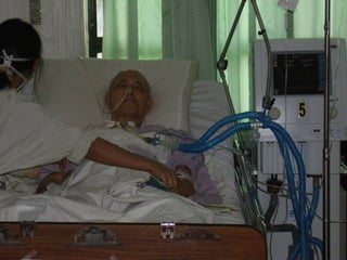
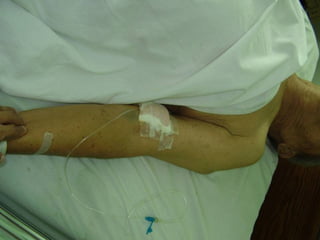
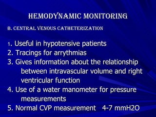
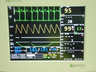
PHYSIOLOGIC MONITORING CELSO M. FIDEL, MD, FPCS, FPSGS Diplomate Philippine Board of Surgery
 
Parameters Monitored Hemodynamic Monitoring RESPIRATORY Monitoring Renal Monitoring Neurologic monitoring Metabolic monitoring temperature monitoring
Hemodynamic Monitoring 1. Provides information as to the C-P status of the patient 2. Traditional clinical assessment are usually unreliable 3.  Major changes in the cardiovascular status may not be clinically obvious 4.  Invasive techniques must be utilized
Hemodynamic Monitoring A.  Arterial Catheterization 1. Indications   a. Need for continuous blood    pressure monitoring.   b. Need for frequent arterial blood    sampling.
Hemodynamic Monitoring Conditions that need continuous and accurate BP monitoring: i. Shock states ii. Hypertensive crisis iii. Surgery in high risk patients iv. Use of potent vasoactive or  inotropic drugs v. Controlled hypotensive anesthesia vi. Situations that may lead to rapid changes in cardiac function  Arterial Catheterization
Hemodynamic Monitoring 2. contraindications No ABSOLUTE  contraindication to arterial catheterization RELATIVE contraindications are: a.  Bleeding problems (hemophilia)   b. Anticoagulant therapy   c. Presence of a vascular prosthesis   d. Local infection Arterial Catheterization
Hemodynamic Monitoring 3. Sites of catheterization a . R adial artery b. Axillary artery c. Femoral artery d. Dorsalis pedis artery e. Superficial femoral artery f.  Brachial artery Arterial Catheterization
Hemodynamic Monitoring 3. Sites of Catheterization a. radial artery   - dual blood supply   - most commonly used site   - simple canulation   - low complication rate   - modified “Allen’s” test – assess ulnar artery   - Doppler technique, plethysmography,  pulse oximetry
Hemodynamic Monitoring 3. Sites of Catheterization b. axillary artery - for long term monitoring - large size - close proximity to the aorta - deep location - technical difficulty in insertion - located near neurovascular structures
Hemodynamic Monitoring 3. Sites of Catheterization c. femoral artery -  large size and superficial location - prone to atherosclerosis - difficult to keep clean
Hemodynamic Monitoring 3. Sites of Catheterization d. dorsalis pedis e. superficial temporal artery -  surgical exposure is required - neurologic complications observed
Hemodynamic Monitoring 3. Sites of Catheterization f. brachial artery   - for short term use only   - median nerve contracture    (Volkman’s contracture)
Hemodynamic Monitoring   Arterial Catheterization 4.   Complications of arterial catheterization a. failure to cannulate b. hematoma   c. disconnection from monitoring system
Hemodynamic Monitoring   Arterial Catheterization 4.   Complications of arterial catheterization d. infection - catheters in place for more than 4 days - surgical insertion - local inflammation e. retrograde cerebral embolization   f. A-V fistula / pseudoaneurysm   g. severe pain, distal necrosis
Hemodynamic Monitoring B. Central Venous Catheterization 1. Indications a. access for fluid therapy b. access for drug infusion c. parenteral nutrition d. CVP monitoring
Hemodynamic Monitoring B. Central Venous Catheterization 1. Indications   e. other indications - aspirate air embolism - placement of cardiac  pacemaker / vena cava filters - hemodialysis access
 
Hemodynamic Monitoring B. Central Venous Catheterization 1 . Useful in hypotensive patients 2. Tracings for arrythmias 3. Gives information about the relationship  between intravascular volume and right  ventricular function 4. Use of a water manometer for pressure  measurements 5. Normal CVP measurement  4-7 mmH2O
Central Venous Pressure Monitoring    In seriously ill patients the  vital  problem  is  determination of the  proper amount  of fluids  and blood requirements necessary to  MAINTAIN an optimal blood volume  in the:    Preoperative    Operative    Postoperative Hemodynamic Monitoring
Hemodynamic Monitoring Central Venous Pressure Monitoring     CVP Monitoring is a reliable procedure  to evaluate properly and promptly  optimal fluid  and  blood requirement   in these patients .    The procedure removes much of the  guess work in rapid restoration and  maintenance of adequate circulation  w/o fear of overloading the heart
Hemodynamic Monitoring Central Venous Pressure Monitoring 2. Sites of catheterization a. subclavian vein - easiest to cannulate - pneumothorax most common complication - difficult to control bleeding
Hemodynamic Monitoring Central Venous Pressure Monitoring 2. Sites of catheterization b. internal jugular vein   - lower risk of pneumothorax   - arterial puncture most common complication c. external jugular vein d. basilic vein
Hemodynamic Monitoring Central Venous Pressure Monitoring  (  .    CVP measured anywhere in the SVC or IVC or  their immediate tributaries>>>Innominate,  and the Common Iliac Veins    It is determined by a complex interaction of:    Blood Volume    Cardiac Pump Action    Vascular Tone    Serves as index of circulating blood volume  relative to the Cardiac Pump Action
Hemodynamic Monitoring Central Venous Pressure Monitoring  (     CVP or the pressure in the Right Atrium &  adjacent Caval system will reflect ability of  the Cardiac Pump Action to handle the  returning blood volume at that particular time.    Indications:    When Massive blood replacement  is  instituted rapidly in rapid exsanguinating  type of bleeding.
Hemodynamic Monitoring Central Venous Pressure Monitoring (    Indications:    In Acute blood volume deficit in cases  operated for strangulating type of Intestinal  Obstruction where rapid fluid replacement is  indicated    In obscure cases of Shock immediately post- op whether hypovolemic due to internal  bleeding or nonhypovolemic  from  Myocardial  Infarction.
Hemodynamic Monitoring Central Venous Pressure Monitoring (    Indications:    In elderly patients with limited cardiac  reserve undergoing difficult, time  consuming operations.    In surgical patients with anuria due to  possible renal shutdown.
Hemodynamic Monitoring Central Venous Pressure Monitoring  Basic Facts about CVP .    Normal CVP is about 4 to 7 cm    Low CVP  0-3 circulating blood vol. is  below  the normal blood volume the  heart can handle.    High CVP 8-20 (more than the heart can  handle)
Hemodynamic Monitoring Central Venous Pressure Monitoring    Technique    Cannulation of the Superior Vena Cava  through Basilic or Cephalic Veins.    A polyethelene tube size French 8 and 42  inches long is inserted at the Basilic Vein just  above the elbow and pushed up to 20 inches.
Hemodynamic Monitoring Central Venous Pressure Monitoring    Technique    Connect an Intravenous administration set to the venous catheter through which IV fluid,  may be administered.    A Manometer is connected to IV set w/ a  three way stopcock. Zero point should be at  level of the Atrium or approximately at  Mid-axillary line
Hemodynamic Monitoring Central Venous Pressure Monitoring Complications 1. catheter malposition 2. dysrythmmias 3. embolization 4. vascular injury 5. cardiac, pleural, mediastinal, neurologic  injury
Respiratory Monitoring Monitoring ventilation and gas exchange * Indications 1. Decision making for the need of    mechanical ventilation. 2. Assessment of response to therapy. 3. Optimize ventilatory management. 4. Decision to wean from ventilator.
Respiratory Monitoring Monitoring ventilation and gas exchange A. Ventilation monitoring 1. Tidal volume – volume of air moved in or  out  of the lung in  a single breath 2. Vital capacity – maximal volume at expiration  after a maximal inspiration 3. Minute volume – total volume of air leaving  the lung each minute 4. Phsiologic dead space – the portion of tidal volume that does not  participate in in gas exchange a. anatomic dead space b. phsiologic dead space
Respiratory Monitoring Monitoring ventilation and gas exchange A. Ventilation monitoring   4. Phsiologic dead space – the portion of tidal  volume that does not participate in in gas  exchange a. anatomic dead space b. phsiologic dead space
Respiratory Monitoring Monitoring ventilation and gas exchange B. Gas Monitoring - Reported as directly measured partial  pressures (PO2 and PCO2) - Use of pulse oximeters for continuous  measurements 1.  Blood gas analysis  – information about: a. efficacy of gas exchange b. adequacy of alveolar ventilation c. acid – base status
Respiratory Monitoring Monitoring ventilation and gas exchange B. Gas Monitoring 2. Capnography  - graphic display of CO2 concentration in wave form  - available systems a. infrared analysis b. mass spectrometry c. Raman scattering d. disposable colorimetric devices e. semi-quantitive measurement on the end- tidal CO2 concentration
Respiratory Monitoring B. Gas Monitoring 3 . Pulse oximetry reliable, real time estimation of the arterial Hgb saturation -  wide clinical acceptance
 
Respiratory Monitoring Monitoring ventilation and gas exchange B. Gas Monitoring 4. Gastric Tonometry - relatvely non-invasive  monitoring of    adequacy of aerobic metabolism in    organs whose superficial mucosal    lining is extremely vulnerable to low    flow changes and hypoxemia
Renal  Monitoring The kidney is an excellent monitor of  adequacy of perfusion Prevention of renal failure Predict drug clearance and proper dose  adjustment
Renal  Monitoring A. Urine Output - Commonly monitored but may be misleading - Normal urine output    0.5 ml/kg/hour - Correlates with glomerular filtration rate (GFR) - High output may not accurately reflect GFR  ex. Diabetes Insipidus - May be affected by other factors
Renal  Monitoring B. Glomerular Function Tests   1. Blood Urea Nitrogen (BUN) a. Affected by GFR and urea production b. Increased in TPN, GI bleeding, trauma,  sepsis, steroid use c. Lowered in starvation, liver disease d. Not a reliable test
Renal  Monitoring 2. Plasma Creatinine a. More accurate than BUN b. Directly proportional to creatinine production c. Inversely related to GFR d. Not affected by protein or nitrogen  production or rate of fluid  flow through  tubules e. Related to muscle mass
Renal  Monitoring 3. Creatinine Clearance a. used if values of plasma creatinine are  affected by muscle disease b. serial determination of urine is done and  is currently the most reliable method  of assessing GFR
Renal  Monitoring C. Tubular Function Tests 1. The most reliable test to distinguish pre-   renal azotemia from tubular necrosis 2. Requires simultaneous collected urine  and blood samples
Neurologic Monitoring Early recognition of cerebral  dysfunction Facilitate prompt intervention  and treatment
Neurologic Monitoring A. Intracranial Pressure Monitoring 1. Cerebral Perfusion Pressure - >70mmHg 2. Glasgow Coma Scale
Neurologic Monitoring B. Electrophysiologic Monitoring   Reflects spontaneous and on- going electrical activity in the  brain
Neurologic Monitoring   C. Trans-cranial Ultrasound Monitors cerebral blood flow Detects vasospasm
Neurologic Monitoring   D. Jugular Venous Oximetry   Measures relationship of blood flow  to O2 consumption
Metabolic Monitoring A. Caloric Demands B. Respiratory Quotient of Food
Temperature Monitoring A. Rectal    B. Middle Ear   C. Esophageal
THANK YOU!!!

Physiologic Monitoring

  • 1.
    PHYSIOLOGIC MONITORING CELSOM. FIDEL, MD, FPCS, FPSGS Diplomate Philippine Board of Surgery
  • 2.
  • 3.
    Parameters Monitored HemodynamicMonitoring RESPIRATORY Monitoring Renal Monitoring Neurologic monitoring Metabolic monitoring temperature monitoring
  • 4.
    Hemodynamic Monitoring 1.Provides information as to the C-P status of the patient 2. Traditional clinical assessment are usually unreliable 3. Major changes in the cardiovascular status may not be clinically obvious 4. Invasive techniques must be utilized
  • 5.
    Hemodynamic Monitoring A. Arterial Catheterization 1. Indications a. Need for continuous blood pressure monitoring. b. Need for frequent arterial blood sampling.
  • 6.
    Hemodynamic Monitoring Conditionsthat need continuous and accurate BP monitoring: i. Shock states ii. Hypertensive crisis iii. Surgery in high risk patients iv. Use of potent vasoactive or inotropic drugs v. Controlled hypotensive anesthesia vi. Situations that may lead to rapid changes in cardiac function Arterial Catheterization
  • 7.
    Hemodynamic Monitoring 2.contraindications No ABSOLUTE contraindication to arterial catheterization RELATIVE contraindications are: a. Bleeding problems (hemophilia) b. Anticoagulant therapy c. Presence of a vascular prosthesis d. Local infection Arterial Catheterization
  • 8.
    Hemodynamic Monitoring 3.Sites of catheterization a . R adial artery b. Axillary artery c. Femoral artery d. Dorsalis pedis artery e. Superficial femoral artery f. Brachial artery Arterial Catheterization
  • 9.
    Hemodynamic Monitoring 3.Sites of Catheterization a. radial artery - dual blood supply - most commonly used site - simple canulation - low complication rate - modified “Allen’s” test – assess ulnar artery - Doppler technique, plethysmography, pulse oximetry
  • 10.
    Hemodynamic Monitoring 3.Sites of Catheterization b. axillary artery - for long term monitoring - large size - close proximity to the aorta - deep location - technical difficulty in insertion - located near neurovascular structures
  • 11.
    Hemodynamic Monitoring 3.Sites of Catheterization c. femoral artery - large size and superficial location - prone to atherosclerosis - difficult to keep clean
  • 12.
    Hemodynamic Monitoring 3.Sites of Catheterization d. dorsalis pedis e. superficial temporal artery - surgical exposure is required - neurologic complications observed
  • 13.
    Hemodynamic Monitoring 3.Sites of Catheterization f. brachial artery - for short term use only - median nerve contracture (Volkman’s contracture)
  • 14.
    Hemodynamic Monitoring Arterial Catheterization 4. Complications of arterial catheterization a. failure to cannulate b. hematoma c. disconnection from monitoring system
  • 15.
    Hemodynamic Monitoring Arterial Catheterization 4. Complications of arterial catheterization d. infection - catheters in place for more than 4 days - surgical insertion - local inflammation e. retrograde cerebral embolization f. A-V fistula / pseudoaneurysm g. severe pain, distal necrosis
  • 16.
    Hemodynamic Monitoring B.Central Venous Catheterization 1. Indications a. access for fluid therapy b. access for drug infusion c. parenteral nutrition d. CVP monitoring
  • 17.
    Hemodynamic Monitoring B.Central Venous Catheterization 1. Indications e. other indications - aspirate air embolism - placement of cardiac pacemaker / vena cava filters - hemodialysis access
  • 18.
  • 19.
    Hemodynamic Monitoring B.Central Venous Catheterization 1 . Useful in hypotensive patients 2. Tracings for arrythmias 3. Gives information about the relationship between intravascular volume and right ventricular function 4. Use of a water manometer for pressure measurements 5. Normal CVP measurement 4-7 mmH2O
  • 20.
    Central Venous PressureMonitoring  In seriously ill patients the vital problem is determination of the proper amount of fluids and blood requirements necessary to MAINTAIN an optimal blood volume in the:  Preoperative  Operative  Postoperative Hemodynamic Monitoring
  • 21.
    Hemodynamic Monitoring CentralVenous Pressure Monitoring  CVP Monitoring is a reliable procedure to evaluate properly and promptly optimal fluid and blood requirement in these patients .  The procedure removes much of the guess work in rapid restoration and maintenance of adequate circulation w/o fear of overloading the heart
  • 22.
    Hemodynamic Monitoring CentralVenous Pressure Monitoring 2. Sites of catheterization a. subclavian vein - easiest to cannulate - pneumothorax most common complication - difficult to control bleeding
  • 23.
    Hemodynamic Monitoring CentralVenous Pressure Monitoring 2. Sites of catheterization b. internal jugular vein - lower risk of pneumothorax - arterial puncture most common complication c. external jugular vein d. basilic vein
  • 24.
    Hemodynamic Monitoring CentralVenous Pressure Monitoring ( .  CVP measured anywhere in the SVC or IVC or their immediate tributaries>>>Innominate, and the Common Iliac Veins  It is determined by a complex interaction of:  Blood Volume  Cardiac Pump Action  Vascular Tone  Serves as index of circulating blood volume relative to the Cardiac Pump Action
  • 25.
    Hemodynamic Monitoring CentralVenous Pressure Monitoring (  CVP or the pressure in the Right Atrium & adjacent Caval system will reflect ability of the Cardiac Pump Action to handle the returning blood volume at that particular time.  Indications:  When Massive blood replacement is instituted rapidly in rapid exsanguinating type of bleeding.
  • 26.
    Hemodynamic Monitoring CentralVenous Pressure Monitoring (  Indications:  In Acute blood volume deficit in cases operated for strangulating type of Intestinal Obstruction where rapid fluid replacement is indicated  In obscure cases of Shock immediately post- op whether hypovolemic due to internal bleeding or nonhypovolemic from Myocardial Infarction.
  • 27.
    Hemodynamic Monitoring CentralVenous Pressure Monitoring (  Indications:  In elderly patients with limited cardiac reserve undergoing difficult, time consuming operations.  In surgical patients with anuria due to possible renal shutdown.
  • 28.
    Hemodynamic Monitoring CentralVenous Pressure Monitoring Basic Facts about CVP .  Normal CVP is about 4 to 7 cm  Low CVP 0-3 circulating blood vol. is below the normal blood volume the heart can handle.  High CVP 8-20 (more than the heart can handle)
  • 29.
    Hemodynamic Monitoring CentralVenous Pressure Monitoring  Technique  Cannulation of the Superior Vena Cava through Basilic or Cephalic Veins.  A polyethelene tube size French 8 and 42 inches long is inserted at the Basilic Vein just above the elbow and pushed up to 20 inches.
  • 30.
    Hemodynamic Monitoring CentralVenous Pressure Monitoring  Technique  Connect an Intravenous administration set to the venous catheter through which IV fluid, may be administered.  A Manometer is connected to IV set w/ a three way stopcock. Zero point should be at level of the Atrium or approximately at Mid-axillary line
  • 31.
    Hemodynamic Monitoring CentralVenous Pressure Monitoring Complications 1. catheter malposition 2. dysrythmmias 3. embolization 4. vascular injury 5. cardiac, pleural, mediastinal, neurologic injury
  • 32.
    Respiratory Monitoring Monitoringventilation and gas exchange * Indications 1. Decision making for the need of mechanical ventilation. 2. Assessment of response to therapy. 3. Optimize ventilatory management. 4. Decision to wean from ventilator.
  • 33.
    Respiratory Monitoring Monitoringventilation and gas exchange A. Ventilation monitoring 1. Tidal volume – volume of air moved in or out of the lung in a single breath 2. Vital capacity – maximal volume at expiration after a maximal inspiration 3. Minute volume – total volume of air leaving the lung each minute 4. Phsiologic dead space – the portion of tidal volume that does not participate in in gas exchange a. anatomic dead space b. phsiologic dead space
  • 34.
    Respiratory Monitoring Monitoringventilation and gas exchange A. Ventilation monitoring 4. Phsiologic dead space – the portion of tidal volume that does not participate in in gas exchange a. anatomic dead space b. phsiologic dead space
  • 35.
    Respiratory Monitoring Monitoringventilation and gas exchange B. Gas Monitoring - Reported as directly measured partial pressures (PO2 and PCO2) - Use of pulse oximeters for continuous measurements 1. Blood gas analysis – information about: a. efficacy of gas exchange b. adequacy of alveolar ventilation c. acid – base status
  • 36.
    Respiratory Monitoring Monitoringventilation and gas exchange B. Gas Monitoring 2. Capnography - graphic display of CO2 concentration in wave form - available systems a. infrared analysis b. mass spectrometry c. Raman scattering d. disposable colorimetric devices e. semi-quantitive measurement on the end- tidal CO2 concentration
  • 37.
    Respiratory Monitoring B.Gas Monitoring 3 . Pulse oximetry reliable, real time estimation of the arterial Hgb saturation - wide clinical acceptance
  • 38.
  • 39.
    Respiratory Monitoring Monitoringventilation and gas exchange B. Gas Monitoring 4. Gastric Tonometry - relatvely non-invasive monitoring of adequacy of aerobic metabolism in organs whose superficial mucosal lining is extremely vulnerable to low flow changes and hypoxemia
  • 40.
    Renal MonitoringThe kidney is an excellent monitor of adequacy of perfusion Prevention of renal failure Predict drug clearance and proper dose adjustment
  • 41.
    Renal MonitoringA. Urine Output - Commonly monitored but may be misleading - Normal urine output  0.5 ml/kg/hour - Correlates with glomerular filtration rate (GFR) - High output may not accurately reflect GFR ex. Diabetes Insipidus - May be affected by other factors
  • 42.
    Renal MonitoringB. Glomerular Function Tests 1. Blood Urea Nitrogen (BUN) a. Affected by GFR and urea production b. Increased in TPN, GI bleeding, trauma, sepsis, steroid use c. Lowered in starvation, liver disease d. Not a reliable test
  • 43.
    Renal Monitoring2. Plasma Creatinine a. More accurate than BUN b. Directly proportional to creatinine production c. Inversely related to GFR d. Not affected by protein or nitrogen production or rate of fluid flow through tubules e. Related to muscle mass
  • 44.
    Renal Monitoring3. Creatinine Clearance a. used if values of plasma creatinine are affected by muscle disease b. serial determination of urine is done and is currently the most reliable method of assessing GFR
  • 45.
    Renal MonitoringC. Tubular Function Tests 1. The most reliable test to distinguish pre- renal azotemia from tubular necrosis 2. Requires simultaneous collected urine and blood samples
  • 46.
    Neurologic Monitoring Earlyrecognition of cerebral dysfunction Facilitate prompt intervention and treatment
  • 47.
    Neurologic Monitoring A.Intracranial Pressure Monitoring 1. Cerebral Perfusion Pressure - >70mmHg 2. Glasgow Coma Scale
  • 48.
    Neurologic Monitoring B.Electrophysiologic Monitoring Reflects spontaneous and on- going electrical activity in the brain
  • 49.
    Neurologic Monitoring C. Trans-cranial Ultrasound Monitors cerebral blood flow Detects vasospasm
  • 50.
    Neurologic Monitoring D. Jugular Venous Oximetry Measures relationship of blood flow to O2 consumption
  • 51.
    Metabolic Monitoring A.Caloric Demands B. Respiratory Quotient of Food
  • 52.
    Temperature Monitoring A.Rectal B. Middle Ear C. Esophageal
  • 53.