2. INTRODUCTION
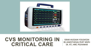
Most ICUs monitor ECG, HR, BP SpO2
This approach might be inappropriate in the ICU
No monitoring device can improve patient outcomes unless it is
coupled to a treatment e.g.
Hemorrhage often manifests as tachycardia and HTN with low SpO2.
Neither B-blockers nor vasodilators are indicated in the initial management
of such patients
Thus, it is not enough to monitor the patient closely, but also interpret in
the context of the pathophysiology and stage of the patient’s disease
2
3. Purposes
To detect an impending CVS
crisis before organ damage
To monitor the response to
CVS therapy
To titrated treatments to
specific CVS responses
To differentiate causes of
hemodynamic instability
Effectiveness
Accuracy of technology
Healthcare professional’s
ability to diagnose
Effective treatment of the
underlying diseases for
which the monitoring is used
3
4. COMMON VARIABLES
Variables
Arterial blood pressure (BP)
Heart Rate (HR)
Oxygen saturation (SpO2)
Central venous pressure (CVP)
Pulmonary artery occlusion
pressure
Cardiac output (CO)
Mixed venous oxygen saturation
Mode of
measurement
Invasive and non-invasive
Non invasive
Non invasive
Invasive
Invasive
Invasive
Invasive
4
5. COMMON VARIABLES
•Threshold values exist such that values above or
below them may reflect CVS compromise
•That usually not tolerated for prolonged intervals
without resulting in end-organ dysfunction and/or
death e.g.
•Bradycardia (HR < 45/min)
•Tachycardia (HR > 130/min)
•Hypotension (mean BP < 65 mm Hg)
•Hypertension (mean BP > 180 mm Hg)
5
6. SIMPLISTIC STATEMENTS
1. Tachycardia is never a good thing
2. Hypotension is always pathological
3. There is no such thing as a normal cardiac output
4. CVP is only elevated in disease
5. Peripheral edema is of cosmetic concern
6
7. HEART RATE MONITORING
• “Finger on pulse” is the easiest, quickest and most accurate
method to assess heart rate
• ECG is most common method to detect heart rate in OT, by
measurement of R-R interval
• ECG can be confounded by electrosurgical instruments, power
line noises, twitching and fasciculation, and fluid warmer
• Beside these pulse oximeter and stethoscope are also used for
HR monitoring
7
8. ARTERIAL BLOOD PRESSURE
MONITORING
Non invasive (indirect method)
o Manual
o Automated
oManual Intermittent techniques
o Automated Intermittent techniques
o Automated Continuous techniques
Invasive (Direct method)
8
9. NIBP (MANUAL)
• Sphygmomanometer was used for SBP 1st time by Riva and Rocci in
1896 (palpatory method)
• Karotkoff in 1905 described measurement of diastolic as well
(auscultatory method)
• Size of cuff should be 40% to 80% of circumference of arm
• Too large can still be accepted but loose give low reading
• Too small will give high reading
• Pressure should be released slowly to assess Karotkoff sounds
properly
• Very low frequency sounds (25-50 Hz) produced by turbulent blood
flow 9
10. NIBP (AUTOMATED)
• Intermittent based oscillatory method, 1st described by Marey
in 1876
• Assess MAP most accurately and SBP and DBP are derived
(DBP least reliable)
• This method is unreliable, and its use other than upper arm is
not validated
• Complications may occur due to continuous use in patients
like:
• Coagulopathies
• Arterial and venous insufficiency
• Thrombolytic therapy
10
12. IBP/DIRECT BP MONITORING
• IBP monitoring is an ideal standard method for BP monitoring
• Provide timely and crucial information
• Although it have various complications and need expertise
• Arterial cannulation can be done in radial, ulnar, brachial, axillary
or femoral artery
• Before cannulation, confirm collateral supply (Allen’s test)
12
14. IBP/DIRECT BP MONITORING
• More central the artery, more will be the chances
of embolism
• In radial artery cannulation hyperextension is
avoided to prevent median nerve injury
• In femoral artery cannulation must be done below
the inguinal ligament
14
15. INDICATIONS FOR ARTERIAL
CANNULATION
• Need for continuous, real-time and beat to beat BP
monitoring
• Repeated blood sampling
• Failure of indirect arterial blood pressure
measurement
• Supplementary diagnostic information from the
waveform
• Determination of volume responsiveness from SBP
and PP variation 15
16. COMPONENT
S
• Intra-arterial cannula
• Coupling system (stopcock)
• Pressure transducer
• Infusion flushing system
• Signal processor, amplifier and
display
16
18. LEVELLING &ZEROING
Zeroing:
• For a pressure transducer to read accurately, atmospheric
pressure must be discounted
• This is done by exposing the transducer to atmospheric
pressure and calibrating the pressure reading to zero
Levelling:
• The pressure transducer must be set at the appropriate level
• Patient’s heart, at the 4th intercostal space, in the mid-axillary
line
18
20. CENTRAL VENOUS PRESSURE
MONITORING
•CVP is the pressure measured at the junction of the superior venae
cava and the right atrium
•It reflects the relationship of blood volume to the capacity of the
venous system
•Normal CVP in an awake ,spontaneously breathing patient =1-
7mmHg OR 5-10 cmH2O
•Mechanical ventilation 3-5 cmH2O higher
20
21. CVP MONITORING
• Veins should be:
• Right/left internal jugular
• Right/left subclavian
• Femoral
• Most commonly used size is 7 French, 20cm catheter
with 18g introducer needle and a guide wire
21
23. INDICATIONS FOR CVP
• Central venous pressure
monitoring
• Temporary hemodialysis
• Drug administration
• Concentrated vasoactive
drugs
• Chemotherapy
• Prolonged antibiotic
therapy (e.g., endocarditis)
• Rapid infusion of fluids
• Major surgery
• Trauma
• Inadequate peripheral
intravenous access
• Sampling for repeated
tests
• Total parenteral
nutrition
23
24. a = Atrial contraction
c = Isovolumic
ventricular
contraction
x = Atrial relaxation
v = filling of the atria
y = ventricular filling
h = Diastolic plateau
24
28. PULMONARY ARTERY
CATHETER
Also called as Swan Ganz catheter
It measure pulmonary artery pressure
This is measured by inserting a catheter into the pulmonary
artery
The mean pressure is typically 9 - 18 mmHg
It is used for direct and indirect measurement of different
parameters
28
31. PAC KIT
Standard PAC is 7.0, 7.5 or 8.0 French in circumference
and 110 cm in length divided in 10 cm intervals
It also include:
1. A syringe that can be filled with only 1.5 mL of air to
prevent over inflation of the balloon
2. A long plastic sheath that is used to maintain sterility of
the PAC as it is advanced and withdrawn
31
32. PAC KIT
PAC has 4-5 lumens:
1. Temperature thermistor to measure pulmonary
blood temperature
2. Proximal port for CVP monitoring, fluid and drug
administration
3. Distal port at catheter tip for PAP monitoring
4. Variable infusion port (VIP) for fluid and drug
administration
5. Balloon at catheter tip
32
33. PREPARATION & INSERTION
• Patient has to be monitored with continuous ECG
throughout the procedure
• Supine position regardless of the approach
• Aseptic precautions must be employed
• Cautions should be taken while cannulation via IJV/
Subclavian vein
• Local infiltration
• Check balloon integrity by inflating with 1.5ml of air
33
34. PREPARATION & INSERTION
• Check lumens patency by flushing with saline 0.9%
• Pass catheter through sheath with tip curved towards the heart
• Once tip of catheter passed through introducer sheath, inflate
balloon at level of right ventricle
• Advance the catheter through right atrium and ventricle into
pulmonary artery and wedge position can be monitored by
changes in pressure trace
• After acquiring wedge pressure, deflate balloon
34
36. INDICATIONS
Diagnostic:
• Differentiation among causes of shock
• Differentiation between mechanisms of pulmonary
edema
• Evaluation of pulmonary hypertension
• Diagnosis of pericardial tamponade
• Diagnosis of right to left intracardiac shunts
• Unexplained dyspnea 36
37. INDICATIONS
Therapeutic:
• Management of perioperative patients with unstable
status
• Management of complicated myocardial infarction
• Management of patients following cardiac surgery/high
surgery
• Guide to pharmacologic therapy
• Assess response to pulmonary hypertension specific 37
38. CONTRAINDICATIONS
Absolute:
• Infection at insertion site
• Presence of RV assist
device
• Insertion during CPB
• Lack of consent
Relative:
• Coagulopathy
• Thrombocytopenia
• Electrolyte disturbances
• Severe Pulmonary HTN
38