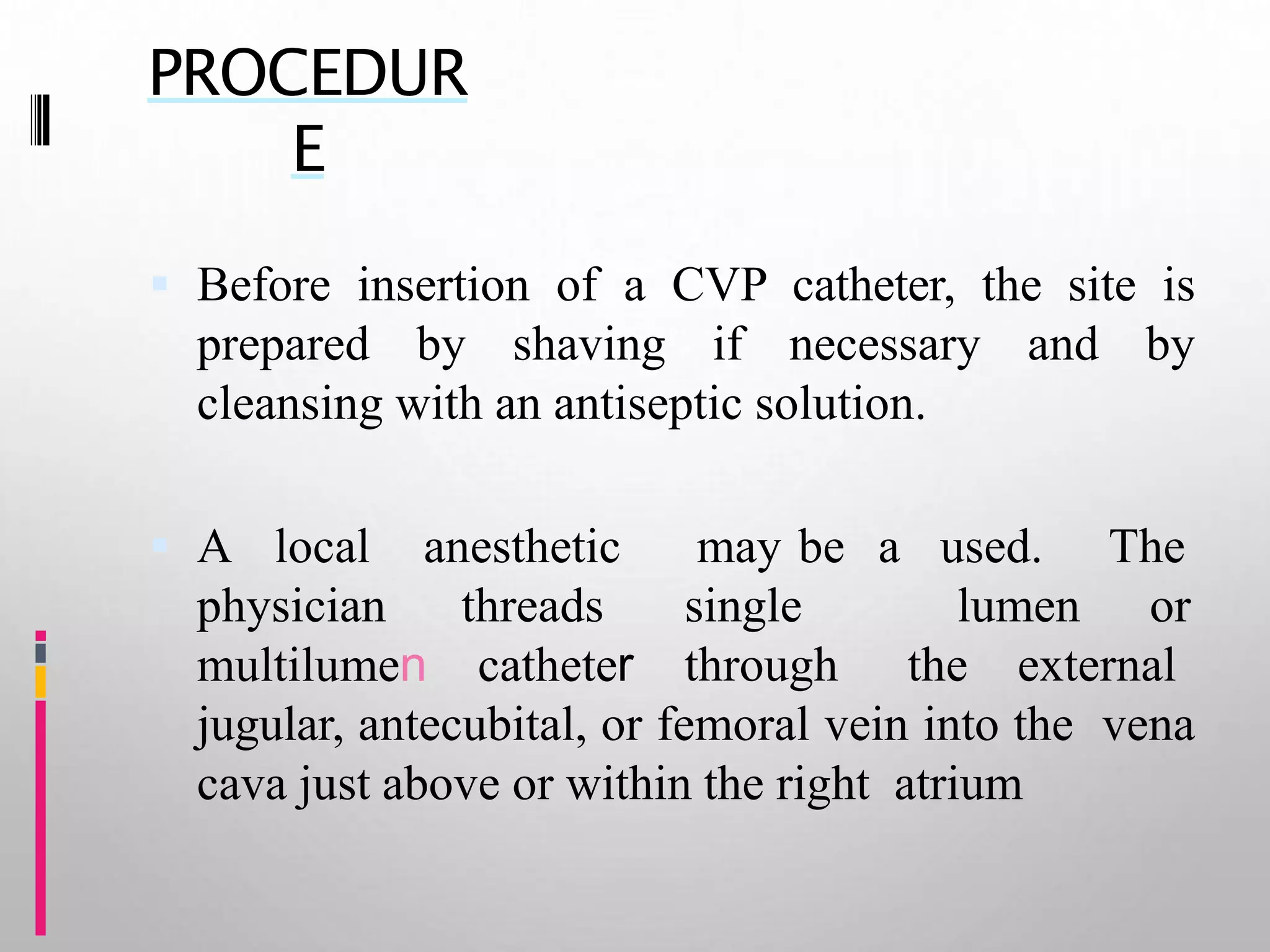
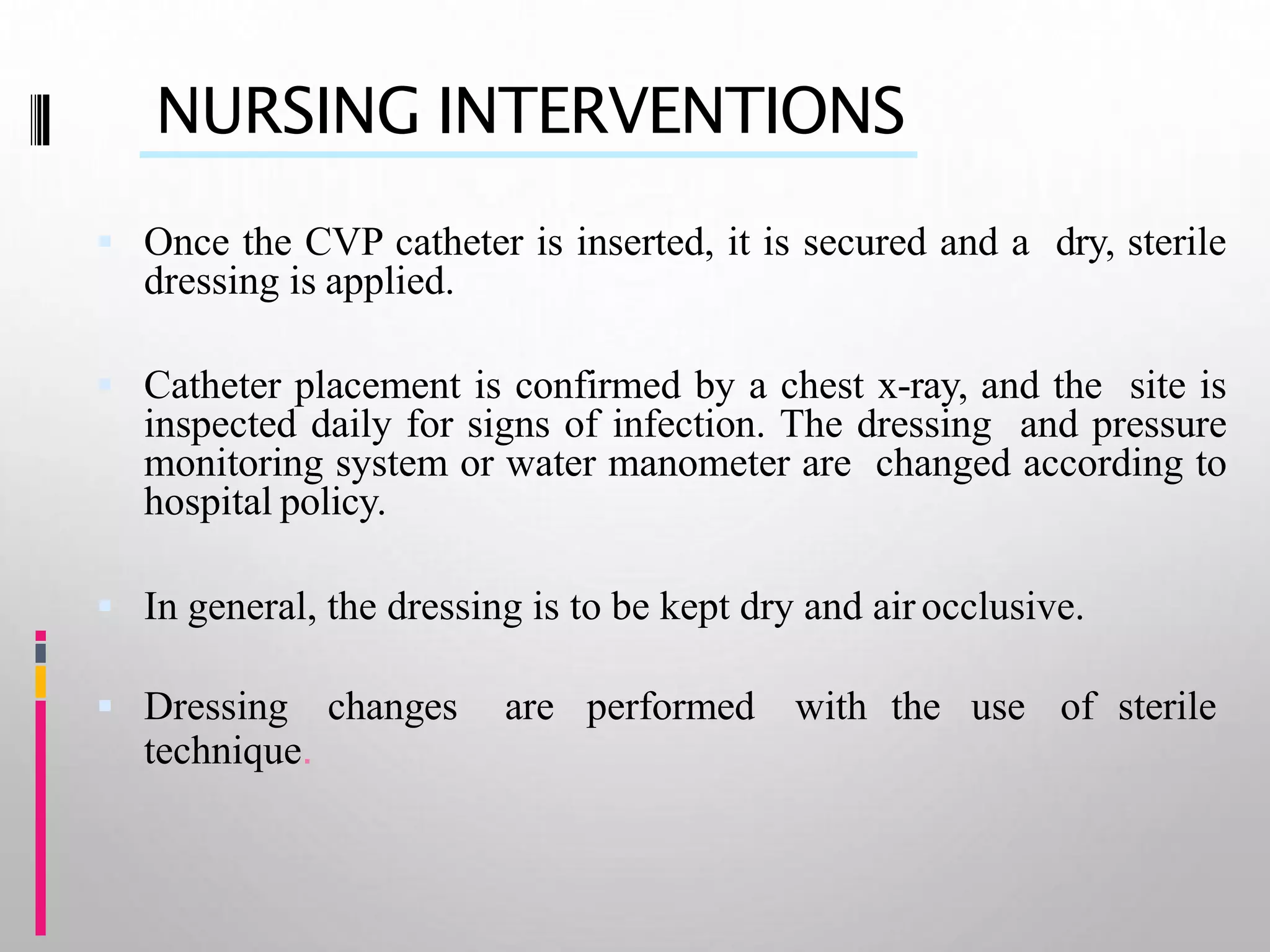
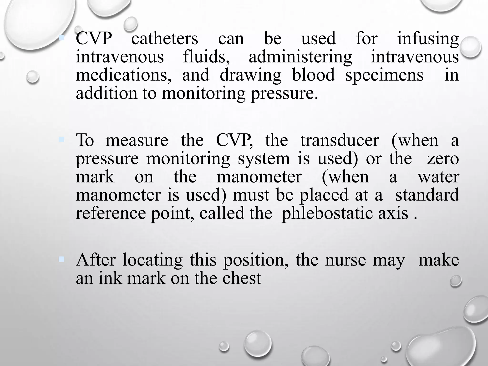
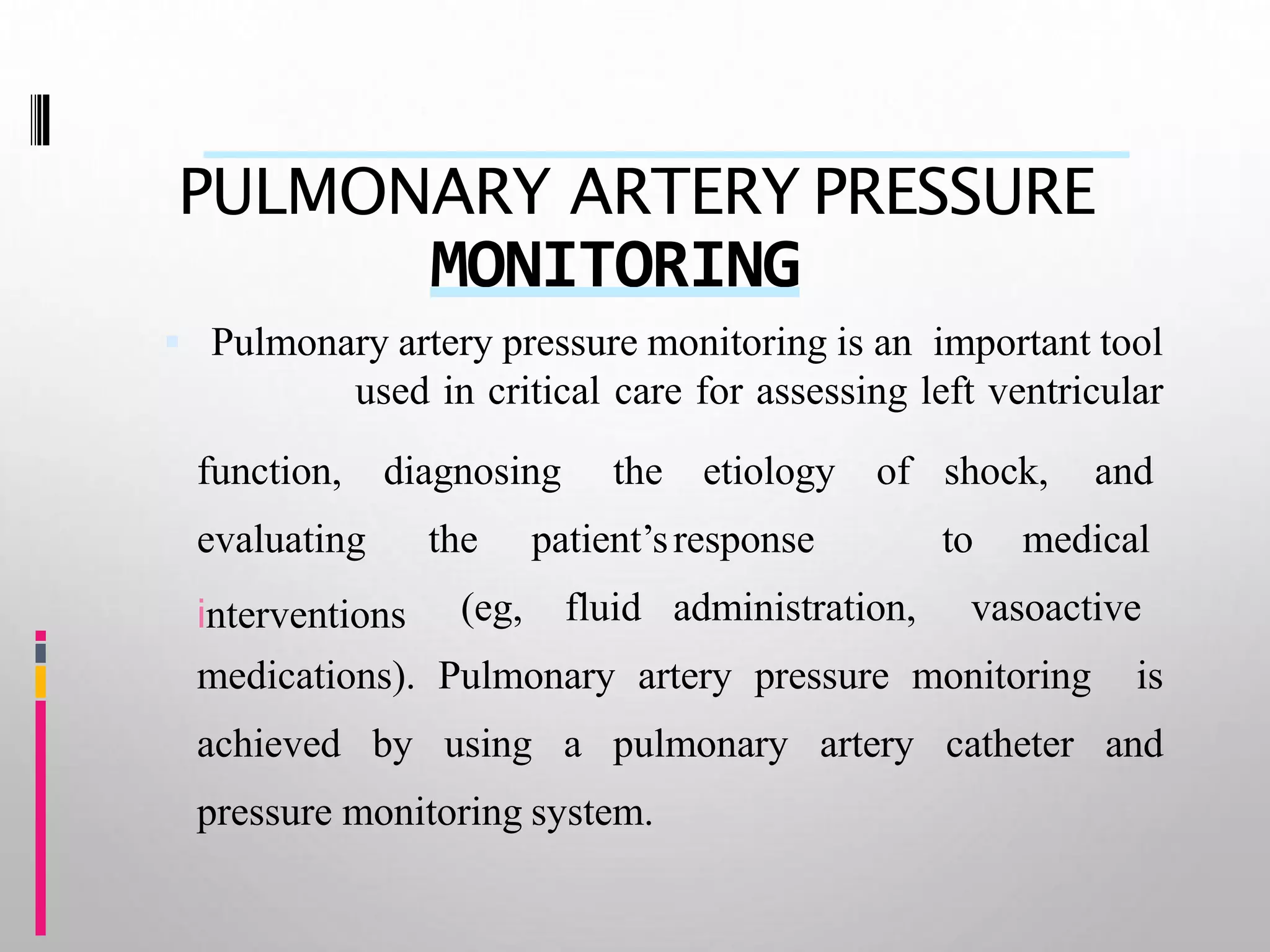
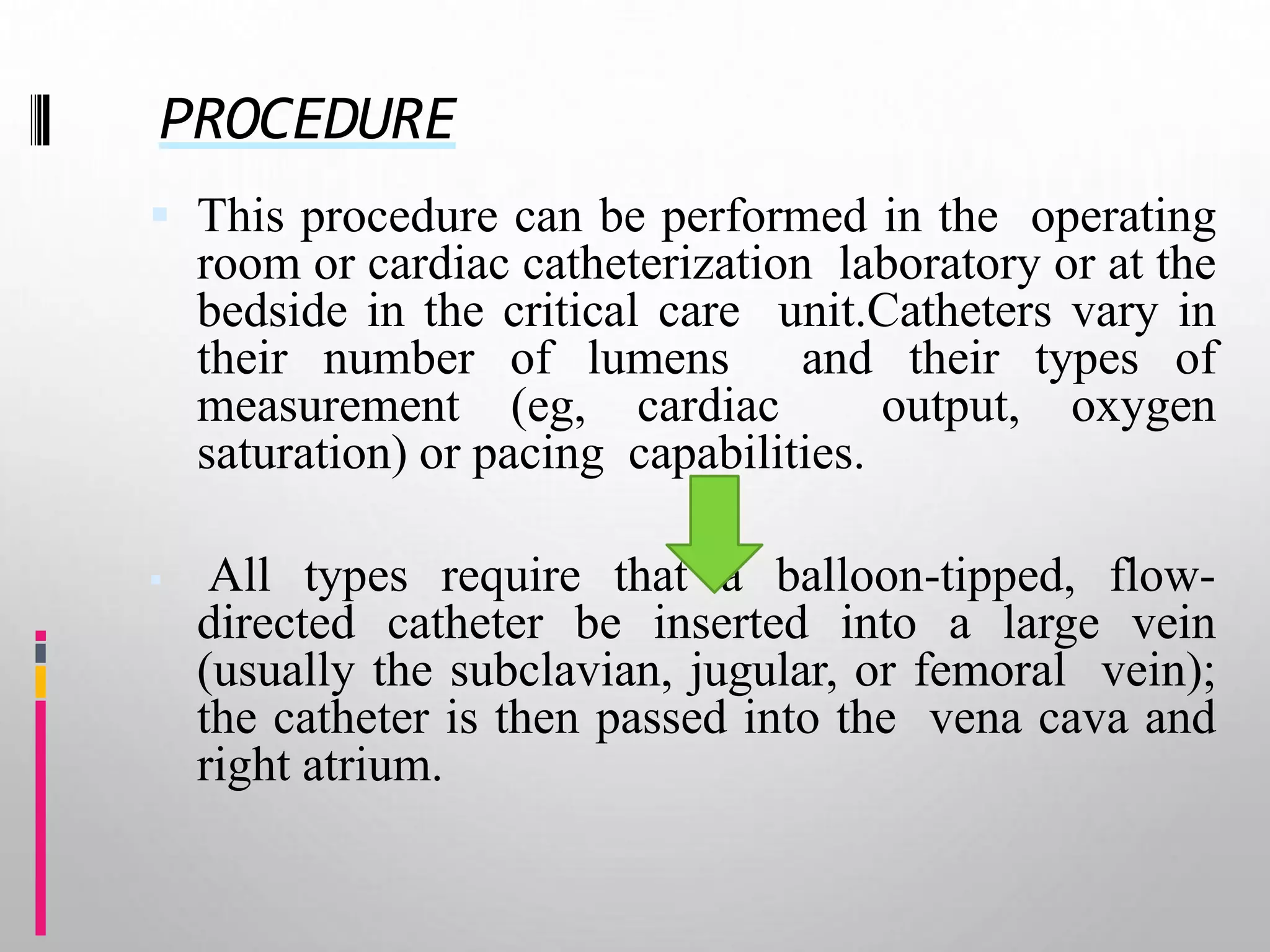
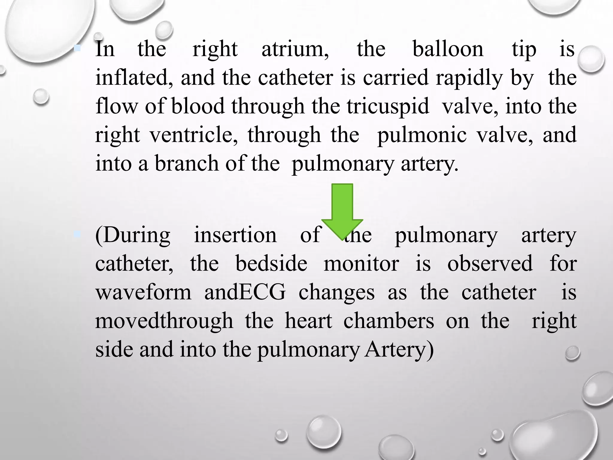
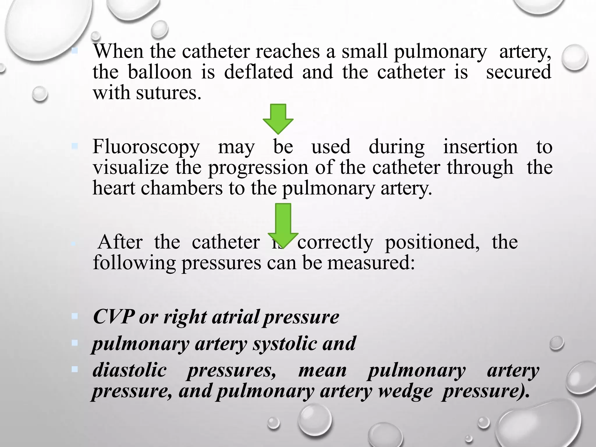
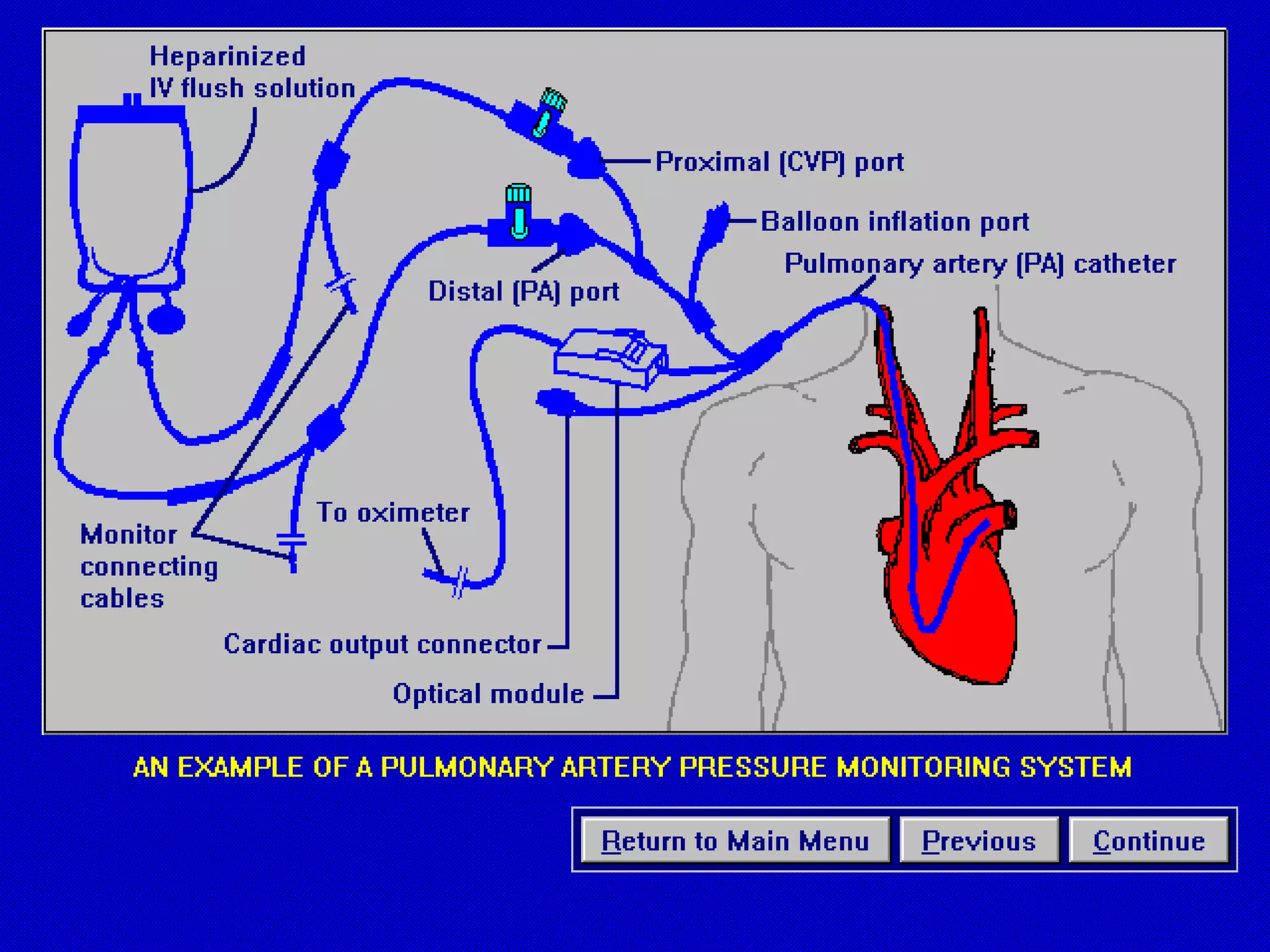
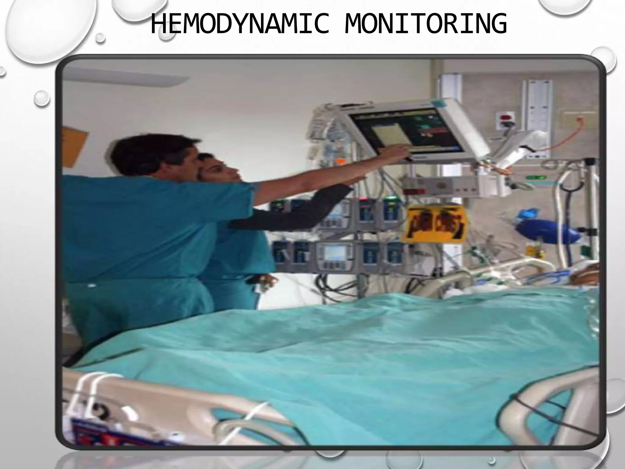
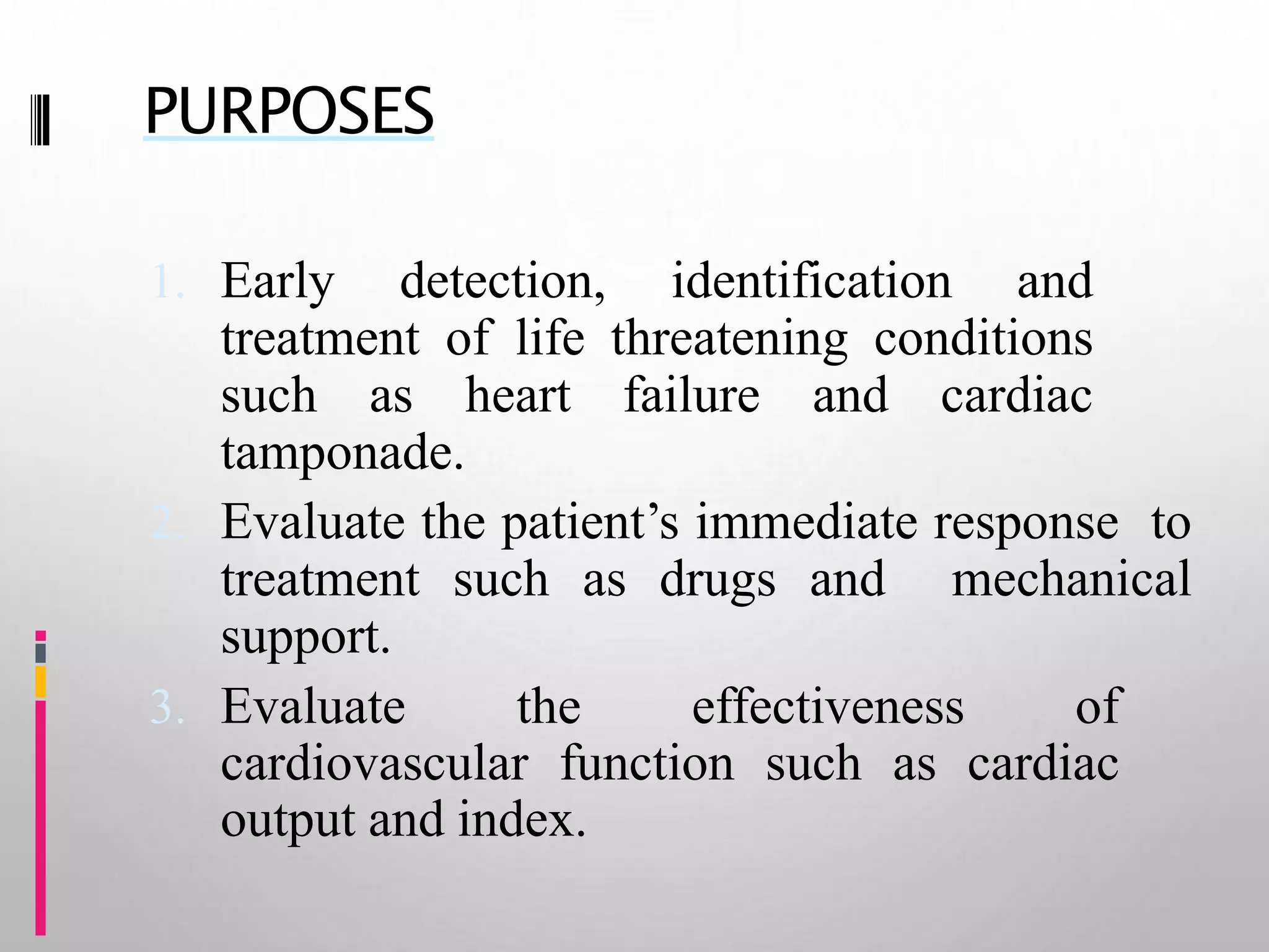
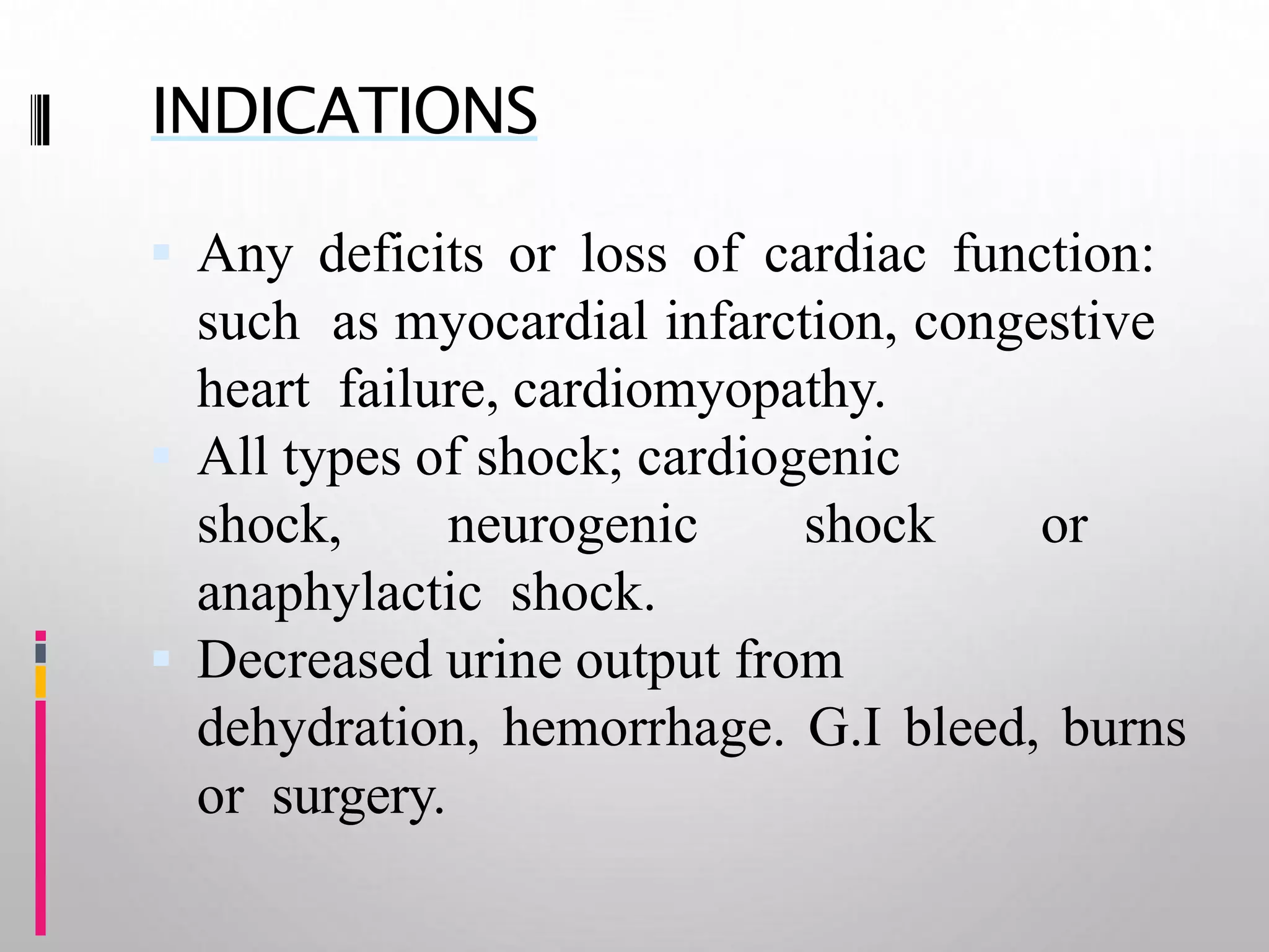
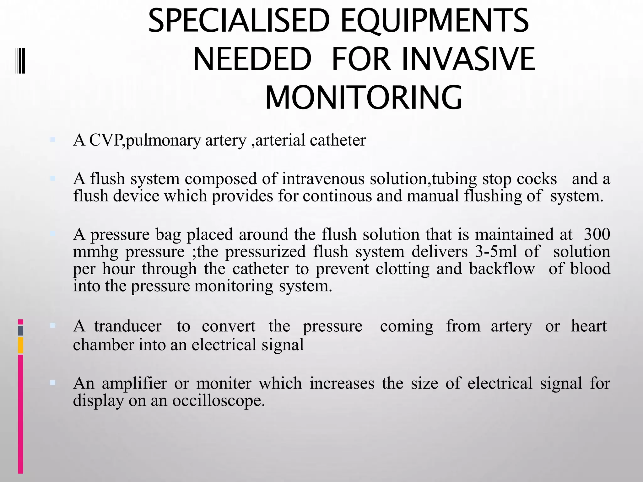
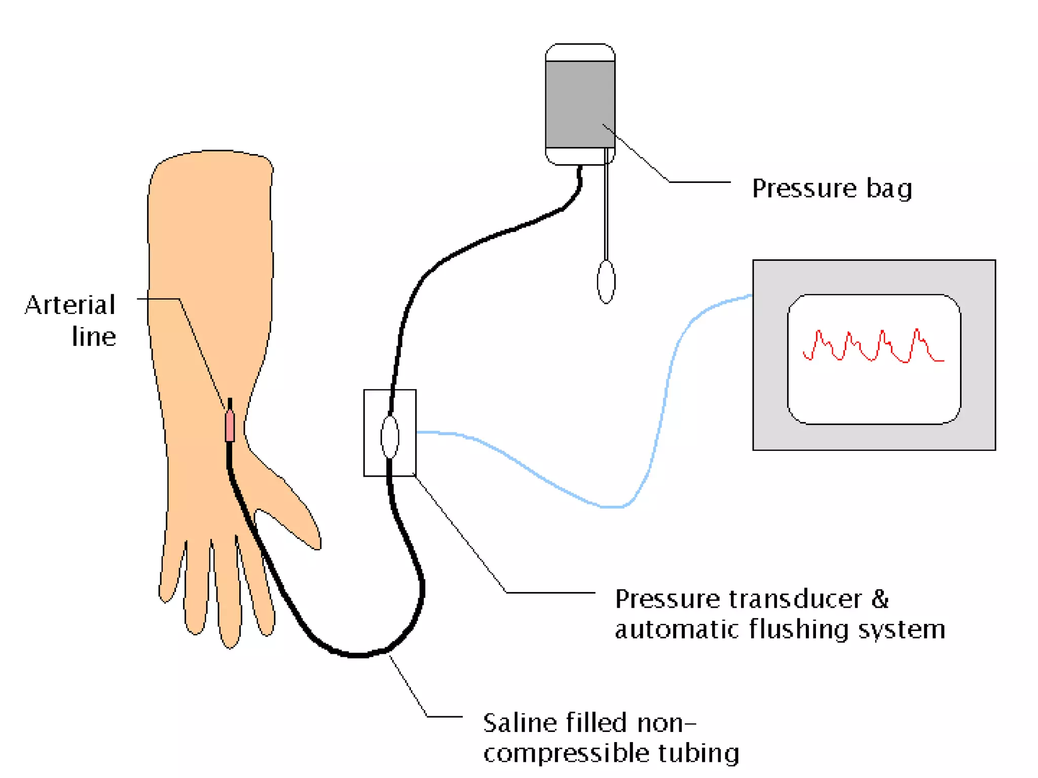
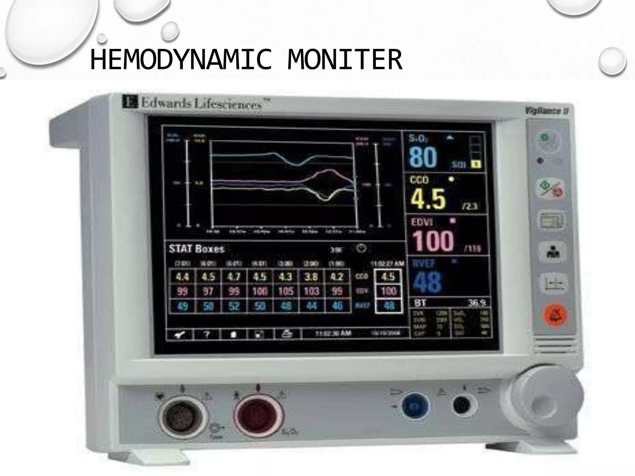
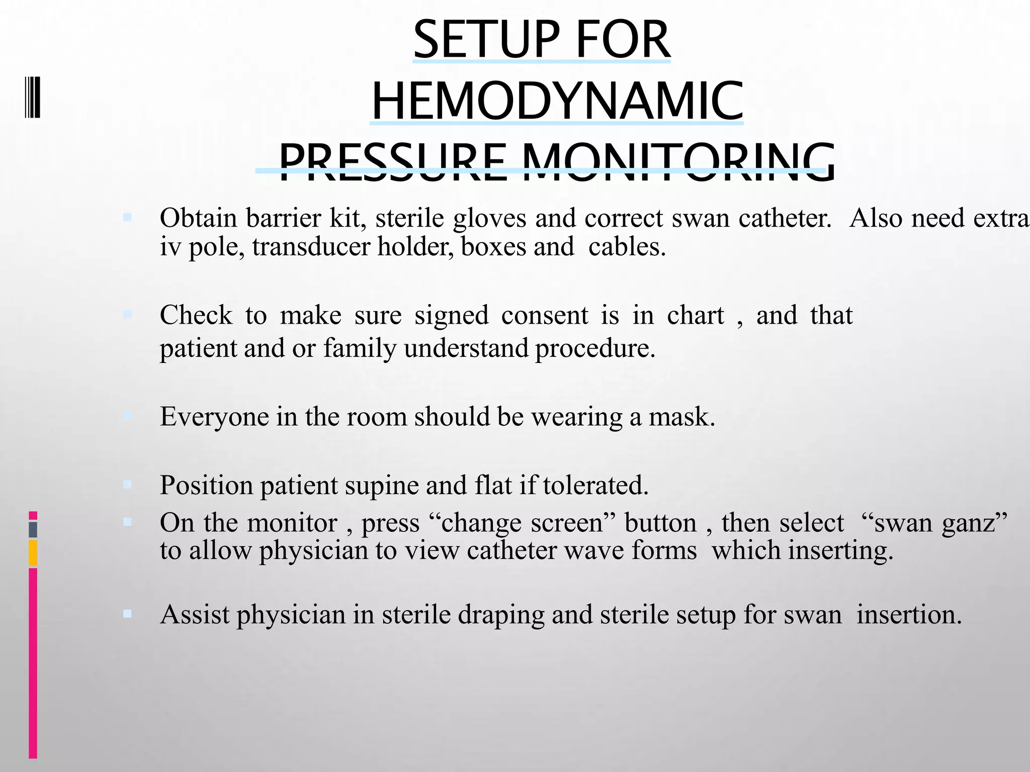
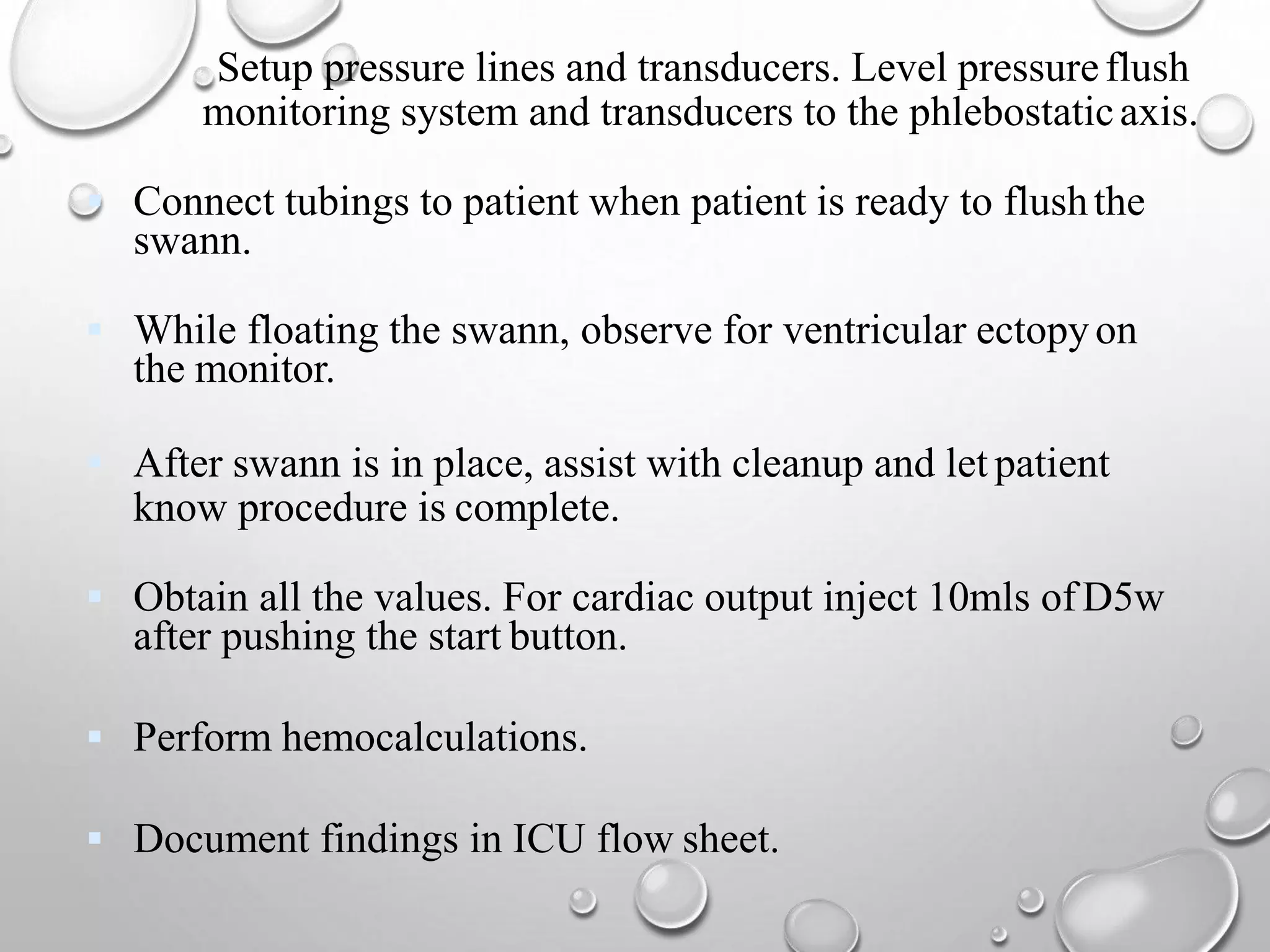
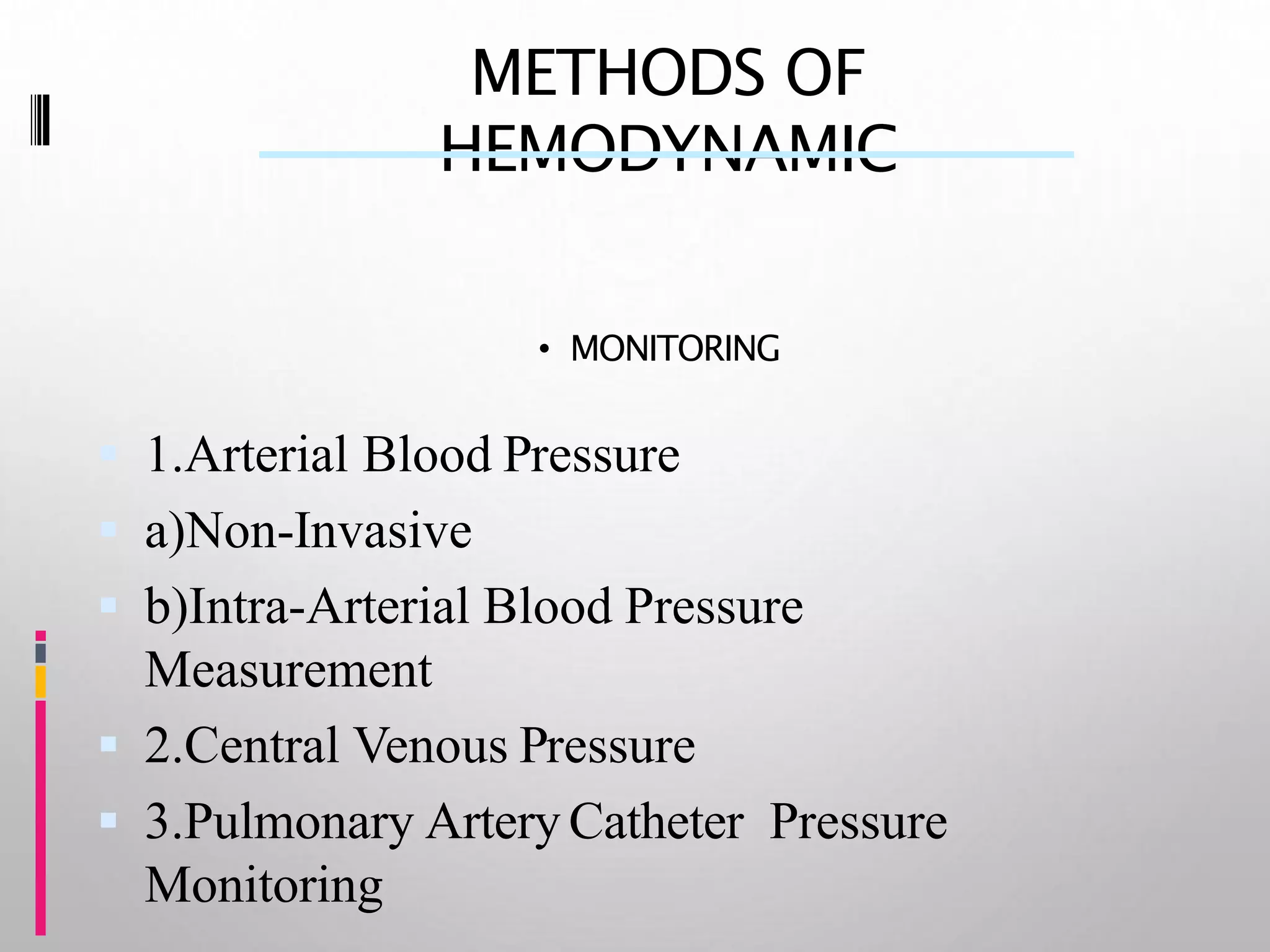
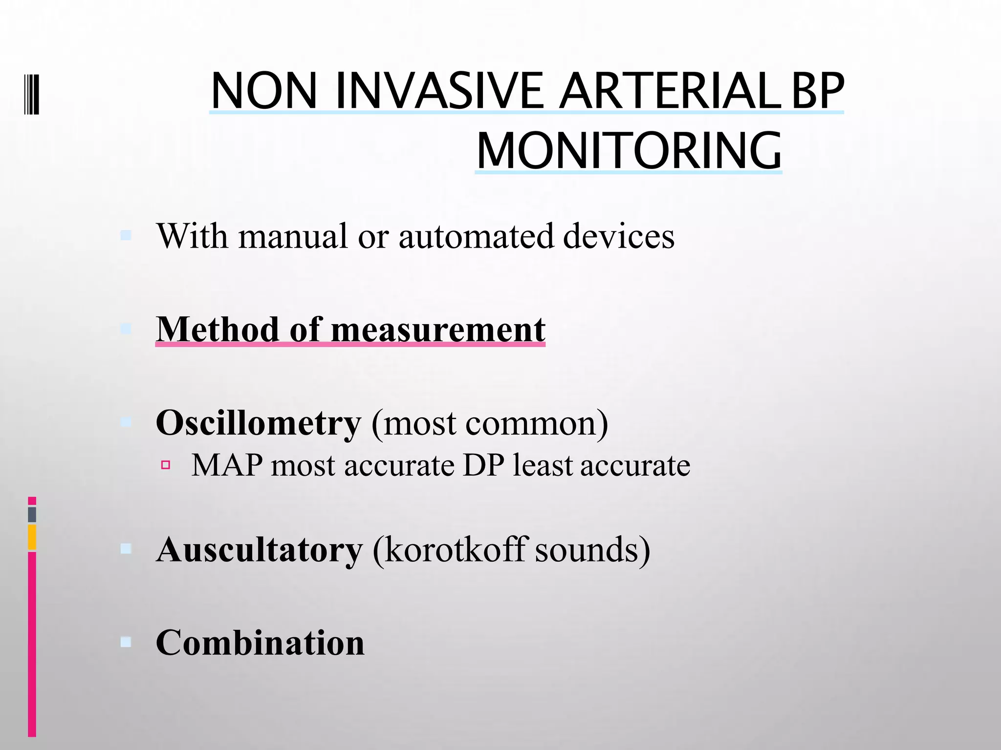
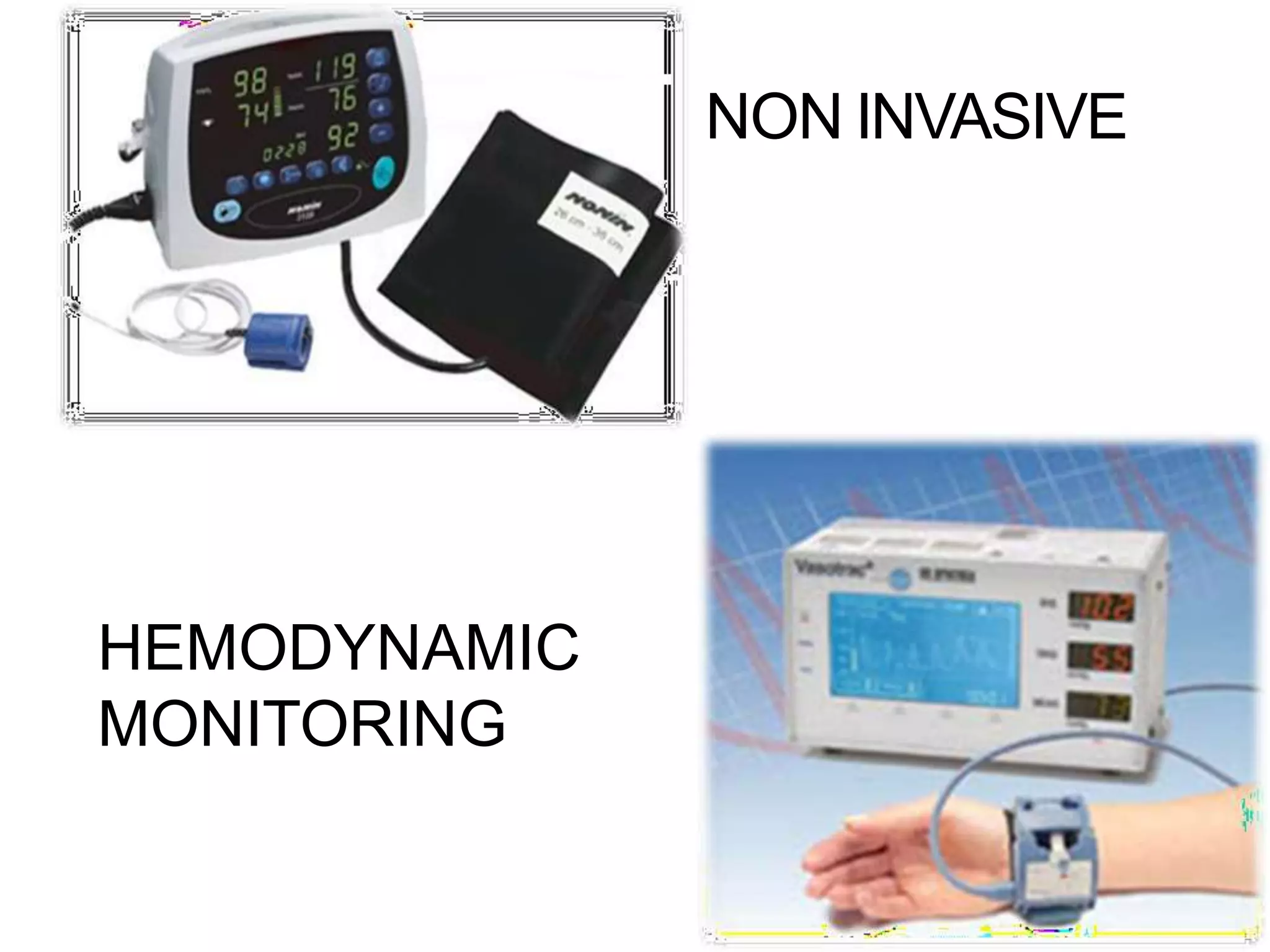
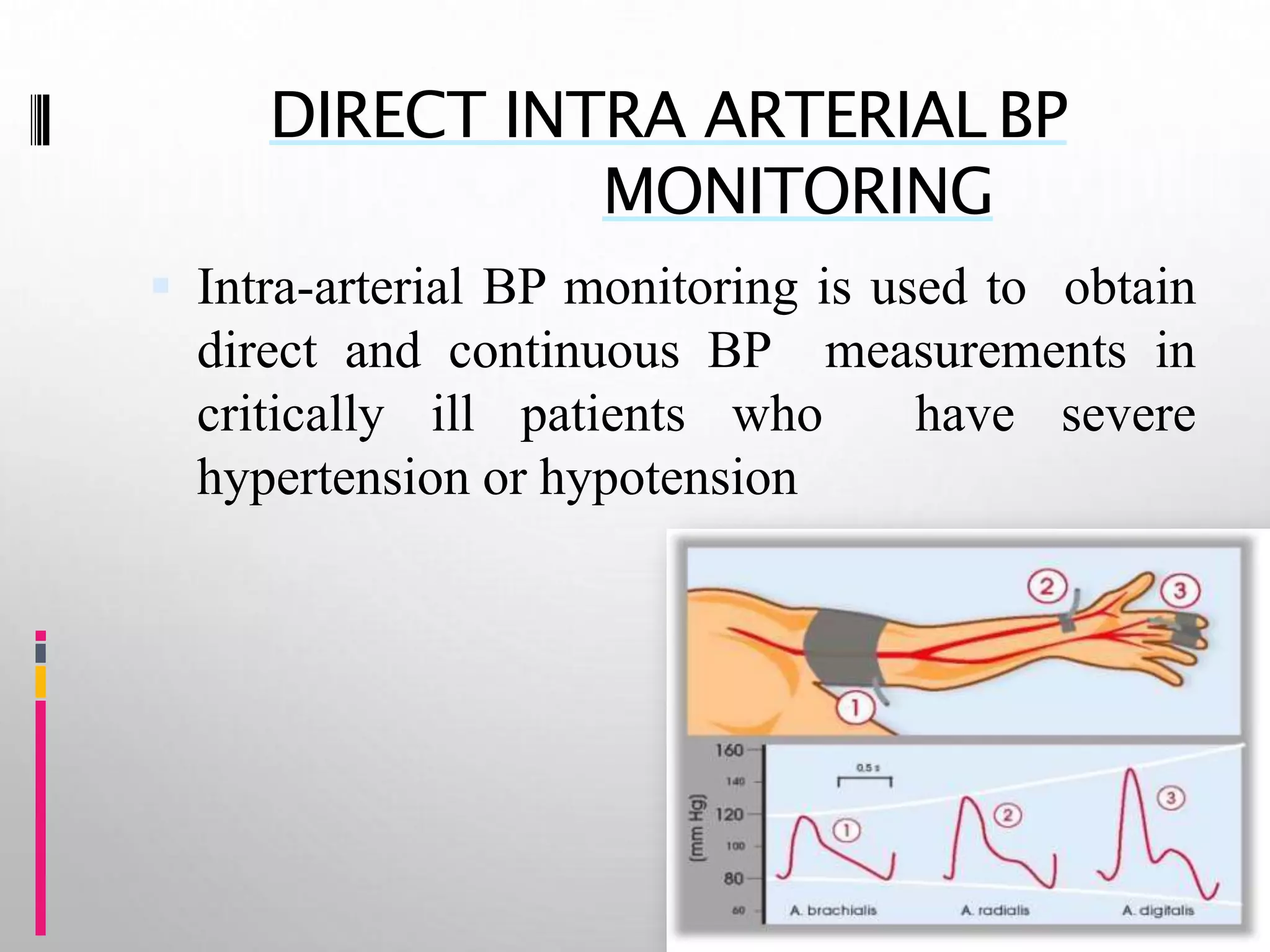
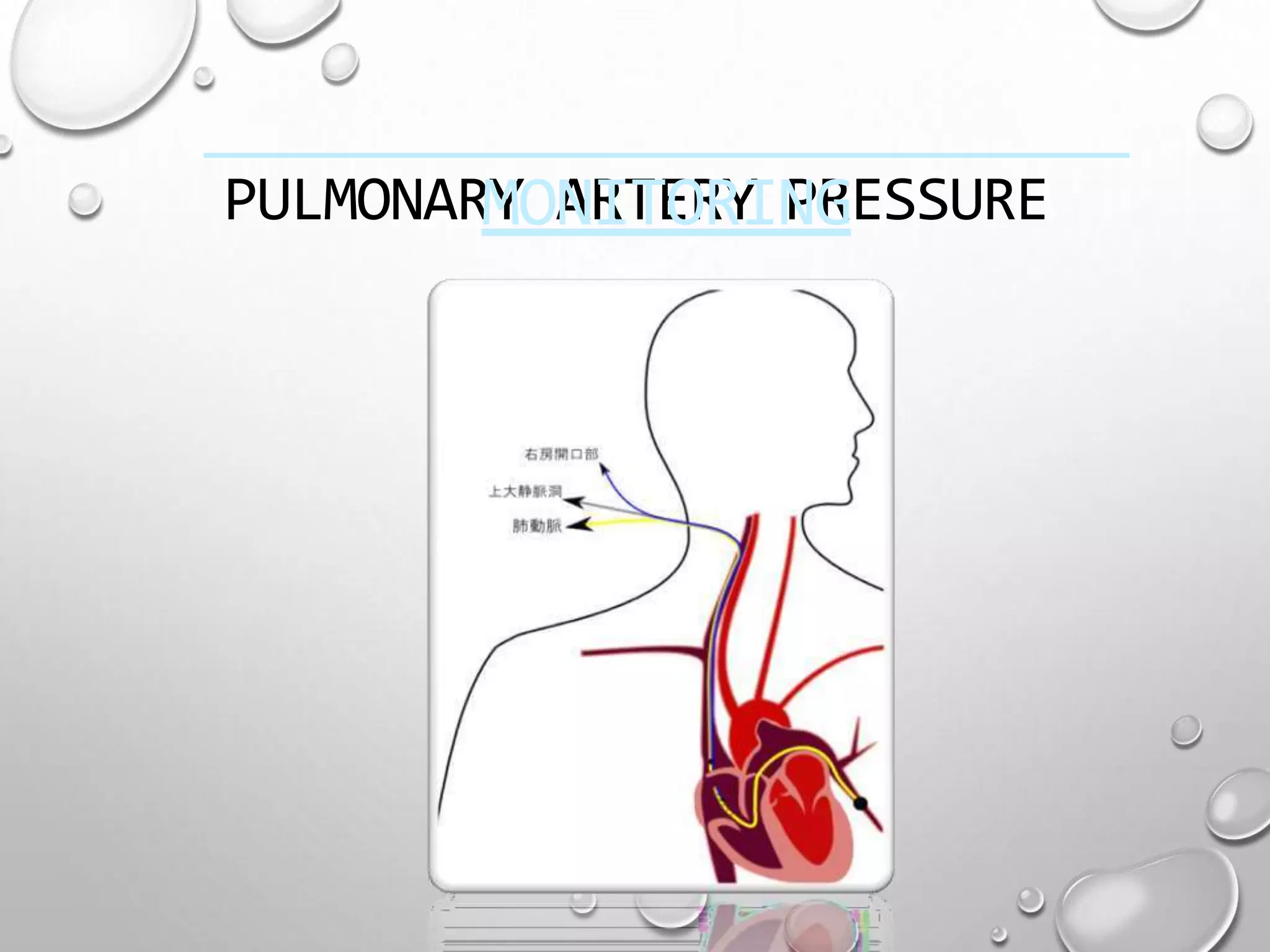
This document discusses hemodynamic monitoring, which refers to measuring the pressure, flow, and oxygenation of blood within the cardiovascular system. It is most commonly done through arterial blood pressure monitoring, central venous pressure monitoring, and pulmonary artery catheter pressure monitoring. These invasive monitoring methods provide information about how well the heart is pumping and circulating blood throughout the body. The document outlines the purposes, indications, equipment, procedures, complications, and nursing responsibilities for each of these hemodynamic monitoring methods.
Comprehensive presentation on intra arterial blood pressure with a good insight into the the basic physics and brief look into the risks and complications.
Hemodynamic monitoring- Hemodynamic monitoring refers to the measurement of pressure, flow and oxygenation within the cardiovascular system. Hemodynamic monitoring is amandatory process in all the critical care units to assess the patients progress. This presentation is aimed to create an insight on Hemodynamic monitoring.
This is a very simple presentation prepared for nurses. It will help nurses to understand the need of monitoring and the available methods. The presentation has been constructed on a clinical case base scenario and gradually different methods of monitoring has been introduced.
Intra Aortic Balloon Pump by Rubina Shehzadi RNRubina Shehzadi
An intra-aortic balloon pump (IABP) is a type of therapeutic device which helps heart to pump more blood. You may need it if your heart is unable to pump enough blood for your body. The IABP consists of a thin, flexible tube called a catheter. Attached to the tip of the catheter is a long balloon.
Nursing management of patient with cardiac surgeries.PrashantSalve10
It will be helpful to overview cardiac surgeries like CABG, Valvular surgeries and heart transplant. It also enumerates the nursing diagnoses and its brief description.
continuous or intermittent monitoring of heart activity, generally by electrocardiography, with assessment of the patient's condition relative to their cardiac rhythm.
Hemodynamic monitoring of critically ill patientsV4Veeru25
Hemodynamic monitoring measures the blood pressure inside the veins, heart, and arteries. It also measures blood flow and oxygen proportion in the blood. Monitoring hemodynamic events provides information about the adequacy of a patient's circulation , perfusion, and oxygenation of the tissues and organ systems. The effectiveness of hemodynamic monitoring depends both on available technology and on physician ability to diagnose and effectively treat the disease
The CVP catheter is an important tool used to assess right ventricular function and systemic fluid status. Normal CVP is 2-6 mm Hg. CVP is elevated by : overhydration which increases venous return.
Report Back from SGO 2024: What’s the Latest in Cervical Cancer?bkling
Are you curious about what’s new in cervical cancer research or unsure what the findings mean? Join Dr. Emily Ko, a gynecologic oncologist at Penn Medicine, to learn about the latest updates from the Society of Gynecologic Oncology (SGO) 2024 Annual Meeting on Women’s Cancer. Dr. Ko will discuss what the research presented at the conference means for you and answer your questions about the new developments.
Lung Cancer: Artificial Intelligence, Synergetics, Complex System Analysis, S...Oleg Kshivets
RESULTS: Overall life span (LS) was 2252.1±1742.5 days and cumulative 5-year survival (5YS) reached 73.2%, 10 years – 64.8%, 20 years – 42.5%. 513 LCP lived more than 5 years (LS=3124.6±1525.6 days), 148 LCP – more than 10 years (LS=5054.4±1504.1 days).199 LCP died because of LC (LS=562.7±374.5 days). 5YS of LCP after bi/lobectomies was significantly superior in comparison with LCP after pneumonectomies (78.1% vs.63.7%, P=0.00001 by log-rank test). AT significantly improved 5YS (66.3% vs. 34.8%) (P=0.00000 by log-rank test) only for LCP with N1-2. Cox modeling displayed that 5YS of LCP significantly depended on: phase transition (PT) early-invasive LC in terms of synergetics, PT N0—N12, cell ratio factors (ratio between cancer cells- CC and blood cells subpopulations), G1-3, histology, glucose, AT, blood cell circuit, prothrombin index, heparin tolerance, recalcification time (P=0.000-0.038). Neural networks, genetic algorithm selection and bootstrap simulation revealed relationships between 5YS and PT early-invasive LC (rank=1), PT N0—N12 (rank=2), thrombocytes/CC (3), erythrocytes/CC (4), eosinophils/CC (5), healthy cells/CC (6), lymphocytes/CC (7), segmented neutrophils/CC (8), stick neutrophils/CC (9), monocytes/CC (10); leucocytes/CC (11). Correct prediction of 5YS was 100% by neural networks computing (area under ROC curve=1.0; error=0.0).
CONCLUSIONS: 5YS of LCP after radical procedures significantly depended on: 1) PT early-invasive cancer; 2) PT N0--N12; 3) cell ratio factors; 4) blood cell circuit; 5) biochemical factors; 6) hemostasis system; 7) AT; 8) LC characteristics; 9) LC cell dynamics; 10) surgery type: lobectomy/pneumonectomy; 11) anthropometric data. Optimal diagnosis and treatment strategies for LC are: 1) screening and early detection of LC; 2) availability of experienced thoracic surgeons because of complexity of radical procedures; 3) aggressive en block surgery and adequate lymph node dissection for completeness; 4) precise prediction; 5) adjuvant chemoimmunoradiotherapy for LCP with unfavorable prognosis.
micro teaching on communication m.sc nursing.pdfAnurag Sharma
Microteaching is a unique model of practice teaching. It is a viable instrument for the. desired change in the teaching behavior or the behavior potential which, in specified types of real. classroom situations, tends to facilitate the achievement of specified types of objectives.
Title: Sense of Smell
Presenter: Dr. Faiza, Assistant Professor of Physiology
Qualifications:
MBBS (Best Graduate, AIMC Lahore)
FCPS Physiology
ICMT, CHPE, DHPE (STMU)
MPH (GC University, Faisalabad)
MBA (Virtual University of Pakistan)
Learning Objectives:
Describe the primary categories of smells and the concept of odor blindness.
Explain the structure and location of the olfactory membrane and mucosa, including the types and roles of cells involved in olfaction.
Describe the pathway and mechanisms of olfactory signal transmission from the olfactory receptors to the brain.
Illustrate the biochemical cascade triggered by odorant binding to olfactory receptors, including the role of G-proteins and second messengers in generating an action potential.
Identify different types of olfactory disorders such as anosmia, hyposmia, hyperosmia, and dysosmia, including their potential causes.
Key Topics:
Olfactory Genes:
3% of the human genome accounts for olfactory genes.
400 genes for odorant receptors.
Olfactory Membrane:
Located in the superior part of the nasal cavity.
Medially: Folds downward along the superior septum.
Laterally: Folds over the superior turbinate and upper surface of the middle turbinate.
Total surface area: 5-10 square centimeters.
Olfactory Mucosa:
Olfactory Cells: Bipolar nerve cells derived from the CNS (100 million), with 4-25 olfactory cilia per cell.
Sustentacular Cells: Produce mucus and maintain ionic and molecular environment.
Basal Cells: Replace worn-out olfactory cells with an average lifespan of 1-2 months.
Bowman’s Gland: Secretes mucus.
Stimulation of Olfactory Cells:
Odorant dissolves in mucus and attaches to receptors on olfactory cilia.
Involves a cascade effect through G-proteins and second messengers, leading to depolarization and action potential generation in the olfactory nerve.
Quality of a Good Odorant:
Small (3-20 Carbon atoms), volatile, water-soluble, and lipid-soluble.
Facilitated by odorant-binding proteins in mucus.
Membrane Potential and Action Potential:
Resting membrane potential: -55mV.
Action potential frequency in the olfactory nerve increases with odorant strength.
Adaptation Towards the Sense of Smell:
Rapid adaptation within the first second, with further slow adaptation.
Psychological adaptation greater than receptor adaptation, involving feedback inhibition from the central nervous system.
Primary Sensations of Smell:
Camphoraceous, Musky, Floral, Pepperminty, Ethereal, Pungent, Putrid.
Odor Detection Threshold:
Examples: Hydrogen sulfide (0.0005 ppm), Methyl-mercaptan (0.002 ppm).
Some toxic substances are odorless at lethal concentrations.
Characteristics of Smell:
Odor blindness for single substances due to lack of appropriate receptor protein.
Behavioral and emotional influences of smell.
Transmission of Olfactory Signals:
From olfactory cells to glomeruli in the olfactory bulb, involving lateral inhibition.
Primitive, less old, and new olfactory systems with different path
Ethanol (CH3CH2OH), or beverage alcohol, is a two-carbon alcohol
that is rapidly distributed in the body and brain. Ethanol alters many
neurochemical systems and has rewarding and addictive properties. It
is the oldest recreational drug and likely contributes to more morbidity,
mortality, and public health costs than all illicit drugs combined. The
5th edition of the Diagnostic and Statistical Manual of Mental Disorders
(DSM-5) integrates alcohol abuse and alcohol dependence into a single
disorder called alcohol use disorder (AUD), with mild, moderate,
and severe subclassifications (American Psychiatric Association, 2013).
In the DSM-5, all types of substance abuse and dependence have been
combined into a single substance use disorder (SUD) on a continuum
from mild to severe. A diagnosis of AUD requires that at least two of
the 11 DSM-5 behaviors be present within a 12-month period (mild
AUD: 2–3 criteria; moderate AUD: 4–5 criteria; severe AUD: 6–11 criteria).
The four main behavioral effects of AUD are impaired control over
drinking, negative social consequences, risky use, and altered physiological
effects (tolerance, withdrawal). This chapter presents an overview
of the prevalence and harmful consequences of AUD in the U.S.,
the systemic nature of the disease, neurocircuitry and stages of AUD,
comorbidities, fetal alcohol spectrum disorders, genetic risk factors, and
pharmacotherapies for AUD.
These simplified slides by Dr. Sidra Arshad present an overview of the non-respiratory functions of the respiratory tract.
Learning objectives:
1. Enlist the non-respiratory functions of the respiratory tract
2. Briefly explain how these functions are carried out
3. Discuss the significance of dead space
4. Differentiate between minute ventilation and alveolar ventilation
5. Describe the cough and sneeze reflexes
Study Resources:
1. Chapter 39, Guyton and Hall Textbook of Medical Physiology, 14th edition
2. Chapter 34, Ganong’s Review of Medical Physiology, 26th edition
3. Chapter 17, Human Physiology by Lauralee Sherwood, 9th edition
4. Non-respiratory functions of the lungs https://academic.oup.com/bjaed/article/13/3/98/278874
TEST BANK for Operations Management, 14th Edition by William J. Stevenson, Ve...kevinkariuki227
TEST BANK for Operations Management, 14th Edition by William J. Stevenson, Verified Chapters 1 - 19, Complete Newest Version.pdf
The prostate is an exocrine gland of the male mammalian reproductive system
It is a walnut-sized gland that forms part of the male reproductive system and is located in front of the rectum and just below the urinary bladder
Function is to store and secrete a clear, slightly alkaline fluid that constitutes 10-30% of the volume of the seminal fluid that along with the spermatozoa, constitutes semen
A healthy human prostate measures (4cm-vertical, by 3cm-horizontal, 2cm ant-post ).
It surrounds the urethra just below the urinary bladder. It has anterior, median, posterior and two lateral lobes
It’s work is regulated by androgens which are responsible for male sex characteristics
Generalised disease of the prostate due to hormonal derangement which leads to non malignant enlargement of the gland (increase in the number of epithelial cells and stromal tissue)to cause compression of the urethra leading to symptoms (LUTS
Prix Galien International 2024 Forum ProgramLevi Shapiro
June 20, 2024, Prix Galien International and Jerusalem Ethics Forum in ROME. Detailed agenda including panels:
- ADVANCES IN CARDIOLOGY: A NEW PARADIGM IS COMING
- WOMEN’S HEALTH: FERTILITY PRESERVATION
- WHAT’S NEW IN THE TREATMENT OF INFECTIOUS,
ONCOLOGICAL AND INFLAMMATORY SKIN DISEASES?
- ARTIFICIAL INTELLIGENCE AND ETHICS
- GENE THERAPY
- BEYOND BORDERS: GLOBAL INITIATIVES FOR DEMOCRATIZING LIFE SCIENCE TECHNOLOGIES AND PROMOTING ACCESS TO HEALTHCARE
- ETHICAL CHALLENGES IN LIFE SCIENCES
- Prix Galien International Awards Ceremony
ARTIFICIAL INTELLIGENCE IN HEALTHCARE.pdfAnujkumaranit
Artificial intelligence (AI) refers to the simulation of human intelligence processes by machines, especially computer systems. It encompasses tasks such as learning, reasoning, problem-solving, perception, and language understanding. AI technologies are revolutionizing various fields, from healthcare to finance, by enabling machines to perform tasks that typically require human intelligence.
2. INTRODUCTION
• Critically ill patients require continuos assessment of their
cardiovascular system to diagnose and manage their complex
medical conditions.
• This is most commonly achieved by the use of direct pressure
monitoring systems,often refered to as hemodynamic monitoring.
• Heart function is the main focus of hemodynamic studies.
Hemodynamic pressure monitoring provides information about
blood volume , fluid balance and how well the heart is pumping.
• Nurses are responsible for the collection measurement and
interpretation of these dynamic patient status parameters.
4. HEMODYNAMICS
Hemodynamics
circulate blood
are the
through
forces
the
Specifically, hemodynamics is the
which
body.
term
used to describe the intravascular pressure and
flow that occurs when the heart muscle
contracts and pumps blood throughout the
body.
6. PURPOSES
1. Early detection, identification and
treatment of life threatening conditions
such as heart failure and cardiac
tamponade.
2. Evaluate the patient’s immediate response to
treatment such as drugs and mechanical
support.
3. Evaluate the effectiveness of
cardiovascular function such as cardiac
output and index.
7. INDICATIONS
Any deficits or loss of cardiac function:
such as myocardial infarction, congestive
heart failure, cardiomyopathy.
All types of shock; cardiogenic
shock, neurogenic shock or
anaphylactic shock.
Decreased urine output from
dehydration, hemorrhage. G.I bleed, burns
or surgery.
8. SPECIALISED EQUIPMENTS
NEEDED FOR INVASIVE
MONITORING
A CVP,pulmonary artery ,arterial catheter
A flush system composed of intravenous solution,tubing stop cocks and a
flush device which provides for continous and manual flushing of system.
A pressure bag placed around the flush solution that is maintained at 300
mmhg pressure ;the pressurized flush system delivers 3-5ml of solution
per hour through the catheter to prevent clotting and backflow of blood
into the pressure monitoring system.
A tranducer to convert the pressure coming from artery or heart
chamber into an electrical signal
An amplifier or moniter which increases the size of electrical signal for
display on an occilloscope.
11. SETUP FOR
HEMODYNAMIC
PRESSURE MONITORING
Obtain barrier kit, sterile gloves and correct swan catheter. Also need extra
iv pole, transducer holder, boxes and cables.
Check to make sure signed consent is in chart , and that
patient and or family understand procedure.
Everyone in the room should be wearing a mask.
Position patient supine and flat if tolerated.
On the monitor , press “change screen” button , then select “swan ganz”
to allow physician to view catheter wave forms which inserting.
Assist physician in sterile draping and sterile setup for swan insertion.
12. Setup pressure lines and transducers. Level pressureflush
monitoring system and transducers to the phlebostaticaxis.
Connect tubings to patient when patient is ready to flushthe
swann.
While floating the swann, observe for ventricular ectopyon
the monitor.
After swann is in place, assist with cleanup and let patient
know procedure is complete.
Obtain all the values. For cardiac output inject 10mls ofD5w
after pushing the start button.
Perform hemocalculations.
Document findings in ICU flow sheet.
16. NON INVASIVE ARTERIALBP
MONITORING
With manual or automated devices
Method of measurement
Oscillometry (most common)
MAP most accurate DP least accurate
Auscultatory (korotkoff sounds)
Combination
18. LIMITATIONS
Cuff must be placed correctly and must be
appropriately sized
flow
Auscultatory method is very inaccurate
(Korotkoff sound is difficult to hear)
Significant underestimation in low
(shock)
Oscillometric also mostly in accurate
( >5mmhg off directly recorded pressures)
19. DIRECT INTRA ARTERIAL BP
MONITORING
Intra-arterial BP monitoring is used to obtain
direct and continuous BP measurements in
critically ill patients who have severe
hypertension or hypotension
20. PROCEDURE
Once an arterial site is selected (radial, brachial,
femoral, or dorsalis pedis), collateral circulation
to the area must be confirmed before the catheter
is placed. This is a safety precaution to prevent
compromised arterial perfusion to the area distal
to the arterial catheter insertion site. If no
collateral circulation exists and the cannulated
artery became occluded,
ischemia and infarction of the area distal to that
artery could occur.
Collateral circulation to the hand can be checked
by the Allen test
21. With the Allen test, the nurse compresses the
radial and ulnar arteries simultaneously and asks
the patient to make a fist, causing the hand to
blanch.
After the patient opens the fist, the nurse
releases the pressure on the ulnar artery
while maintaining pressure on the radial
artery. The patient’s hand will turn pink if the
ulnar artery is patent.
22. COMPLICATION
S
Local destruction with distal ischemia
external hemorrhage
massive ecchymosis
dissection
air embolism
blood loss
pain
arteriospasm and
infection.
23. NURSING INTERVENTIONS
Before insertion of a catheter, the site is prepared
by shaving if necessary and by cleansing with an
antiseptic solution. A local anesthetic may be
used.
Once the arterial catheter is inserted, it is secured
and a dry, sterile dressing is applied.
The site is inspected daily for signs of infection.
The dressing and pressure monitoring system or
water manometer are changed according to
hospital policy.
24. In general, the dressing is to be kept dry and air
occlusive.
Dressing changes are performed with the use of sterile
technique.
Arterial catheters can be used for infusing intravenous
fluids, administering intravenous medications, and drawing
blood specimens in addition to monitoring pressure.
To measure the arterial pressure, the transducer (when a
pressure monitoring system is used) or the zero mark on the
manometer (when a water manometer is used) must be
placed at a standard reference point, called the phlebostatic
axis .
After locating this position, the nurse may make an ink mark
on the chest
25. CENTRAL VENOUS PRESSURE
MONITORING
The CVP, the pressure in the vena cava or
right atrium, is used to assess right ventricular
function and venous blood return to the right
side of the heart. The CVP can be
continuously measured by connecting either a
catheter positioned in the vena cava or the
proximal port of a pulmonary artery catheter
to a pressure monitoring system
26. PROCEDUR
E
Before insertion of a CVP catheter, the site is
prepared by shaving if necessary and by
cleansing with an antiseptic solution.
A local
physician
anesthetic
threads
used. The
lumen or
multilumen catheter
may be a
single
through the external
jugular, antecubital, or femoral vein into the vena
cava just above or within the right atrium
27. NURSING INTERVENTIONS
Once the CVP catheter is inserted, it is secured and a dry, sterile
dressing is applied.
Catheter placement is confirmed by a chest x-ray, and the site is
inspected daily for signs of infection. The dressing and pressure
monitoring system or water manometer are changed according to
hospital policy.
In general, the dressing is to be kept dry and airocclusive.
Dressing changes are performed with the use of sterile
technique.
28. CVP catheters can be used for infusing
intravenous fluids, administering intravenous
medications, and drawing blood specimens in
addition to monitoring pressure.
To measure the CVP, the transducer (when a
pressure monitoring system is used) or the zero
mark on the manometer (when a water
manometer is used) must be placed at a standard
reference point, called the phlebostatic axis .
After locating this position, the nurse may make
an ink mark on the chest
29. PULMONARY ARTERY PRESSURE
MONITORING
Pulmonary artery pressure monitoring is an important tool
used in critical care for assessing left ventricular
function, diagnosing the etiology of shock, and
evaluating
interventions
the patient’sresponse to medical
(eg, fluid administration, vasoactive
medications). Pulmonary artery pressure monitoring is
achieved by using a pulmonary artery catheter and
pressure monitoring system.
31. PULMONARY ARTERY
CATHETER
Development of the balloon-tipped
flow directed catheter has enabled
continuous direct monitoring
Pulmonary artery catheter
of PA pressure
known as “swan- ganz catheter”.
33. INSERTION OF PAC
PA monitoring must be carried out in a critical
care unit under careful scrutiny of an experienced
nursing staff.
Before insertion of the catheter , explain to the
client that;
The procedure may be uncomfortable but not
painful.
A local anesthetic will be given at the catheter
insertion site. Support of the critically ill client at
this time helps promote cooperation and lessen
anxiety.
34. PROCEDURE
This procedure can be performed in the operating
room or cardiac catheterization laboratory or at the
bedside in the critical care unit.Catheters vary in
their number of lumens and their types of
measurement (eg, cardiac output, oxygen
saturation) or pacing capabilities.
All types require that a balloon-tipped, flow-
directed catheter be inserted into a large vein
(usually the subclavian, jugular, or femoral vein);
the catheter is then passed into the vena cava and
right atrium.
35. In the right atrium, the balloon tip is
inflated, and the catheter is carried rapidly by the
flow of blood through the tricuspid valve, into the
right ventricle, through the pulmonic valve, and
into a branch of the pulmonary artery.
(During insertion of the pulmonary artery
catheter, the bedside monitor is observed for
waveform andECG changes as the catheter is
movedthrough the heart chambers on the right
side and into the pulmonary Artery)
36. When the catheter reaches a small pulmonary artery,
the balloon is deflated and the catheter is secured
with sutures.
Fluoroscopy may be used during insertion to
visualize the progression of the catheter through the
heart chambers to the pulmonary artery.
After the catheter is correctly positioned, the
following pressures can be measured:
CVP or right atrial pressure
pulmonary artery systolic and
diastolic pressures, mean pulmonary artery
pressure, and pulmonary artery wedge pressure).
37.
38. NORMAL
RESULTS
Normal pulmonary artery pressure is 25/9
mm Hg, with a mean pressure of 15 mm Hg.
Pulmonary capillary wedge pressure is a
mean pressure and is normally 4.5 to 13 mm
Hg.
39. NURSING INTERVENTIONS
Catheter site care is essentially the same as for a CVP
catheter. As in measuring CVP, the transducer must be
positioned at the phlebostatic axis to ensure accurate
readings .
The nurse who obtains the wedge reading ensures that the
catheter has returned to its normal position in the pulmonary
artery by evaluating the pulmonary artery pressure
waveform.
The pulmonary artery diastolic reading and the wedge
pressure reflect the pressure in the ventricle at end- diastole
and are particularly important to monitor in critically ill
patients, because they are used to evaluate left ventricular
filling pressures (preload)
40. At end-diastole, when the mitral valve is
open, the wedge pressure is the same as the
pressure in the left atrium and the left
ventricle, unless the patient has mitral valve
disease or pulmonary hypertension.
Critically ill patients usually require higher
left ventricular filling pressures to optimize
cardiac output. These patients may need to
have their wedge pressure maintained as high
as 18 mm Hg.
41. COMPLICATION
S
Infection
pulmonary artery rupture
pulmonary thromboembolism
pulmonary infarction
catheter kinking,
dysrhythmias, and
air embolism.
42. DERIVED PARAMETERS
Cardiac o/p measurements may be combined with systemic arterial,
venous, and PAP determinations to calculate a number of variables
useful in assessing the overall hemodynamic status of the patient.
They are,
Cardiac index = Cardiac output / Body surfacearea
Systemic vascular resistance = [(Mean arterial pressure -
resistance CVP or rt atrial pressure)/Cardiac output] x80
Pulmonary vascular resistance = [(PAP - PAWP) / Cardiac vascular
resistance output] x 80
Mixed venous oxygen saturation (SvO2)
(SvO2 = SaO2 - [VO2 / (1.36 x Hb x CO)]
(6)
43. NURSING RESPONSIBILITIES
Site Care and Catheter Safety:
A sterile dressing is placed over the insertion site and the
catheter is taped in place. The insertion site should be
assessed for infection and the dressing changed every 72
hours and prn.
The placement of the catheter, stated in centimeters,
should be documented and assessed every shift.
The integrity of the sterile sleeve must be maintained so
the catheter can be advanced or pulled back without
contamination.
The catheter tubing should be labeled and all the
connections secure. The balloon should always be
deflated and the syringe closed and locked unless you are
taking a PCWP measurement
44. PATIENTACTIVITYAND
POSITIONING:
Many physicians allow stable patients who have
PA catheters, such as post CABG patients, to
getout of bed and sit. The nurse must position
the patient in a manner that avoids
dislodging the catheter.
Proper positioning during hemodynamic
readings will ensure accuracy.
45. DYSRHYTHMIA
PREVENTION:
Continuous EKG monitoring is essential
while the PAcatheter is in place.
Do not advance the catheter unless the
balloon is inflated.
Antiarrhythmic medications should be
readily available to treat lethal
dysrhythmias.
46. MONITORING WAVEFORMS FOR
PROPER CATHETERPLACEMENT:
The nurse must be vigilant in assessing the
patient for proper catheter placement. If the
PA waveform suddenly looks like the RV or
PCWP waveform, the catheter may have
become misplaced. The nurse must implement
the proper procedures for correcting the
situation.
47. MONITORING
HEMODYNAMIC VALUES
FOR RESPONSETO
TREATMENTS:
The purpose of the PA catheter is to assist
healthcare team members in assessing the
patient’s condition and response to treatment.
Therefore, accurate documentation of
values before and after treatment changes is
necessary.
48. ASSESSING THE PATIENT FOR
COMPLICATIONS ASSOCIATED WITH THE
PA-CATHETER:
Occluded ports
Balloon rupture caused by overinflating the balloon or
frequent use of the balloon.
Pneumothorax - may occur during initial placement.
Dysrhythmias - caused by catheter migration
Air embolism - caused by balloon rupture or air in the
infusion line.
Pulmonary thromboembolism - improper flushing
technique, non-heparinized flush solution.
Pulmonary artery rupture - perforation during placement,
overinflation of the balloon, overuse of the balloon.
Pulmonary infarction - caused by the catheter migrating into
the wedge position, the balloon left inflated, or thrombus
formation around the catheter which causes an occlusion.
49. CONCLUSION
Hemodynamics is the forces involved in blood circulation. Hemodynamic
monitoring started with the estimation of heart rate using the simple skill
of 'finger on the pulse' and then moved on to more and more sophisticated
techniques like stethoscope, sphygmomanometer, ECG etc.
The status of critically ill patients can be assessed either from non-invasive
single parameter indicators or various invasive techniques that provide
multi- parameter hemodynamic measurements.
As a result, comprehensive data can be provided for the clinician to
proactively address hemodynamic crisis and safely manage the patient
instead of reacting to late indicators of hemodynamic instability