Introduction
• Generalized term
•Sensory nerves, motor nerves, or both
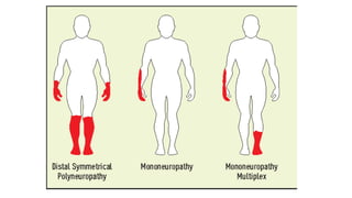
• May affect one nerve (mononeuropathy), several nerves together (polyneuropathy) or
several nerves not contiguous (Mononeuropathy multiplex)
• cell body (e.g., neuronopathy or ganglionopathy), myelin (myelinopathy), and the
axon (axonopathy)
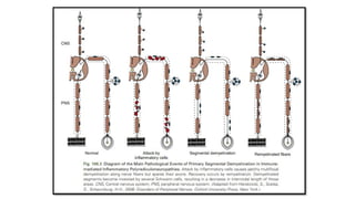
Mononeuropathy
• Local process:
–Direct trauma
– Compression or entrapment
– Vascular lesions
– Neoplastic compression or infiltration
– CARPAL TUNNEL SYNDROME
12.
Mononeuropathy Multiplex
• Simultaneous/sequential damage to multiple noncontiguous nerves.
• Ischemia caused by vasculitis
• Microangiopathy in diabetes mellitus
• Less common causes : Granulomatous, leukemic, or neoplastic infiltration,
Hansen's disease (leprosy) and sarcoidosis.
13.
Polyneuropathy
• Characterized bysymmetrical, distal motor and sensory deficits that have a
graded increase in severity distally and by distal attenuation of reflexes,
• Rarely predominantly proximal:(E.g: acute intermittent porphyria).
• The sensory deficits generally follow a length-dependent stocking-glove pattern
• DIABETES
15.
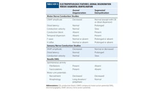
Axonopathies
• By farthe majority of the toxic, metabolic and endocrine causes
• NCVs: CMAPs ↓ 80% lower limit of normal w/o or min velocity or distal motor
latency change.
• Legs>> arms.
• EMG: Signs of denervation (acute, chronic) and reinnervation
16.
Myelinopathies
• Unusual bycomparison with axonopathies
• Clues
– hypertrophic nerves on exam, global areflexia, weakness without wasting, motor >> sensory
deficits
• NCS
– Distal motor latency prolonged (>125% ULN), Conduction velocities slowed (<80% LLN)
May have conduction block
• EMG
– Reduced recruitment w/o much denervation
20.
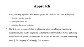
Approach
• In approachinga patient with a neuropathy, the clinician has three main goals:
– identify where the lesion is,
– identify the cause, and
– determine the proper treatment.
• The first goal is accomplished by obtaining a thorough history, neurologic
examination, and electrodiagnostic and other laboratory studies. While gathering
this information, seven key questions are asked, the answers to which can usually
identify the category of pathology that is present.
24.
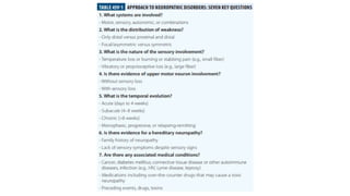
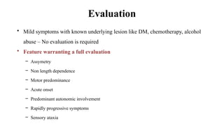
Evaluation
• Mild symptomswith known underlying lesion like DM, chemotherapy, alcohol
abuse – No evaluation is required
• Feature warranting a full evaluation
– Assymetry
– Non length dependence
– Motor predominance
– Acute onset
– Predominant autonomic involvement
– Rapidly progressive symptoms
– Sensory ataxia
History
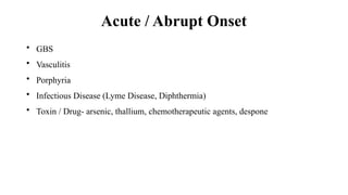
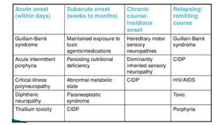
• The temporalcourse of a neuropathy varies, based on the etiology.
– With trauma or ischemic infarction, the onset will be acute, with the most severe symptoms at
onset.
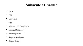
– Inflammatory and some metabolic neuropathies have a subacute course extending over days
to weeks.
– A chronic course over weeks to months is the hallmark of most toxic and metabolic
neuropathies.
29.
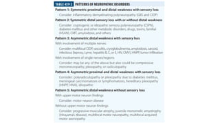
• A chronic,slowly progressive neuropathy over many years occurs with most
hereditary neuropathies or with chronic inflammatory demyelinating
polyradiculoneuropathy (CIDP).
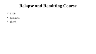
• Neuropathies with a relapsing and remitting course include CIDP, acute
porphyria, Refsum's disease, hereditary neuropathy with liability to pressure
palsies (HNPP), familial brachial plexus neuropathy, and repeated episodes of
toxin exposure.
30.
• Ischemic neuropathiesoften have pain as a prominent feature.
• Small-fiber neuropathies often present with burning pain, lightning-like or
lancinating pain, aching, or uncomfortable paresthesias (dysesthesias).
• Dying-back (distal symmetric axonal) neuropathies initially involve the tips of
the toes and progress proximally in a stocking-glove distribution.
• Peripheral neuropathy can present as restless leg syndrome.
• Proximal involvement may result in difficulty climbing stairs, getting out of a
chair, lifting and bulbar involvement can also be seen
32.
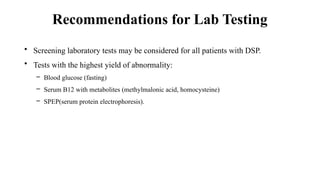
Recommendations for LabTesting
• Screening laboratory tests may be considered for all patients with DSP.
• Tests with the highest yield of abnormality:
– Blood glucose (fasting)
– Serum B12 with metabolites (methylmalonic acid, homocysteine)
– SPEP(serum protein electrophoresis).
33.
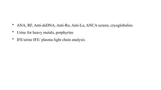
• ANA, RF,Anti-dsDNA, Anti-Ro, Anti-La, ANCA screen, cryoglobulins
• Urine for heavy metals, porphyrins
• IFE/urine IFE/ plasma light chain analysis
34.
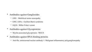
Neuropathies + SerumAutoantibodies
• Antibodies against Gangliosides
– GM1 : Multifocal motor neuropathy
– GM1, GD1a : Guillain-Barré syndrome
– GQ1b : Miller Fisher variant
• Antibodies against Glycoproteins
– Myelin-associated glycoprotein : MGUS
• Antibodies against RNA-binding proteins
– Anti-Hu, antineuronal nuclear antibody 1: Malignant inflammatory polyganglionopathy
Laboratory Evaluation
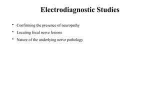
• Thelimitations of EMG/NCS should be taken into account when interpreting the
findings.
– There is no reliable means of studying proximal sensory nerves.
– NCS results can be normal in patients with small-fiber neuropathies
– Lower extremity sensory responses can be absent in normal elderly patients.
• EMG/NCS are not substitutes for a good clinical examination.
38.
Nerve Biopsy
• Invasculitis, amyloid neuropathy, leprosy, CIDP, Inherited disorders of myelin,
and rare axonopathies
• The Sural nerve is selected most commonly
• The superficial peroneal nerve – alternative; :advantage of allowing simultaneous
biopsy of the peroneus brevis muscle through the same incision.
• This combined nerve and muscle biopsy procedure increases the yield of
identifying suspected vasculitis.
40.
Skin Biopsy
• Smallfibre neuropathy
• Very small piece of skin just proximal to ankle is removed.
• Special stains are applied: Qualitative assessment or by careful counting to
determine intraepidermal nerve fibre.