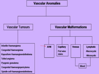
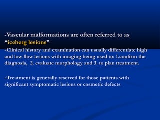
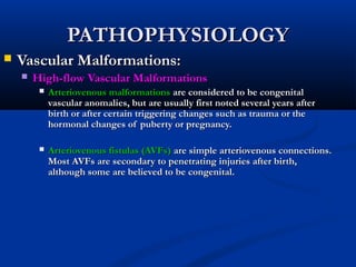
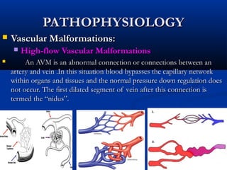
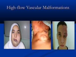
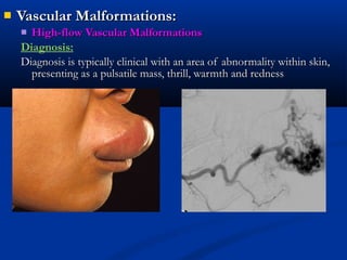
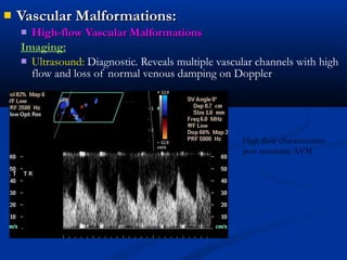
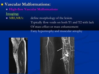
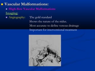
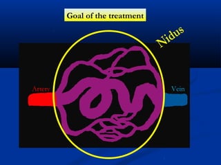
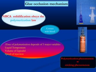
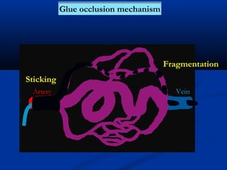
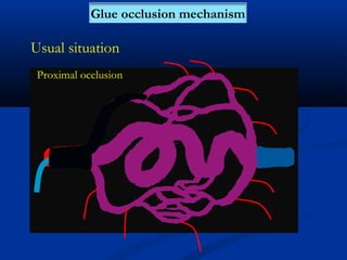
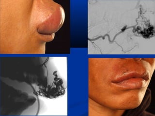
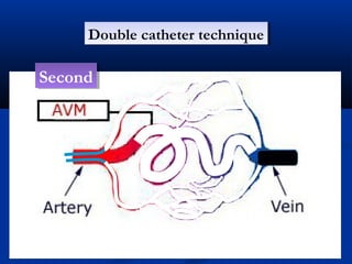
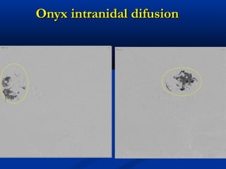
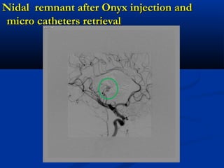
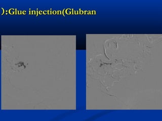
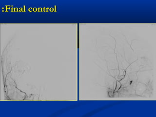
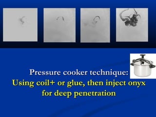
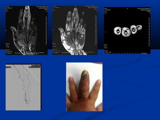
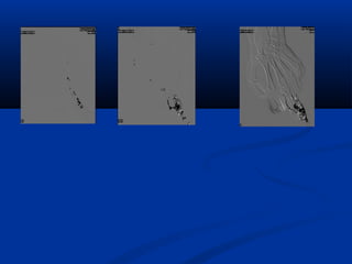
This document discusses the endovascular management of peripheral arteriovenous malformations (AVMs). It defines AVMs as abnormal connections between arteries and veins, bypassing the capillary network. The pathophysiology involves an ectatic capillary bed lacking proper sphincter control. Clinical presentation depends on location and shunting degree, and can include pain, overgrowth, bleeding, and high output cardiac failure in large shunts. Diagnosis is typically clinical and confirmed with imaging showing high flow characteristics. Treatment involves endovascular embolization to occlude arterial feeders using various embolic agents like coils, liquids, and recently the liquid polymer Onyx, which has greater potential to occlude AVMs due to
1. Peripheral AVM: endovascularPeripheral AVM: endovascular
managementmanagement
Dr. Hazem HabboubDr. Hazem Habboub
King Hussein Medical CenterKing Hussein Medical Center
Amman - JordanAmman - Jordan
2. INTRODUCTIONINTRODUCTION
Vascular malformationsVascular malformations are among the most commonare among the most common
congenital abnormalities observed in infants and children.congenital abnormalities observed in infants and children.
Unfortunately, these lesions are also among the mostUnfortunately, these lesions are also among the most
confusing and misunderstood conditions, largely becauseconfusing and misunderstood conditions, largely because
of a history of inconsistent terminology used forof a history of inconsistent terminology used for
classification.classification.
Vascular malformationsVascular malformations are considered a group ofare considered a group of
conditions typified by localized defects in vascularconditions typified by localized defects in vascular
morphogenesis caused by dysfunction in embryogenesismorphogenesis caused by dysfunction in embryogenesis
and vasculogenesisand vasculogenesis
3. Vascular MalformationsVascular Malformations
Diffuse disorder of vascular anomalies.Diffuse disorder of vascular anomalies.
1982 : Mulliken and Glowacki: 2 groups1982 : Mulliken and Glowacki: 2 groups
HaemangiomasHaemangiomas
Vascular MalformationsVascular Malformations
1992: International society for the study of vascular1992: International society for the study of vascular
anomalies-ISSVA- sentinel classificationanomalies-ISSVA- sentinel classification
--Vascular tumorsVascular tumors
-vascular malformations: Divided by Dynamic flow-vascular malformations: Divided by Dynamic flow
characteristic: High flow and Low flow.characteristic: High flow and Low flow.
6. -Vascular malformations are often referred to as-Vascular malformations are often referred to as
““iceberg lesionsiceberg lesions””
--Clinical history andClinical history and examinationexamination can usually differentiate highcan usually differentiate high
and low flow lesions with imaging being used to: 1.confirm theand low flow lesions with imaging being used to: 1.confirm the
diagnosis, 2. evaluate morphology and 3. to plan treatment.diagnosis, 2. evaluate morphology and 3. to plan treatment.
--Treatment is generally reserved for those patients with
significant symptomatic lesions or cosmetic defects
7. PATHOPHYSIOLOGYPATHOPHYSIOLOGY
Vascular Malformations:Vascular Malformations:
High-flow Vascular MalformationsHigh-flow Vascular Malformations
Arteriovenous malformationsArteriovenous malformations are considered to be congenitalare considered to be congenital
vascular anomalies, but are usually first noted several years aftervascular anomalies, but are usually first noted several years after
birth or after certain triggering changes such as trauma or thebirth or after certain triggering changes such as trauma or the
hormonal changes of puberty or pregnancy.hormonal changes of puberty or pregnancy.
Arteriovenous fistulas (AVFs)Arteriovenous fistulas (AVFs) are simple arteriovenous connections.are simple arteriovenous connections.
Most AVFs are secondary to penetrating injuries after birth,Most AVFs are secondary to penetrating injuries after birth,
although some are believed to be congenital.although some are believed to be congenital.
8. PATHOPHYSIOLOGYPATHOPHYSIOLOGY
Vascular Malformations:Vascular Malformations:
High-flow Vascular MalformationsHigh-flow Vascular Malformations
An AVM is an abnormal connection or connections between anAn AVM is an abnormal connection or connections between an
artery and vein .In this situation blood bypasses the capillary networkartery and vein .In this situation blood bypasses the capillary network
within organs and tissues and the normal pressure down regulation doeswithin organs and tissues and the normal pressure down regulation does
not occur. The first dilated segment of vein after this connection isnot occur. The first dilated segment of vein after this connection is
termed thetermed the “nidus”.“nidus”.
9. Histological Analysis of AVMsHistological Analysis of AVMs
Histological analysis of AVMs has shed some light onHistological analysis of AVMs has shed some light on
their pathogenesistheir pathogenesis
Examination of specimens revealed that a nidus is madeExamination of specimens revealed that a nidus is made
up of a bed of dilated capillaries.up of a bed of dilated capillaries.
As the lesion matures, the degree of ectasia increases, andAs the lesion matures, the degree of ectasia increases, and
the development of venous dilation and arterialthe development of venous dilation and arterial
hypertrophy becomes apparent.hypertrophy becomes apparent.
The primary abnormality or nidus, therefore, appears toThe primary abnormality or nidus, therefore, appears to
be anbe an ectatic capillary bed.ectatic capillary bed.
Arterial hypertrophy and venous dilation are secondaryArterial hypertrophy and venous dilation are secondary
phenomena that result from the increase flow across thephenomena that result from the increase flow across the
nidus.nidus.
10. Histological Analysis of AVMsHistological Analysis of AVMs
(continued(continued((
Because the nidus is simply an ectatic capillary bed and because theBecause the nidus is simply an ectatic capillary bed and because the
precapillary sphincters regulate the blood flow through theprecapillary sphincters regulate the blood flow through the
capillary bed, we believe that arteriovenous malformations resultcapillary bed, we believe that arteriovenous malformations result
from an abnormality at the level of thefrom an abnormality at the level of the precapillary sphincterprecapillary sphincter..
An absence of autonomic nerve supply to the sphincters, anAn absence of autonomic nerve supply to the sphincters, an
absence of the actual sphincters, or some deficiency in theabsence of the actual sphincters, or some deficiency in the
neuroreceptors at this level will result in free flow across thatneuroreceptors at this level will result in free flow across that
particular capillary bed.particular capillary bed.
In time, the vessels in the bed dilate, and eventually the areaIn time, the vessels in the bed dilate, and eventually the area
supplying the arteries enlarge and the veins dilate.supplying the arteries enlarge and the veins dilate.
This absence of capillary sphincter control may be absolute orThis absence of capillary sphincter control may be absolute or
relative, hence the variation in age of presentation and speed ofrelative, hence the variation in age of presentation and speed of
progression.progression.
11. AVMs Growth and BleedingAVMs Growth and Bleeding
CycleCycle??
Some people are born with the nidus. As years go by, it tends toSome people are born with the nidus. As years go by, it tends to
enlarge as the pressure of the arterial vessels cannot be handled byenlarge as the pressure of the arterial vessels cannot be handled by
the veins that drain out of it.the veins that drain out of it.
Most of these malformations bleed between the ages of 10-55;Most of these malformations bleed between the ages of 10-55;
after 55, the chances of bleeding diminishes rapidly. Before 55,after 55, the chances of bleeding diminishes rapidly. Before 55,
the likelihood of hemorrhaging is between 3-4% per year (with athe likelihood of hemorrhaging is between 3-4% per year (with a
death incidence of about 1%).death incidence of about 1%).
Once a patient has hemorrhaged, the risk of having another oneOnce a patient has hemorrhaged, the risk of having another one
may approach 20% during the first year, and will gradually lessenmay approach 20% during the first year, and will gradually lessen
to about 3-4% over the next few years.to about 3-4% over the next few years.
12. What Are Some AVM StatisticsWhat Are Some AVM Statistics
AMVs affect approximately 300,000 Americans.AMVs affect approximately 300,000 Americans.
In the Netherlands between 1980 and 1990, the annual incidenceIn the Netherlands between 1980 and 1990, the annual incidence
of symptomatic AVMs was 1.1 per 100,000 population.of symptomatic AVMs was 1.1 per 100,000 population.
They occur equally in males and females from all ethnic and racialThey occur equally in males and females from all ethnic and racial
backgrounds.backgrounds.
They are more prevalent in late childhood (over 9 years of age)They are more prevalent in late childhood (over 9 years of age)
than early childhood, although they can occur at any age.than early childhood, although they can occur at any age.
More than 50% present with intracranial AVMs.More than 50% present with intracranial AVMs.
About 12% of the affected population will present with symptomsAbout 12% of the affected population will present with symptoms
that vary greatly in severity.that vary greatly in severity.
Each year about 1% of those with AVMs will die as a direct resultEach year about 1% of those with AVMs will die as a direct result
of the AVM.of the AVM.
13. PATHOPHYSIOLOGYPATHOPHYSIOLOGY
Vascular Malformations:Vascular Malformations:
High-flow Vascular MalformationsHigh-flow Vascular Malformations
Classification:
Hudart Classification
-Type 3 is the commonest(>60%). And most difficult to treat.
Type 1 Arteriovenous No more than 3
separate arteries
Type 2 Arteriolovenous Multiple arteries shunt
to a single vein
Type 3 Arteriolovenulous Multiple shunts
between arteries and
venules. Multiple nidi
14. Vascular Malformations:Vascular Malformations:
High-flow Vascular MalformationsHigh-flow Vascular Malformations
Clinical features:
Depends on the region of involvement & degree of shunting
Pain
Overgrowth
Bleeding
High cardiac output: less common, large AV shunt
Rapid growth over short time,
Swelling ,especially after trauma.
Schobinger
Schobinger classification
Type 1 Queiscent-stable
Type2 Growing
Type3 Symptomatic:Pain ,bleeding,functional
problems
Type4 Decompensating,high flow cardiac output
15. SIGNS AND SYMPTOMSSIGNS AND SYMPTOMS
Vascular Malformations:Vascular Malformations:
High-flow Vascular MalformationsHigh-flow Vascular Malformations
Arteriovenous malformations (AVMs) are generally present in neonates atArteriovenous malformations (AVMs) are generally present in neonates at
birth, but they often suddenly become obvious when the patient is olderbirth, but they often suddenly become obvious when the patient is older
because of various stimuli such as trauma, pregnancy, or puberty. There arebecause of various stimuli such as trauma, pregnancy, or puberty. There are
four recognized stages of AVMs:four recognized stages of AVMs:
Stage IStage I lesion has a pinkish-bluish stain and warmth.lesion has a pinkish-bluish stain and warmth.
Stage IIStage II, the lesion has pulsations, thrill, and bruit., the lesion has pulsations, thrill, and bruit.
Stage IIIStage III, the patient has dystrophic skin changes, ulceration, bleeding,, the patient has dystrophic skin changes, ulceration, bleeding,
and pain.and pain.
Stage IVStage IV, the patient has high-output cardiac failure., the patient has high-output cardiac failure.
17. Vascular Malformations:Vascular Malformations:
High-flow Vascular MalformationsHigh-flow Vascular Malformations
Diagnosis:
Diagnosis is typically clinical with an area of abnormality within skin,Diagnosis is typically clinical with an area of abnormality within skin,
presenting as a pulsatile mass, thrill, warmth and rednesspresenting as a pulsatile mass, thrill, warmth and redness
18. Vascular Malformations:Vascular Malformations:
High-flow Vascular MalformationsHigh-flow Vascular Malformations
Imaging:
Ultrasound: Diagnostic. Reveals multiple vascular channels with high
flow and loss of normal venous damping on Doppler
hhhhhh
High flow characteristics
post traumatic AVM
19.
20.
21. TREATMENTTREATMENT
High-flow Malformations:High-flow Malformations:
Surgical treatment:Surgical treatment: Small, superficial arteriovenous
malformations can be removed surgically. However
according to Szilagy (editor of Journal of vascular
surgery)” with few exceptions ,AVM cure by surgical
means is impossible”. Out of 82 patients with AVM ,
only 18 were suitable for operation. At FU, 6 were
improved and 12 were worse.
Embolization:Embolization: It has been the only feasible treatmentIt has been the only feasible treatment
option for most arteriovenous malformations.option for most arteriovenous malformations.
Embolization, which closes off the arterial feeders ofEmbolization, which closes off the arterial feeders of
the malformation, is generally effective in arteriovenousthe malformation, is generally effective in arteriovenous
malformations to stabilize the malformation.malformations to stabilize the malformation.
22.
23. Vascular Malformations:Vascular Malformations:
High-flow Vascular MalformationsHigh-flow Vascular Malformations
Treatment
No agreement on the protocols for treatment. Best method to access
and treat AVM.
The most single dominator for treatment is operator experience.
A number of access route is available:
-Trans arterial: most common used with grade 2,3&4
-direct stick: with type 1, slow venous flow
-Transvenous: if there is a single draining vein, small nidus
24. Vascular Malformations:Vascular Malformations:
High-flow Vascular MalformationsHigh-flow Vascular Malformations
Treatment
Embolic agents:
No unified agreement on agent of choice.
Depends on: Experience. Availability, location, morphology
Wide range of agents :
Particulate – Gel foam. PVA .EmbospheresParticulate – Gel foam. PVA .Embospheres
Coils – 035”.018”.controlled releaseCoils – 035”.018”.controlled release
Liquids – Alcohol.Alcohol/Lipiodol.GlueLiquids – Alcohol.Alcohol/Lipiodol.Glue
Sclerosants – STD.PolidocanolSclerosants – STD.Polidocanol
GlueGlue
Detachable balloonsDetachable balloons
Occlusion devices. PlugsOcclusion devices. Plugs
27. Vascular Malformations:Vascular Malformations:
High-flow Vascular MalformationsHigh-flow Vascular Malformations
Treatment
Onyx: Ethylene vinyl alcohol copolymer, relatively new non
adhesive liquid agent that contains tantalum for radio
opacity.
It precipitates on contact with aqueous solution.
It is the most useful for type2,3 and 4.
in our practice >90% of AVMs are treated with Onyx. As
the Flow dynamics of onyx has the greatest potential to
occlude the AVM nidus
29. Glue occlusion mechanism
NBCA solidification obeys the
polymerization law
Contact
with blood
Time of polymerization depends of 3 major variables:
Liquid Temperature
Volume of Lipiodol
Speed of injection
Polymerization phenomenon
=
sticking phenomenon
38. AVM of the hand. Slow venous filling. Direct stick with Foam.
Marked reduction in nidus filling.
Resolution of symptoms
39. Challenges of the treatment:Challenges of the treatment:
Strategy.Strategy.
Number of sessions: 6-8 weeksNumber of sessions: 6-8 weeks
when to stop at each sessionwhen to stop at each session
40. Techniques to improve embolizationTechniques to improve embolization
Pressure cooker technique
Dual catheter technique
45. 35 Y.O. Female35 Y.O. Female
Frontal and supraorbital AVMFrontal and supraorbital AVM
3 endovascular treatments using glue in 20003 endovascular treatments using glue in 2000
followed by Radiosurgery (Gama Knife) in 2001followed by Radiosurgery (Gama Knife) in 2001
In 2008, 7 years later, new seizureIn 2008, 7 years later, new seizure
AngiogramAngiogram
58. Pressure cooker technique:Pressure cooker technique:
Using coil+ or glue, then inject onyxUsing coil+ or glue, then inject onyx
for deep penetrationfor deep penetration
59.
60.
61. ConclusionConclusion
--Treatment of peripheral AVM is challenging andTreatment of peripheral AVM is challenging and
requires a multidisciplinary team.requires a multidisciplinary team.
-experience and understanding the capabilities and-experience and understanding the capabilities and
physics of the embolic agent will determine thephysics of the embolic agent will determine the
success of treatment.success of treatment.
-Embolization is proving effective as a stand alone-Embolization is proving effective as a stand alone
option” with the proper use of the controlled liquidoption” with the proper use of the controlled liquid
embolic agents.embolic agents.