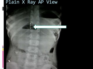
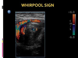
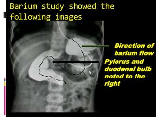
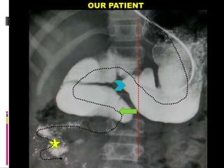
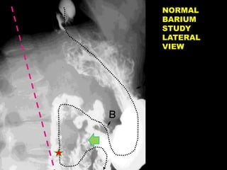
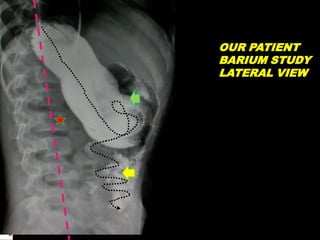
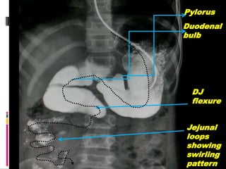
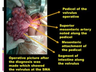
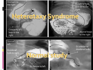
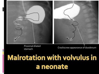
CASE PRESENTATION
Dr akshay gursale
History and clinical
examination
 A 10 yr old female patient comes
to casualty with complaints of
 pain in epigastric region which was
acute in onset since 2-4 days
 Bilious vomiting since 2-4 days
 A lump was felt in the epigastrium
with localised tenderness
 Temperature was slightly raised
 Rest parameters were within normal
limits
Plain X Ray AP View
Doppler on ultrasound
SMV
SMA
Barium study showed the
following images
Pylorus and
duodenal bulb
noted to the
right
Direction of
barium flow
The direction
of barium
flow
NGT in
situ
A NORMAL BARIUM STUDY
OUR PATIENT
NORMAL
BARIUM
STUDY
LATERAL
VIEW
OUR PATIENT
BARIUM STUDY
LATERAL VIEW
Pylorus
Duodenal
bulb
DJ
flexure
Jejunal
loops
showing
swirling
pattern
Following barium studies and
Ultrasound findings a
diagnosis of Malrotation of
Gut with Midgut volvulus was
made.
Final diagnosis
Pedicel of the
volvulus
operative
Superior
mesenteric artery
noted along the
pedicel
Mesenteric
attachment of
the pedicel
Segment of
intestine along
the volvulus
Operative picture after
the diagnosis was
made which showed
the volvulus at the SMA
TAKE HOME MESSAGE
Upper gastrointestinal
barium studies are not
obsolete
One can make a
FINAL DIAGNOSIS
on base of
sonography and
barium studies
alone
Compare with
normal
appearances of
upper GI barium
series to diagnose
MALROTATION
MIDGUT VOLVULUS
 EMBRYOLOGY OF ROTATIONOF GUT
 Gut develops from yolk sac which is further divided
into 3 parts
 Foregut supplied by Coeliac trunk upto mid 1/3 of
duodenum
 Midgut supplied by superior mesenteric artery upto
distal transverse colon
 And hindgut supplied by inferior mesenteric artery
upto anal canal
 The intestine upto 4 weeks is a straight
tubular structure
 By 12weeks it grows rapidly by some complex
steps involving a rotation of 270 degrees and
fixation in normal position in abdomen
 First duodenum rotates 90 deg
counterclockwise to the right of SMA while
colon 90 deg to the left of SMA
 Then midgut herniated through umbilical
cord and duodenum go another 90 deg
counterclockwise rotation but colon
undergoes no rotation
 By 10 week the bowel returns to the
abdominal cavity and the duodenum
undergoes the final 90 deg counterclockwise
rotation until duodeno-jejunal junction is to
the left of spine and the colon rotates by 180
deg until the caecum is in right lower
quadrant
 This rotation produces a long mesenteric
attachment for the bowel
Salient features of rotation
of gut
 Duodenum describes the c loop with
concavity to patients left and the third part of
duodenum to left of midline
 SMA runs in front of 3rd part of duodenum
 The mesentery run along posteriorly from the
ligament of trietz in left upper quadrant to
caecum in right lower quadrant preventing its
torsion
 The ascending colon is fixed in right side of
abdomen and desending colon in left side of
abdomen
 Malrotation is usually daignosed in upto 75%
cases in newborns and upto 90% cases by 1st
year
 In individuals with malrotation, the mesenteric
attachment of the midgut, particularly the
portion from the duodenojejunal junction to the
cecum, is abnormally short and is therefore
prone to twist counterclockwise around the
superior mesenteric artery and vein.
 This condition, known as midgut volvulus, may
cause intermittent abdominal distention and
pain or acute bowel necrosis.
Duodenal bulb
with DJ to the
right
Jejunal loops
to the right
Stomach to
the right
Concavity of C
loop to the
right
Stomach to
left
Duodenal
bulb to right
DJ flexure to left
DJ inferior to
duodenal bulb
 The normal position of the duodenojejunal
junction is to the left of the left-sided pedicles of
the vertebral body at the level of the duodenal
bulb on frontal views and posterior
(retroperitoneal) on lateral views.
 In children with acute duodenal obstruction, the
upper GI series may depict a Z-shaped
configuration of the duodenum in the presence
of obstructing peritoneal bands or a corkscrew-
shaped duodenum in the presence of volvulus .
 In children who have bowel malrotation without
volvulus, the upper GI series shows an abnormal
position of the duodenojejunal junction and of
the ligament ofTreitz
DJ flexure with duodunal
bulb to right
Duodenal bulb with jejunal
loops to right
Proximal dilated
stomach
Crockscrew appearance of duodenum
Normal position of
duodenal bulb and c loop of
duodenum
Abnormal position of DJ
flexure

Midgut volvulus

  • 1.
  • 2.
    History and clinical examination A 10 yr old female patient comes to casualty with complaints of  pain in epigastric region which was acute in onset since 2-4 days  Bilious vomiting since 2-4 days  A lump was felt in the epigastrium with localised tenderness  Temperature was slightly raised  Rest parameters were within normal limits
  • 3.
    Plain X RayAP View
  • 4.
  • 6.
    Barium study showedthe following images Pylorus and duodenal bulb noted to the right Direction of barium flow
  • 7.
  • 8.
  • 9.
  • 10.
  • 11.
  • 12.
  • 13.
    Following barium studiesand Ultrasound findings a diagnosis of Malrotation of Gut with Midgut volvulus was made. Final diagnosis
  • 14.
    Pedicel of the volvulus operative Superior mesentericartery noted along the pedicel Mesenteric attachment of the pedicel Segment of intestine along the volvulus Operative picture after the diagnosis was made which showed the volvulus at the SMA
  • 15.
    TAKE HOME MESSAGE Uppergastrointestinal barium studies are not obsolete One can make a FINAL DIAGNOSIS on base of sonography and barium studies alone Compare with normal appearances of upper GI barium series to diagnose MALROTATION
  • 16.
    MIDGUT VOLVULUS  EMBRYOLOGYOF ROTATIONOF GUT  Gut develops from yolk sac which is further divided into 3 parts  Foregut supplied by Coeliac trunk upto mid 1/3 of duodenum  Midgut supplied by superior mesenteric artery upto distal transverse colon  And hindgut supplied by inferior mesenteric artery upto anal canal
  • 17.
     The intestineupto 4 weeks is a straight tubular structure  By 12weeks it grows rapidly by some complex steps involving a rotation of 270 degrees and fixation in normal position in abdomen  First duodenum rotates 90 deg counterclockwise to the right of SMA while colon 90 deg to the left of SMA  Then midgut herniated through umbilical cord and duodenum go another 90 deg counterclockwise rotation but colon undergoes no rotation
  • 18.
     By 10week the bowel returns to the abdominal cavity and the duodenum undergoes the final 90 deg counterclockwise rotation until duodeno-jejunal junction is to the left of spine and the colon rotates by 180 deg until the caecum is in right lower quadrant  This rotation produces a long mesenteric attachment for the bowel
  • 20.
    Salient features ofrotation of gut  Duodenum describes the c loop with concavity to patients left and the third part of duodenum to left of midline  SMA runs in front of 3rd part of duodenum  The mesentery run along posteriorly from the ligament of trietz in left upper quadrant to caecum in right lower quadrant preventing its torsion  The ascending colon is fixed in right side of abdomen and desending colon in left side of abdomen
  • 21.
     Malrotation isusually daignosed in upto 75% cases in newborns and upto 90% cases by 1st year  In individuals with malrotation, the mesenteric attachment of the midgut, particularly the portion from the duodenojejunal junction to the cecum, is abnormally short and is therefore prone to twist counterclockwise around the superior mesenteric artery and vein.  This condition, known as midgut volvulus, may cause intermittent abdominal distention and pain or acute bowel necrosis.
  • 22.
    Duodenal bulb with DJto the right Jejunal loops to the right Stomach to the right Concavity of C loop to the right Stomach to left Duodenal bulb to right DJ flexure to left DJ inferior to duodenal bulb
  • 23.
     The normalposition of the duodenojejunal junction is to the left of the left-sided pedicles of the vertebral body at the level of the duodenal bulb on frontal views and posterior (retroperitoneal) on lateral views.  In children with acute duodenal obstruction, the upper GI series may depict a Z-shaped configuration of the duodenum in the presence of obstructing peritoneal bands or a corkscrew- shaped duodenum in the presence of volvulus .  In children who have bowel malrotation without volvulus, the upper GI series shows an abnormal position of the duodenojejunal junction and of the ligament ofTreitz
  • 24.
    DJ flexure withduodunal bulb to right Duodenal bulb with jejunal loops to right
  • 25.
  • 26.
    Normal position of duodenalbulb and c loop of duodenum Abnormal position of DJ flexure