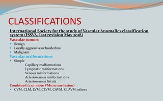
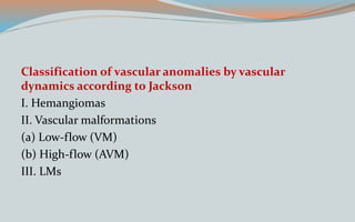
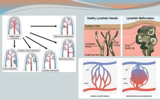
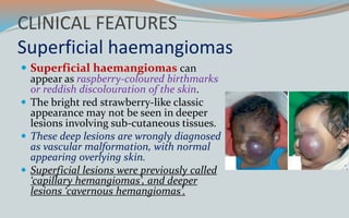
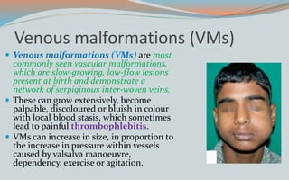
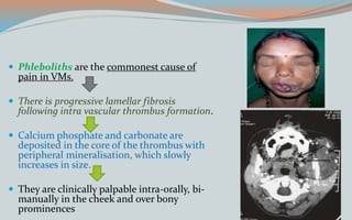
Hierarchy of management that covers different levels of management
Â
VASCULAR MALFORMATION AND ITS MANAGEMENT.pptx
1. Presented by
Dr. DEBRAJ SAMANTA
3rd year PGT
DEPARTMENT OF ORAL AND MAXILLOFACIAL SURGERY
HIDSAR, WEST BENGAL
2. INTRODUCTION
ī Vascular malformations and tumors
comprise a wide heterogenous
spectrum of lesions that involves all
part of the body and can cause
significant morbidity and even
mortality in both adults and children.
ī Vascular anomalies have aâ dynamic
lifecycle.â
ī Vascular anomalies are a âgroupâ of
different entities of varied
etiopathological which may not share
the same treatment approaches.
3. HISTORY
ī The first scientifically documented
âsurgery under an ether anesthetic agentâ
by Morton in 1846 done in Boston,
Massachusetts USA, was for excision of a
âlow flowâ vascular malformation.
ī Rudolph Virchow, in 1863 first
categorized vascular anomalies by
microscopic architectural patterns.
ī From earlier use words âPort wine stain,â
âAngiomas,â etc. Virchowâs attempted an
acceptable Cellular classification as
angioma simplex, angioma cavernosum
and angioma racemosum.
4. CLASSIFICATIONS
International Society for the study of Vascular Anomalies classification
system (ISSVA, last revision May 2018)
Vascular tumors
ī Benign
ī Locally aggressive or borderline
ī Malignant
Vascular malformations
ī Simple
Capillary malformations
Lymphatic malformations
Venous malformations
Arteriovenous malformations
Arteriovenous fistula
Combined (2 or more VMs in one lesion)
ī CVM, CLM, LVM, CLVM, CAVM, CLAVM, others
5. Classification of vascular anomalies by vascular
dynamics according to Jackson
I. Hemangiomas
II. Vascular malformations
(a) Low-flow (VM)
(b) High-flow (AVM)
III. LMs
6. Classification of CNS vascular
malformations
ī Arteriovenous shunts
Classical AV Malformations
Pial AV fistulas
Dural AV shunts
Galenic shunts
ī Cavernous malformations
ī Capillary telangiectasias
ī Venous malformations
Developmental venous
Anomalies
ī Mixed malformations
Classification of non-CNS
vascular malformations
ī Haemangiomas (proliferative
lesions or tumours)
ī Vascular malformations (non-
proliferative)
o High flow
âĸ Arteriovenous malformations
o Low flow:
âĸ Capillary malformations
âĸ Venous malformations
âĸ Lymphatic malformations
âĸ Mixed malformations
9. Pathogenesis
ī There are 3 stages in the
life cycle of
haemangiomas-
īProliferative phase-0â1
year of age
īInvoluting phase-1â5
years of age
īInvoluted phase-more
than 5 years of age
10.
11. CLINICAL FEATURES
Superficial haemangiomas
ī Superficial haemangiomas can
appear as raspberry-coloured birthmarks
or reddish discolouration of the skin.
ī The bright red strawberry-like classic
appearance may not be seen in deeper
lesions involving sub-cutaneous tissues.
ī These deep lesions are wrongly diagnosed
as vascular malformation, with normal
appearing overlying skin.
ī Superficial lesions were previously called
âcapillary hemangiomasâ, and deeper
lesions âcavernous hemangiomasâ.
12. Venous malformations (VMs)
ī Venous malformations (VMs) are most
commonly seen vascular malformations,
which are slow-growing, low-flow lesions
present at birth and demonstrate a
network of serpiginous inter-woven veins.
ī These can grow extensively, become
palpable, discoloured or bluish in colour
with local blood stasis, which sometimes
lead to painful thrombophlebitis.
ī VMs can increase in size, in proportion to
the increase in pressure within vessels
caused by valsalva manoeuvre,
dependency, exercise or agitation.
13. ī Phleboliths are the commonest cause of
pain in VMs.
ī There is progressive lamellar fibrosis
following intra vascular thrombus formation.
ī Calcium phosphate and carbonate are
deposited in the core of the thrombus with
peripheral mineralisation, which slowly
increases in size.
ī They are clinically palpable intra-orally, bi-
manually in the cheek and over bony
prominences
14. Capillary malformations (CMs)
ī Capillary malformations (CMs), can be
seen in the neonatal period and should
not be mistaken with infantile
haemangiomas (IHs).
ī They occur in around 0.3% of children
presenting with vascular anomalies.
ī CMs usually present congenitally as a flat,
red or purple cutaneous patch with
asymmetric borders.
ī They are painless and do not bleed
spontaneously.
15. Lymphatic malformations (LMs)
ī Lymphatic malformations
(LMs) are usually congenital
and not always detected until
later in life.
ī LMs are slow-flow lesions and
can be
īmacro-cystic (>2 cm)
īmicro-cystic (<2 cm)or
mixed, depending on the size of
predominant cysts within them.
16. ī Macro-cystic LMs can show
expansion and compression of
adjacent anatomical structures in the
head and neck causing dysphagia,
dyspnea and masticatory problems.
ī Micro-cystic LMs were previously
called lymphangiomas and
demonstrate an infiltrative nature.
ī LMs with local haemorrhage and
infection can cause significant pain.
17. Arterio-venous malformations (AVMs)
ī Fast-flow arterio-venous malformations
(AVMs), usually present with a pulsatile local
swelling.
ī These AVMs along with some slow-flow
lesions are at an increased risk of
haematological complications like
coagulopathies due to disturbances in
haemostasis and thrombosis.
ī Blood pools in abnormal slow-flow vessels,
resulting in activation of the coagulation
system, resulting in a process known as
âlocalized intravascular coagulopathyâ (LIC),
which shows low fibrinogen, elevated D-dimer
levels and mild thrombocytopaenia
18. Syndromes Associated with Vascular
Anomalies
ī Syndromes Associated with Vascular Tumors
ī Syndromes Associated with Capillary Malformations
ī Syndromes Associated with Low-flow Venous
Malformations, Lymphatic Malformations or Mixed
Malformations
ī Syndromes Associated with High-flow Vascular
Malformations
20. PHACE syndrome
ī Freiden et al. in 1996 reported PHACE
syndrome, an acronym that
encompassed-
īPosterior fossa malformations
īHaemangiomas
īArterial anomalies
īCardiovascular anomalies
īEye anomalies, and sternal defects
21. ī The haemangiomas are infantile
haemangiomas, often occurring in the face
and neck, segmental in distribution and
over 5 cm in diameter.
ī Posterior fossa anomalies include Dandy-
Walker malformations and ventricular
dilatation.
ī Cardiac anomalies include coarctation of
the aorta, atrial septal defect, and
ventricular septal defects.
ī Eye anomalies include cataract, glaucoma,
microphthalmos, and optic nerve
hypoplasia.
23. KasabachâMerritt Syndrome/
Phenomenon
ī Kasabach and Merritt in 1940 reported
a case of capillary haemangioma with
extensive purpura.
ī It was associated with coagulation
abnormalities, reduced platelets
and anaemia, causing
haemorrhage, infection and multi-
organ failure, leading to death in 12â
24% of patients.
ī Current management includes
excision of the lesion, steroids,
interferon, vincristine and
radiotherapy.
25. SturgeâWeber Syndrome
ī SturgeâWeber Syndrome (SWS) is
characterised by a dermal capillary
malformation occurring in association
with vascular malformations of the
leptomeninges and the eye.
ī It was initially described by Sturge in
1879 and further characterised by
Weber in 1922.
26. ī It typically includes a triad of-
īFacial dermal capillary
malformation
īIpsilateral central nervous
syndrome vascular
malformations (leptomeningeal
angiomatosis)
īVascular malformations of the
choroid of the eye associated
with glaucoma.
27. Physical signs of SWS
īPort-wine birthmark
īMacrocephaly
īOcular manifestations
īSoft-tissue hypertrophy
īHemiparesis
īVisual loss
īHemianopsia
28. Diagnosis
ī In young patients, examination under
anesthesia or deep sedation is
necessary to confirm the diagnosis of
glaucoma. Careful assessment in
each eye of IOP, corneal diameter,
cycloplegic refraction, axial length,
and optic nerve cupping, as well as
gonioscopic examination, is
mandatory.
ī Cerebrospinal fluid (CSF) protein
may be elevated, presumably
secondary to microhemorrhage.
29. Management
Medical care in SWS includes-
ī anticonvulsants for seizure control,
ī symptomatic and prophylactic therapy for headache,
ī glaucoma treatment to reduce IOP, and
ī laser therapy for the PWB.
30. KlippelâTrenaunay Syndrome
ī KlippelâTrenaunay Syndrome (KTS)
was initially reported by Klippel and
Trenaunay in 1900.
īcapillary malformation of the
lower limbs
īcongenital varicose veins or
venous malformations associated
with abnormal, dilated âlateral
megaveinsâ that develop in the
lateral aspect of the affected
limbs
ī bone and soft tissue hypertrophy
resulting from overgrowth.
31. ī The capillary malformation may be
distributed in a confluent geographic
pattern or randomly on the affected limb
and adjacent trunk.
ī Geographic patterns are often located in the
lateral aspect of the thigh, knee and lower leg
and can be a predictor of associated lymphatic
malformation and complications of KTS.
ī Life-threatening complications include deep
vein thrombosis, pulmonary embolism, sepsis
and coagulopathy
32. RubinsteinâTaybi Syndrome
ī Rubinstein and Taybi in 1963
reported a condition characterised
by mental retardation, growth
retardation, broad thumbs, large
toes and a typical facial
appearance
33. ī Skin manifestations included capillary
malformations, hirsutism, keloid and
excessive scar formation, and cafÊ au lait
macules.
ī Oral manifestations include thin upper lip,
small oral opening, retro/micrognathia and
high arched narrow palate.
ī Cleft uvula and palate can be part of the
syndrome.
ī Dental anomalies include hypodontia,
hyperdontia and natal teeth.
ī Talon cusps were present in 73% of patients and
92% of permanent teeth.
37. MANAGEMENT
ī Abdominal wall defects are common in newborns with BWS
and may require surgical treatment.
ī These defects can range in severity from omphalocele (most
serious) to umbilical hernia and diastasis recti (least
serious).
ī An omphalocele is a congenital malformation in which a
newborn's intestines, and sometimes other abdominal organs,
protrude out of the abdomen through the umbilicus.
ī Newborns with an omphalocele typically require surgery to place
the abdominal contents back into the abdomen in order to prevent
serious infection or shock.
38. ī Neonatal hypoglycemia, low blood glucose in the first
month of life, occurs in about half of children with BWS.
ī Most of these hypoglycemic newborns are asymptomatic and
have a normal blood glucose level within days.
ī untreated persistent hypoglycemia can lead to permanent
brain damage.
ī Such children may require tube feedings, oral
hyperglycemic medicines, or a partial pancreatectomy
39. ī Macroglossia, a large tongue, is a very common (>90%)
and prominent feature of BWS. Infants with BWS and
macroglossia typically cannot fully close their mouth in
front of their large tongue, causing it to protrude out.
ī These children are often managed by a multidisciplinary
craniofacial team.
ī These teams include speech and language therapists,
craniofacial and paediatric plastic surgeons, and
orthodontists who decide the appropriateness and timing of
tongue reduction surgery.
40. ī Nevus flammeus (port-wine stain) is a flat, red
birthmark caused by a capillary (small blood vessel)
malformation.
ī Nevus flammeus is benign and commonly does not require any
treatment.
ī Hemihypertrophy (hemihyperplasia) is an abnormal
asymmetry between the left and right sides of the body
occurring when one part of the body grows faster than normal.
ī Hemihypertrophy can also cause various orthopedic problems,
so children with significant limb hemihyperplasia should be
evaluated and followed by an orthopedic surgeon.
42. Blue Rubber Bleb Nevus Syndrome
ī Gascoyne in 1860 reported a vascular
malformation in the parotid region
complicated by haemorrhage of the
gastrointestinal (GI) tract.
ī Bean in 1958 reported additional
cases and reviewed the literature and
coined the term âblue rubber bleb
nevus syndromeâ to describe the
classical skin and gastrointestinal
tract findings.
43. ī The typical findings are compressible blue
subcutaneous venous malformations
ranging in size from 0.1 cm to 5.0 cm.
ī They are often present at birth or early
childhood and become more obvious
and numerous as the patients become
older.
ī They can be present in any part of the
skin and mucosa, including the scalp, but
are more commonly located in the trunk
and extremities.
44. ī Gastrointestinal venous
malformations most commonly
affect the small intestine and
colon, though the entire bowel
may be affected.
ī Lesions may also be found in the
orbit, genitourinary tract and
central nervous system.
ī GI lesions may present as chronic
anaemia, abdominal pain, rectal
bleeding and intussusception.
45. Parkes Weber Syndrome
ī Parkes Weber in 1907 described a
vascular lesion with
hemihypertrophy. It is characterised
by-
īovergrowth of the limb (usually lower)
with diffuse arteriovenous fistulas
and shunts.
īThe arteriovenous fistulas often
develop around puberty and can be
complicated by high-output
congestive cardiac failure.
īLymphatic anomalies and
lymphoedema may be present.
46. Proteus Syndrome
ī Wiedemann et al. in 1983 described a
disorder characterised by-
ī hypertrophy of the hands and feet,
pigmented nevi, hemihypertrophy, skull
and visceral abnormalities.
ī They named it Proteus syndrome after
the Greek god, who could change his
shape.
ī The characteristic facial features of
dolichocephaly, long face, down
slanting palpebral fissures, low nasal
bridge and open bite have also been
reported.
47. Maffucci Syndrome
ī Initially described by Maffucci in 1881, the
disorder is characterised by
enchondromatosis and multiple low flow
vascular (mainly venous and rarely
lymphatic) malformations.
ī It often manifests in early childhood with
multiple superficial and deep venous
malformations in the extremities.
ī Oral and intra-abdominal venous and
lymphatic malformations have also been
reported
ī Diagnosis is based on the clinical features and
regular follow-up is mandatory to monitor the
endochondromas and detect malignant
transformation. The venous malformations are
managed conservatively, unless symptomatic.
48. GorhamâStout Syndrome
ī Initially reported by Jackson in 1838, Gorham
and Stout in 1955 reported 24 cases characterised
by vascular malformations, intraosseous
vascular malformations and osteolysis.
ī This syndrome has also been termed as
disappearing bone disease, phantom bone
disease and diffuse skeletal
haemangiomatosis.
ī Lymphatic malformations are most common,
though capillary and venous malformations
have been reported.
ī Cutaneous malformations are uncommon.
ī Truncal bone and upper extremities are most
commonly affected.
ī Surgery, radiotherapy and bisphosphonates
have been used in its management
49. Syndromes Associated with High-flow
Vascular Malformations
ī RenduâOslerâWeber Syndrome (Hereditary
Haemorrhagic Telangiectasia)
ī BonnetâDechumeâBlanc Syndrome or WyburnâMason
Syndrome
50. RenduâOslerâWeber Syndrome (Hereditary
Haemorrhagic Telangiectasia)
ī Rendu (1896), Osler 1901) and Weber
(1907)described a hereditary disorder
characterised by dilated skin and mucosal
blood vessels leading to epistaxis and
haemorrhage into the digestive tract
(hereditary haemorrhagic telangiectasiaâ
HHT).
ī It is an autosomal dominant disorder
affecting one to two per 100,000 people
and belongs to the group of diseases with
abnormal transforming growth factor-
beta.
52. Management of local lesions has included
ī cautery
ī laser photocoagulation
ī sclerotherapy.
ī Bevacizumab, when injected sub-mucosally has also been
shown to be of benefit.
53. BonnetâDechumeâBlanc Syndrome or
WyburnâMason Syndrome
ī Bonnet et al. in 1937 and Wyburn et al. in 1943
reported a disorder characterised by cerebral
arteriovenous malformation usually
involving the midbrain, ipsilateral retinal
vascular malformation and a red stain in the
face, often misinterpreted as a capillary
malformation.
ī BonnetâDechumeâBlanc syndrome and Wyburn-
Mason syndrome are generally considered to be
synonymous.
ī The cutaneous vascular malformation is not a
consistent feature and when present can be
unilateral and involves the skin innervated by the
trigeminal nerve or central involving the mid-
forehead, glabella, nose and upper lip.
56. Radiological Diagnosis of Head and
Neck Vascular Anomalies
īUltrasound is the first choice for an initial evaluation in
most cases. It has a good spatial resolution for superficial
lesions.
īDoppler study can measure blood flow direction and
volume in real time, and essential for differentiating low
flow from high-flow vascular malformations.
īWhen ultrasound findings are equivocal, MRI is the next
step. MRI is a favorable exam for most clinicians when
evaluating very large lesions or lesions within specific
locations (infraorbital or intracranial lesion)
57. Ultrasound image-Venous
malformation of submandibular
region showing multiple cystic
spaces
Coloured Doppler
Ultrasound image of AVM of
neck showing multiple
cystic spaces filled with
colour (flow towards the
transducer seen in âredâ and
away in âblueâ, lighter
shades of colour depict
higher velocity)
CT with
contrast,
Right
mandible
Type-IV lesion
T2-weighted
MRI, left
parapharyn
geal venous
malformati
on
58. AVM of right mandible ((a) Contrast MR
angiography image, (b) T2-weighted MRI
image).
59. Infantile Hemangioma
ī In the proliferative phase, it presents as a
well-defined lobular mass with high
vascular activity. In the involuting phase,
fatty replacement occurs with decreased
vascular activity.
ī Ultrasound demonstrates a well-defined
soft tissue mass with variable internal
echogenicity.
ī Color Doppler is very helpful in showing
the characteristic high vascularity with
both arterial and venous flow.
ī Hypervascularity in proliferative phase is
defined as more than 5 vessels per square
centimeter with low resistive index
hyperechoic subcutaneous soft
tissue mass overlying the right
lambdoid suture
60. ī MRI is mainly used for lesions involving
critical organs (i.e., infraorbital or
intracranial extension) or treatment
planning.
ī In the proliferative phase, lesions are well-
defined with low signal intensity on T1-
weighted image (T1WI), high signal
intensity on T2-weighted image (T2WI),
and intense contrast enhancement.
ī In the involuting phase, there is high signal
intensity on T1WI due to fatty replacement.
61. Congenital Hemangioma
ī Both ultrasound and MRI findings of congenital
hemangioma are similar to infantile hemangioma,
characterized by soft tissue mass with hypervascularity.
ī It often contains internal foci of calcifications or
hemorrhage
62. Low-flow Vascular Malformations
Venous Malformation
īVenous malformation usually presents as an asymptomatic soft
tissue mass. In imaging study, it appears as a well-defined
lobulated lesion.
īUltrasound is very useful for the evaluation of superficial
lesions. It shows a characteristic hypoechoic cystic lesion with
sinusoidal chambers and hyperechoic foci (phleboliths).
īMRI demonstrates a lobulated mass with iso-to- low signal
intensity on T1WI, high signal intensity on T2WI due to its
cystic nature, delayed contrast enhancement, and signal voids
suggestive of phleboliths.
63. Lymphatic Malformation
īThere are two main subtypes of
lymphatic malformation, macrocystic
and microcystic. Each subtype has
distinguished radiologic appearances.
īMacrocystic lymphatic malformation is
a well-defined lobulated soft tissue mass
with multiple septations and fluidâfluid
level.
īMicrocystic lesion is typically an ill-
defined, infiltrative, solid mass that
involves multiple tissue planes
CT images show soft tissue mass
with serpiginous enhancement
and numerus phleboliths
involving multiple compartments
of the right face
64. High-flow Vascular Malformations
Arteriovenous Malformation(AVM)
ī Ultrasound is usually the first choice for
diagnosis.
ī Tangles of enlarged feeding arteries and
dilated draining veins without discrete soft
tissue mass suggest the diagnosis.
ī The feeding arteries have low resistant
waveform on color Doppler due to
shunting.
ī On MRI, the enlarged feeding arteries and
draining veins are shown as large flow
voids on echospin sequences.
Large arterial venous malformation
within the right frontal parasagittal
white matter with tangles of
enlarged feeding arteries and a large
draining vein
67. Medical Management of Vascular
Lesions
Goals of Treatment
īPrevention or reversal of life-threatening or function-
threatening complications.
īPrevention or minimization of a disfigurement from residual
skin changes.
īMinimization of psychosocial distress for the patient and
family.
īAdequate treatment of ulceration to minimize scarring,
bleeding, infection, and pain.
68. Local Therapies
ī Local pharmacotherapy is primarily used to treat small or
superficial infantile hemangiomas and is not ideal for
treating complex lesions.
ītopical beta-blockers
īCorticosteroids
īimiquimod
69. Topical Beta-blockers
ī Treatment of hemangiomas with systemic propranolol led
to the investigation of topical beta-blocker therapy to treat
infantile hemangiomas.
ī Topical timolol gel-forming solution, 0.5%, can be used to
treat lesions. One drop is applied two to three times per day
for 6 to 12 months or until stable improvement.
ī Topical propranolol 1% or topical timolol 0.5% efficacy.
ī MOA: inhibition of signal transduction pathway by beta-
blockers in the proliferative vascular endothelial cells.
70. ī Propranolol at an Oral dose of
1.0â1.5 mg/kg per day- deep-
seated lesions, multiple periodic
doses with monitoring of
cardiovascular and systemic
effects as mentioned below
during the proliferative phase.
71. ī Beta-blocker adverse reaction of hypoglycemia, hypotension,
bradycardia, bronchial constriction, peripheral cyanosis, EKG
changes, gastrointestinal symptoms (anorexia, diarrhea, vomiting),
sleep changes, drowsiness, skin erythema, and exfoliation are all
cared for appropriately, especially in infants.
ī Cardiovascular and vital sign monitoring must be undertaken in an
inpatient setting (3â7 days), especially when therapy is at the
proliferative phase.
ī Duration of the entire treatment can last between 1 and 8 weeks
during the proliferative phase.
ī Low doses of oral beta-blocker are advised during combination
therapy with topical beta-adrenergic therapy in mitigating adverse
effects.
72. Topical corticosteroids
ī High-potency topical corticosteroids (e.g., clobetasol
propionate cream) have been used in the past for small,
superficial hemangiomas at risk for ulceration or small
periocular lesions.
ī The use of intralesional corticosteroids such as
triamcinolone acetonide 10 to 40 mg/mL is limited to small,
well-localized, deep hemangiomas. Individual doses should
not exceed 3 mg/kg.
ī Adverse effects of long-term use of topical corticosteroids
include skin atrophy, hypopigmentation, and hypertrichosis.
73. Systemic Corticosteroids
ī Treatment with systemic corticosteroids remains a treatment
option for patients with complicated hemangiomas, in whom
treatment with beta-blockers is contraindicated.
ī Dosing The starting dose for prednisolone is 2 to 3 mg/kg per day.
ī A single, morning dose is preferred to minimize adrenal
suppression. A response is usually seen within the first few weeks
ī SIDE EFFECTS: Cushingoid facies, personality changes, delayed
skeletal growth, gastric upset occurs in some cases. Serious
corticosteroid complications, such as aseptic necrosis of the
femoral head, hypertension, osteoporosis, and cataracts, are
infrequent in children
74. Other Systemic Therapies
ī STEROID THERAPY
ī SYSTEMIC STEROIDSâPrednisolone in proliferative
phase
ī LOCAL STEROIDSâTriamcinolone (40 mg/ml),
Betamethasone (6 mg/ml)
ī VINCRISTINE FOR LIFE-THREATENING LESIONS
ī INTERFERON THERAPY- ALPHA 2a and BETA (serious
side effects)
ī Beta-blockers â 2 mg/kg body wt. (propranolol)
76. ī PYM, also known as Bleomycin A5, is the
most commonly used single-drug therapy
for the treatment of cervico-facial
malformations.
ī Absolute ethanol causes alteration in
cellular proteins and hence damages the
endothelium of the vascular wall leading to
obliteration of its lumen.
ī Ethanolamine oleate is an emulsion of
fatty acids, which induces thrombosis and
damages the endothelium.
ī Bleomycin was first used in the treatment
of cystic hygromas. It inhibits DNA
synthesis and has a non-specific
inflammatory reaction on the endothelial
cells.
77. Surgical Management
ī Type-I, superficial lesions require excision of skin or mucosa. Local or
regional flaps have been used in reconstruction of the residual defect.
ī Type-II, sub-mucosal lesions require complete excision after elevation of
skin flaps.
ī Type-III, lymphovenous malformations or venous malformations
involve salivary glands and are excised along with the affected gland.
ī Type-IV, intra-osseous lesions require excision with removal of involved
bone and reconstruction of the residual defect as necessary.
ī Type-V lesions involve deep visceral spaces, such as the parapharyngeal
or infra-temporal fossa and usually require mandibular access osteotomy
for complete exposure and complete removal of the lesion.
90. Superficial Type-I surgical
management
ī Superficial Type-I lesions
mostly derive from the
reticular dermis and is
treated with direct excision
and closure or local-flap
reconstruction. Care is
taken to place incisions
along the skin tension
lines of the face and neck
91. Type I vascular lesion
Debulking (V-Y excision
using satinsky clamps)
92. Type-II lesions surgical management
ī Type-II lesions are approached by raising
skin flaps using the following incisions-
ī Commonly-pre-auricular with either neck skin
crease extension or temporal extension
ī neck crease incision alone
ī nasolabial skin crease incision
ī Mid-face Weber-fergusson incision and coronal
approach.
ī Every attempt is made to maintain a sub-
SMAS Plane. There is always a risk of
thinning the skin flap excessively and care
should be taken to avoid button- holing and
avoiding avascular necrosis.
93.
94. Type II vascular lesion
(a) Preoperative venous malformation
of Right buccal space
(b) Post contrast T2 weighted MRI
image
(c) Excision of lesion
95. Type-III lesions surgical management
ī Type-III lesions are approached the
same way as Type-II. Being deeper in
nature, the gland is sacrificed with
some risk of permanent facial nerve
damage.
ī Use of âcorsetingâ in these cases has
shown good results with complete
recovery of facial nerve weakness over
a period of 1 year.
96.
97. Type III vascular lesion
(a) Preoperative Right submandibular
arterial malformation
(b, c)-T1 weighted MRI image and DSA
image
(d) Excision of submandibular gland
98. Type-IV lesions surgical management
ī Type-IV lesions like other types
are rarely seen on their own and
are usually in combination with
Type I, II or V.
ī These lesions are treated with
curettage or segmenatal resection
of bone followed by reconstruction
99. ī Type IV vascular
lesion Preoperative
Left mandible and
submandibular
arterial malformation
ī Preoperative DSA
image pre and post
embolisation
ī Intraoperative
curettage of mandible
100. ī Bleeding following dental
extraction-Extracorporeal
currettage of high flow
bony vascular lesion and
fixation of mandible
ī Ligation of main feeder
ī extracorporcal currettage
and excision of diseased
bone
101. Type-V lesions surgical management
ī Type-V lesions commonly require
access to osteotomies like
mandibulotomy, segmental or
complete Lefort-I, rhinotomy or
zygomatic swing osteotomies to gain
adequate access.
ī The lesions are uncapsulated and
rarely excised completely but improve
the quality of life and reduce risk of
mortality due to airway obstruction
and haemorrhage.
102. Access is obtained through:
1. Mandibulotomy
2. Maxillary Le Fort Osteotomy
3. Orbital Osteotomy
4. Zygomatic Osteotomy
103. ī Thirty-four-year-old male presented
with throbbing headaches and
intact vision. Clinical appearance
showed orbital dystopia, reducible
swelling in front orbital area with
exophthalmos.
ī CT Angiogram showed vascular
dilated sacs in the supra orbital
tissue with dilated superior
ophthalmic vein.
ī The vascular sacs were injected with
Cyanoacrylate glue under CT
guidance.
ī Through a coronal flap frontal
craniotomy followed by superior
orbitotomy was done to access
and excise the coagulated
vascular sacs.
104. ī Type V vascular lesion
Preoperative Left lateral
pharyngeal venous
malformation
ī Post contrast T1 weighted
image
ī Excision via access
mandibulotomy and ECA
control
ī Miniplate fixation of
mandibulotomy site
107. External Carotid Artery (ECA) Control
ī The ECA of the involved side is exposed through a cervical
incision, usually in the visible skin crease of the neck.
ī The external carotid closer to the carotid bifurcation is
identified at this level. The vessel is snared with a vascular sling
passed through a rubber catheter.
ī Gentle strangulation of the vessel can be accomplished by
advancing the catheter.
108. ī Eighteen-year-old male
patient with swelling of
the angle of the jaw,
mobility and bleeding
from the gums
surrounding his second
molar
ī CT Angio revealed hi-flow
arterial lesion with
feeders from the facial
and lingual branches of
ECA.
109. Corset suturing
ī Corset suturing is a proven
technique in management of
large low-flow venous
malformations of head and neck,
especially in lesions where
complete excision is not
practically possible and also those
in close proximity to important
structures like airway, facial
nerve, spinal accessory nerve,
internal jugular vein
110. PROCEDURE
ī raising a flap in the sub-SMAS or sub-
cutaneous plane depending upon the type of
lesion
ī Placement of a bio-resorbable suture
(polydiaxanone) that runs in a continuous
vertical looping fashion from sub-cutaneous
to deep layer and from one end to another
ī The suturing is advanced at regular and
equidistant intervals to involve the bulk of
lesion resulting in compression of the
vascular spaces and causing obstruction of
the afferent and efferent vessels
111. ī Male patient aged 34 with
a diffuse swelling right
cheek. Mild facial
asymmetry was seen at
birth.
ī Surgical approach through
a preauricular incision
with cervical extension
was done with the
elevation of a skin flap at
the level of the superficial
fascia.
112. Complications
ībleeding, ulcerations and infection.
īSuperficial lesions and ulcerations benefit with the use of
pulsed-dye laser.
īBleeding can usually be managed with compression
dressings.
īIncompletely treated AVMs tend to recur and continue to
grow
113. Recent Advances
ī Vascular-disrupting agents (VDAâs) are a
group of âvascular targetingâ agents that show
selective activity against tumour vascular
networks, causing severe obstruction in their
blood flow and subsequent necrosis.
ī Micro-tubule-depolymerising agents are
the largest group of small molecular weight
VDAs, which include lead compound
disodium combretastatin A-4 3-O-phosphate
(CA-4-P), and are under clinical development
for cancer.
ī VDAs can also interfere with angiogenesis
and can be potentially used as novel drugs for
the treatment of conditions with excessive
angiogenesis, in addition to cancer.
114. Conclusion
ī Vascular anomalies are a diverse group of lesions requiring
knowledge and skill to identify, accurately diagnose and treat
adequately.
ī Most of these lesions present as a complex problem with a
combination of different types and other systemic complicating
features.
ī This demands a multi-disciplinary team comprising
maxillofacial surgeon, vascular surgeon, pediatrician,
reconstructive surgeon, anaesthetist and speech and language
therapist with extensive training, understanding and experience
to tailor an appropriate treatment plan