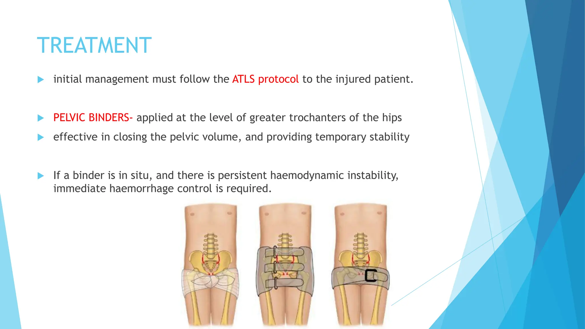
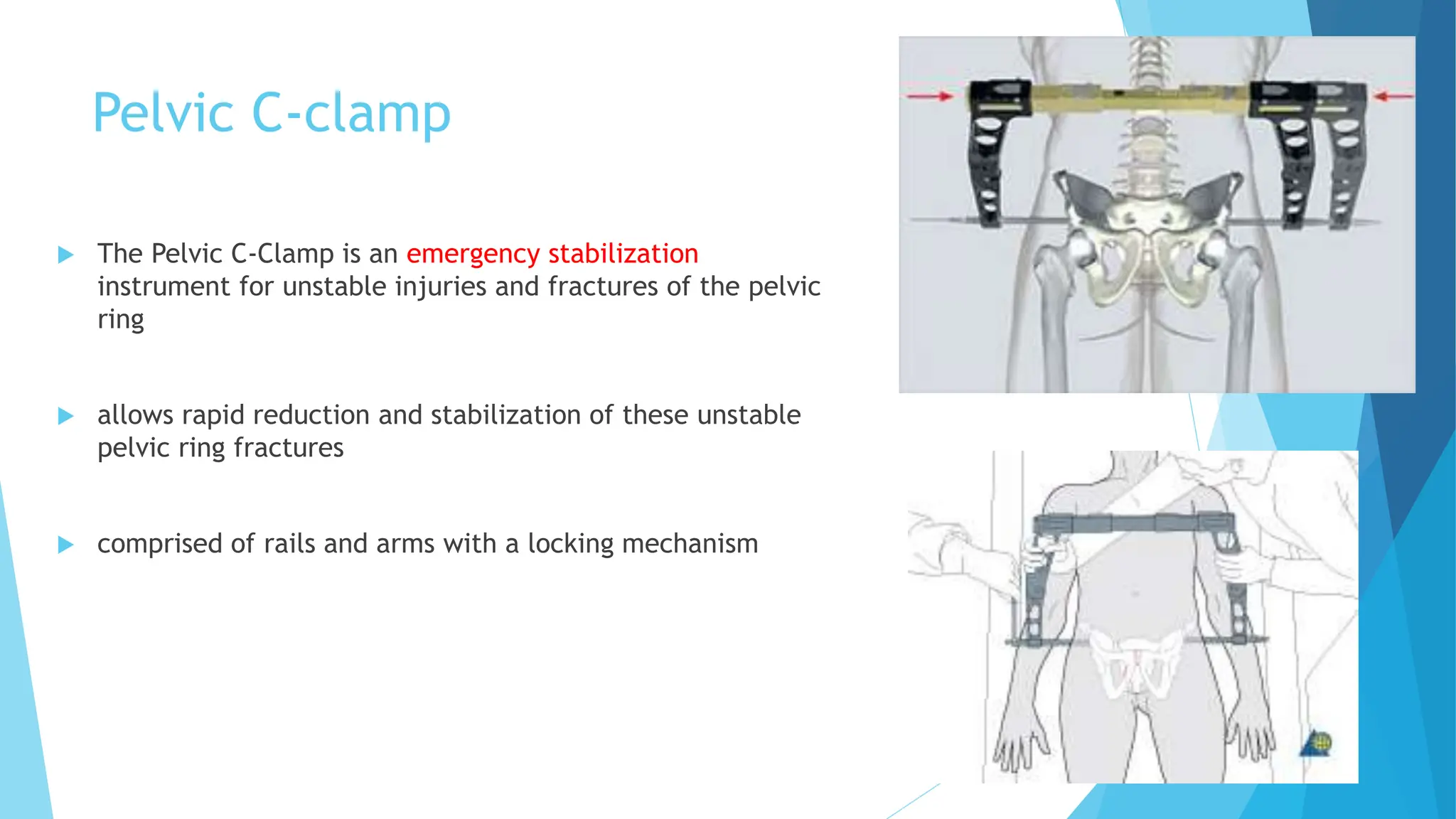
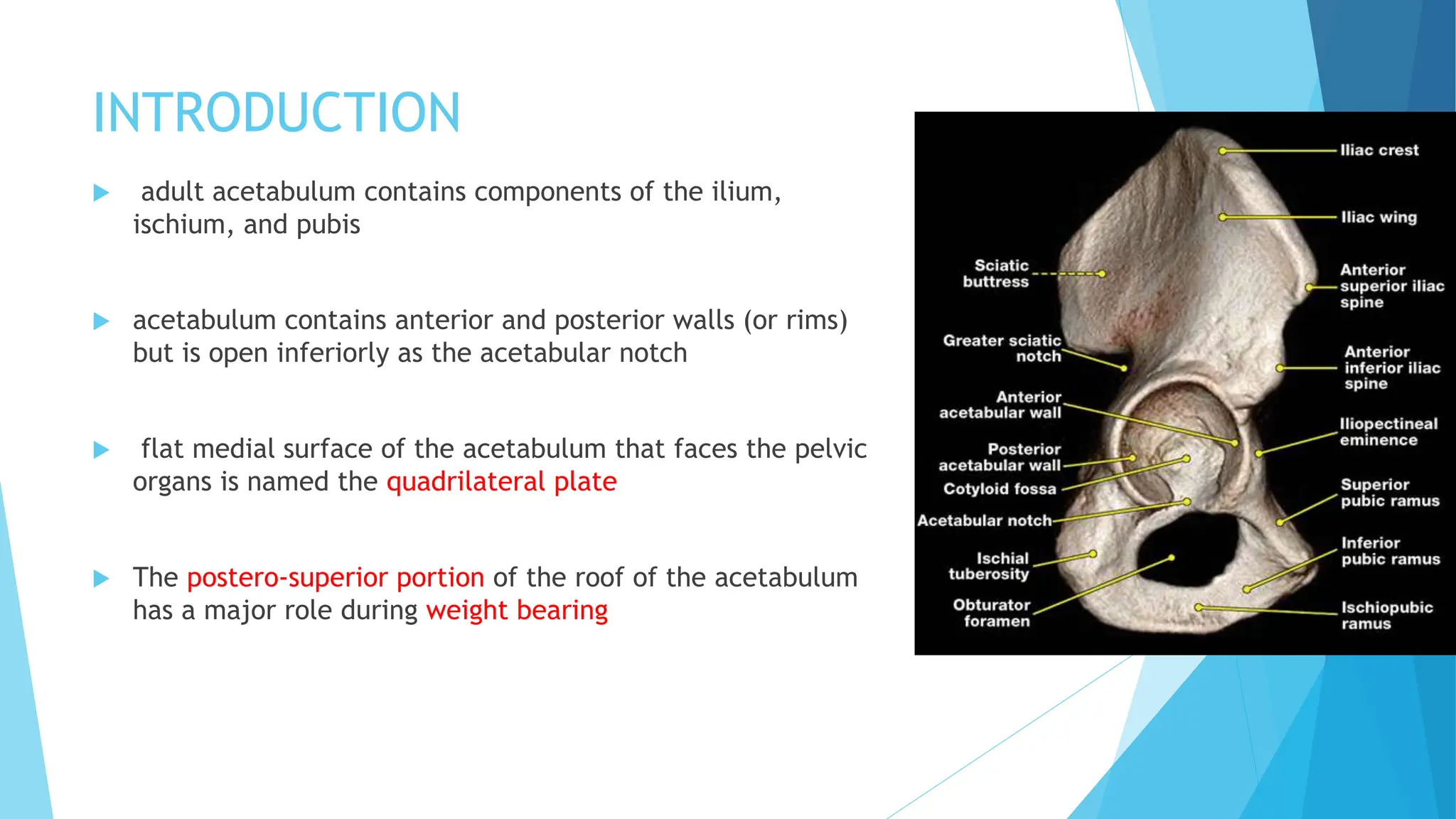
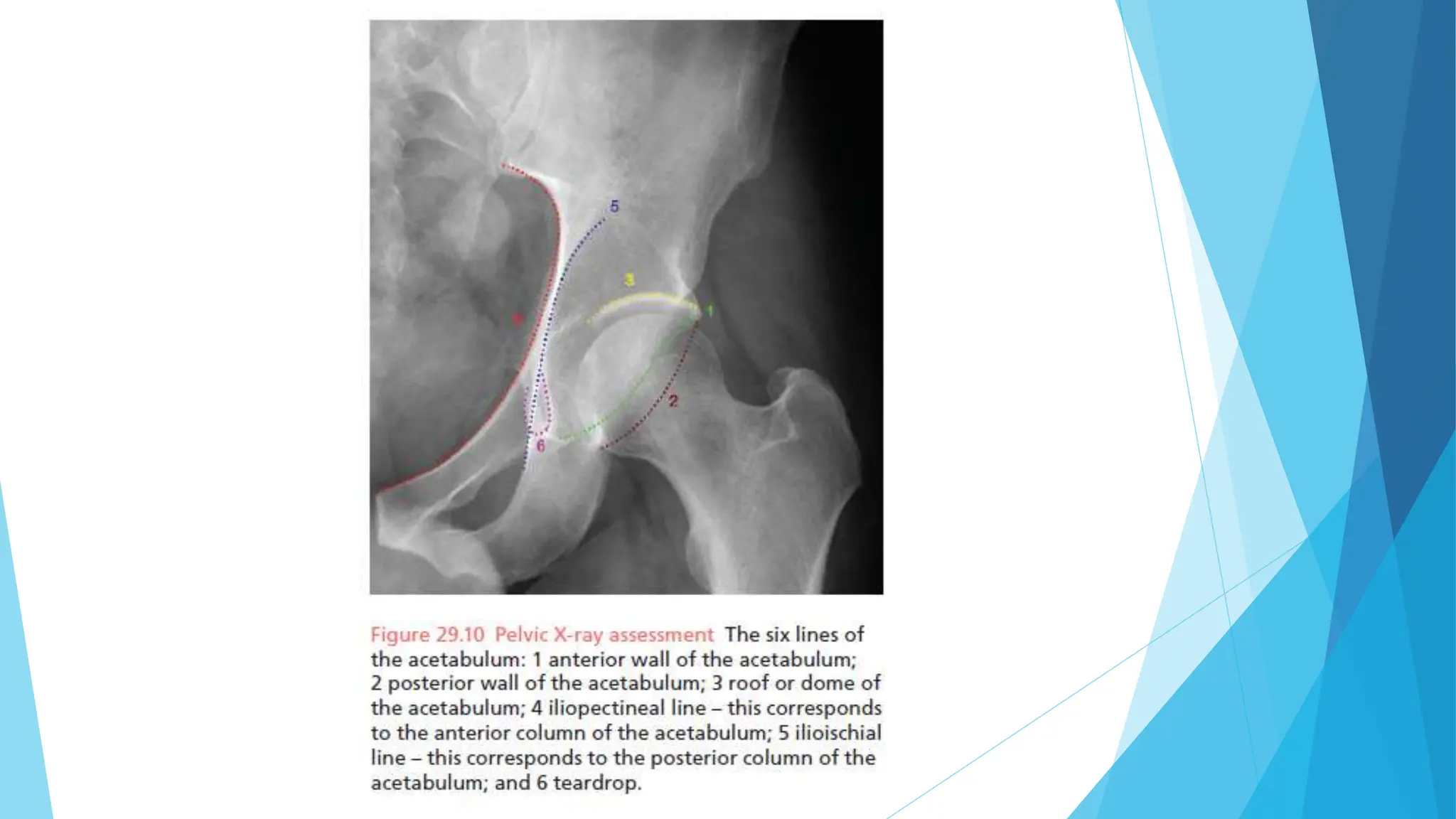
PELVIS & ACETABULUM
INTRODUCTION
 Fractures of the pelvis account for less than 5% of all skeletal injuries
 particularly important due to the potential risk of severe blood loss
 over 10% of these patients will have associated visceral injuries
 in this group the mortality rate is in excess of 10%
ANATOMY
PELVIC RING
2 innominate bones sacrum
 articulating in front at the symphysis pubis
 posteriorly at the sacroiliac joints
 Unlike a hinge joint or a ball-and-socket joint, the pelvic bones do not
articulate as part of a stable construct
 stability of the pelvic ring depends upon the integrity of the strong ligaments
that bind the three segments together
 ANTERIOR STABILITY at symphysis pubis is provided by-
1. superior pubic ligament
2. arcuate pubic ligament
 POSTERIOR STABILITY at sacro-iliac joints is provided by-
Anteriorly Posteriorly
anterior sacroiliac ligaments posterior sacroiliac ligaments
iliolumbar ligaments sacrococcygeal ligaments
Sacrotuberous ligaments
sacrospinous ligaments
Blood vessels in pelvis
 major branches of the common iliac arteries and veins
arise within the pelvis
 internal iliac vessels- supply the pelvic viscera
 external iliac vessels- continue in their journey to supply
the lower limbs
 rich low-pressure venous plexus posteriorly- especially
prone to injury if there is bony disruption around the
sacroiliac joints
 It is bleeding from this plexus that comprises the major
blood loss in pelvic haemorrhage
 L5 and S1 nerve roots- most commonly damaged nerves in pelvic ring injuries
 sciatic nerve- most commonly damaged in acetabular fractures.
 bladder lies behind the symphysis pubis
 urethra is much more mobile and shorter in females, and it is less prone to
injury.
 In severe pelvic injuries the membranous urethra is damaged
FUNCTIONS OF THE PELVIS
 Its primary role is to support the weight of the upper body when sitting and to
transfer this weight to the lower limbs when standing
 serves as an attachment point for trunk and lower limb muscles
 also protects the internal pelvic organs.
CLINICAL ASSESSMENT
 fracture of the pelvis should be suspected in any multiply injured patient
 swelling and bruising of the lower abdomen, the thighs, the perineum, the
scrotum or the vulva
 abdomen should be carefully palpated. Guarding or tenderness suggests the
possibility of intraperitoneal bleeding
 A ruptured bladder should be suspected in patients who do not void or in
whom a bladder is not palpable after adequate fluid replacement
 Neurological examination is very important; there may be damage to the
lumbosacral plexus
IMAGING OF THE BONY PELVIS
 plain anteroposterior (AP) X-ray of the pelvis is obtained at the same time as
the chest X-ray
 carefully Inspected, systematically looking in each of the five zones of injury:
1. The sacroiliac joint area is inspected for any diastasis or sacral fracture.
2. The ilium is inspected for any fracture
3. The teardrop is inspected. correlates to the non-articular floor of the
acetabulum
4. The obturator foramen is inspected for any fracture of the superior or inferior
pubic ramus.
5. The symphysis pubis is examined for any fracture or diastasis.
SPECIALIZED RADIOGRAPHS FOR PELVIS
 1. INLET VIEW
30–40 degrees in a caudal angle
provides an axial view of the sacrum and sacroiliac joints
 2. OUTLET VIEW
30–40 degrees cephalic angle
true anteroposterior view of the sacrum and pubic symphysis areas
Judet views (taken at 30 degrees obliquely)
obturator oblique view iliac oblique view
shows the anterior column
of the acetabulum
shows the posterior column
and anterior wall of the acetabulum.
CT SCANS
 CT scanning provides a detailed anatomical view of the posterior structures,
which are not seen well on conventional radiographs
 Contrast is often also given. This is very helpful in excluding a bladder rupture
or urethral injury
 full ‘trauma CT scan’- This comprises a CT scan of the head, neck, chest,
abdomen and pelvis.
PELVIC FRACTURES-
1. AVULSION FRACTURES
 most common avulsion injuries- anterior inferior iliac spine (rectus femoris
origin) and the ischial tuberosity (hamstring origin)
 Usually seen in sportsmen and women and athletes
 All are essentially muscle injuries, needing only rest for a few days
 If there is a large bony fragment with displacement, however, operative
fixation may be necessary
2. STRESS FRACTURES
 Fractures of the pubic rami are fairly common in osteoporotic bone
 MRI is very helpful for the diagnosis of posterior insufficiency fractures
(around the sacroiliac joints)
 also seen in the superior and inferior pubic rami in slim individuals and long-
distance runners
 Consideration should be given to checking vitamin D levels
 Patients usually heal with rest
PELVIC RING FRACTURES
 usually due to a high-energy injury
 Think of the pelvis as a ‘polo mint’. It is impossible
to break a polo mint in one place. The same
principle applies to the normal bony pelvic ring
 Anteriorly the symphysis pubis or pubic rami will be
disrupted, and posteriorly there will either be a
sacroiliac joint displacement or sacral fracture
CLASSIFICATION OF PELVIC RING
FRACTURES
Young and Burgess
based on the mechanism of injury
predictive of the severity of the injury
(blood loss) and also guides the surgeon
on how to correct any deformity or
displacement of the fracture
Tile classification
provides an assessment of stability
of the pelvis
guides the surgeon as to whether
an injury needs operative fixation.
TREATMENT
 initial management must follow the ATLS protocol to the injured patient.
 PELVIC BINDERS- applied at the level of greater trochanters of the hips
 effective in closing the pelvic volume, and providing temporary stability
 If a binder is in situ, and there is persistent haemodynamic instability,
immediate haemorrhage control is required.
Pubic diastasis before application
of pelvic binder
After application of pelvic
binder
Pelvic C-clamp
 The Pelvic C-Clamp is an emergency stabilization
instrument for unstable injuries and fractures of the pelvic
ring
 allows rapid reduction and stabilization of these unstable
pelvic ring fractures
 comprised of rails and arms with a locking mechanism
Two options exist
Angiography and embolization Immediate transfer to the operating
theatre for pre-peritoneal packing
Operative fixation
 principle of operative fixation- to convert an unstable pelvic ring to a stable
one
 Pubic symphysis diastasis is treated with open reduction and internal fixation
with plates and screws
UROGENITAL INJURIES
 Bladder and urethral injury is the commonest associated injury in pelvic
fractures
 Urethral tears are usually treated conservatively with catheterization for a
few weeks
 If a soft, silicone 16F catheter cannot be passed by a single, gentle attempt, a
suprapubic catheter is required
 Intraperitoneal rupture of the bladder requires emergency laparotomy and
direct repair; extraperitoneal bladder rupture may be treated conservatively
COMPLICATIONS
 Urethral strictures
 Impotence- 30% in pelvic fractures
 Venous thromboembolism
 Nerve injury- L5,S1 roots
 Infection
 Non-union
ACETABULAR
FRACTURES
INTRODUCTION
 adult acetabulum contains components of the ilium,
ischium, and pubis
 acetabulum contains anterior and posterior walls (or rims)
but is open inferiorly as the acetabular notch
 flat medial surface of the acetabulum that faces the pelvic
organs is named the quadrilateral plate
 The postero-superior portion of the roof of the acetabulum
has a major role during weight bearing
 Acetabular fractures occur when the femoral head is driven into the
acetabulum
 Direction of the force determines the fracture pattern
 Displaced fractures result in hip joint incongruency; this will lead to
osteoarthritis
LETOURNEL CLASSIFICATION
 examine the lines on the AP pelvic X-ray
 There are two groups: elemental fractures and associated fractures
AP VIEW
OBTURATOR
OBLIQUE VIEW
ILIAC OBLIQUE VIEW
MANAGEMENT
 goal of treatment-
 restore joint congruency
 provide fracture stability to allow mobilization
 prevent osteoarthritis
 Undisplaced fractures are usually stable and can be managed conservatively
 Patients are mobilized with partial weight-bearing on the affected side for 6
weeks
 If the hip is dislocated, reduction is urgent, followed by the application of
skeletal traction until definitive surgery
 Fractures with more than 2 mm of displacement of the articular surface
should be anatomically reduced and stabilized
 Patients with-
 >3 mm of displacement- poor outcome
 <1 mm displacement- have less progression to osteoarthritis.
 Surgical approaches:
 Ilioinguinal approach
 Kocher-langenbeck approach
COMPLICATIONS
 Heterotopic ossification
 Sciatic nerve palsy
 Avascular necrosis
 Hip abductor dysfunction
 Osteoarthritis
THANK YOU

PELVIS & ACETABULUM Orthopaedics topics slides

  • 1.
  • 2.
    INTRODUCTION  Fractures ofthe pelvis account for less than 5% of all skeletal injuries  particularly important due to the potential risk of severe blood loss  over 10% of these patients will have associated visceral injuries  in this group the mortality rate is in excess of 10%
  • 3.
    ANATOMY PELVIC RING 2 innominatebones sacrum  articulating in front at the symphysis pubis  posteriorly at the sacroiliac joints  Unlike a hinge joint or a ball-and-socket joint, the pelvic bones do not articulate as part of a stable construct  stability of the pelvic ring depends upon the integrity of the strong ligaments that bind the three segments together
  • 4.
     ANTERIOR STABILITYat symphysis pubis is provided by- 1. superior pubic ligament 2. arcuate pubic ligament  POSTERIOR STABILITY at sacro-iliac joints is provided by- Anteriorly Posteriorly anterior sacroiliac ligaments posterior sacroiliac ligaments iliolumbar ligaments sacrococcygeal ligaments Sacrotuberous ligaments sacrospinous ligaments
  • 5.
    Blood vessels inpelvis  major branches of the common iliac arteries and veins arise within the pelvis  internal iliac vessels- supply the pelvic viscera  external iliac vessels- continue in their journey to supply the lower limbs  rich low-pressure venous plexus posteriorly- especially prone to injury if there is bony disruption around the sacroiliac joints  It is bleeding from this plexus that comprises the major blood loss in pelvic haemorrhage
  • 6.
     L5 andS1 nerve roots- most commonly damaged nerves in pelvic ring injuries  sciatic nerve- most commonly damaged in acetabular fractures.
  • 7.
     bladder liesbehind the symphysis pubis  urethra is much more mobile and shorter in females, and it is less prone to injury.  In severe pelvic injuries the membranous urethra is damaged
  • 8.
    FUNCTIONS OF THEPELVIS  Its primary role is to support the weight of the upper body when sitting and to transfer this weight to the lower limbs when standing  serves as an attachment point for trunk and lower limb muscles  also protects the internal pelvic organs.
  • 9.
    CLINICAL ASSESSMENT  fractureof the pelvis should be suspected in any multiply injured patient  swelling and bruising of the lower abdomen, the thighs, the perineum, the scrotum or the vulva  abdomen should be carefully palpated. Guarding or tenderness suggests the possibility of intraperitoneal bleeding  A ruptured bladder should be suspected in patients who do not void or in whom a bladder is not palpable after adequate fluid replacement
  • 10.
     Neurological examinationis very important; there may be damage to the lumbosacral plexus
  • 11.
    IMAGING OF THEBONY PELVIS  plain anteroposterior (AP) X-ray of the pelvis is obtained at the same time as the chest X-ray  carefully Inspected, systematically looking in each of the five zones of injury: 1. The sacroiliac joint area is inspected for any diastasis or sacral fracture. 2. The ilium is inspected for any fracture 3. The teardrop is inspected. correlates to the non-articular floor of the acetabulum 4. The obturator foramen is inspected for any fracture of the superior or inferior pubic ramus. 5. The symphysis pubis is examined for any fracture or diastasis.
  • 13.
    SPECIALIZED RADIOGRAPHS FORPELVIS  1. INLET VIEW 30–40 degrees in a caudal angle provides an axial view of the sacrum and sacroiliac joints  2. OUTLET VIEW 30–40 degrees cephalic angle true anteroposterior view of the sacrum and pubic symphysis areas
  • 15.
    Judet views (takenat 30 degrees obliquely) obturator oblique view iliac oblique view shows the anterior column of the acetabulum shows the posterior column and anterior wall of the acetabulum.
  • 18.
    CT SCANS  CTscanning provides a detailed anatomical view of the posterior structures, which are not seen well on conventional radiographs  Contrast is often also given. This is very helpful in excluding a bladder rupture or urethral injury  full ‘trauma CT scan’- This comprises a CT scan of the head, neck, chest, abdomen and pelvis.
  • 20.
    PELVIC FRACTURES- 1. AVULSIONFRACTURES  most common avulsion injuries- anterior inferior iliac spine (rectus femoris origin) and the ischial tuberosity (hamstring origin)  Usually seen in sportsmen and women and athletes  All are essentially muscle injuries, needing only rest for a few days  If there is a large bony fragment with displacement, however, operative fixation may be necessary
  • 22.
    2. STRESS FRACTURES Fractures of the pubic rami are fairly common in osteoporotic bone  MRI is very helpful for the diagnosis of posterior insufficiency fractures (around the sacroiliac joints)  also seen in the superior and inferior pubic rami in slim individuals and long- distance runners  Consideration should be given to checking vitamin D levels  Patients usually heal with rest
  • 24.
    PELVIC RING FRACTURES usually due to a high-energy injury  Think of the pelvis as a ‘polo mint’. It is impossible to break a polo mint in one place. The same principle applies to the normal bony pelvic ring  Anteriorly the symphysis pubis or pubic rami will be disrupted, and posteriorly there will either be a sacroiliac joint displacement or sacral fracture
  • 25.
    CLASSIFICATION OF PELVICRING FRACTURES Young and Burgess based on the mechanism of injury predictive of the severity of the injury (blood loss) and also guides the surgeon on how to correct any deformity or displacement of the fracture Tile classification provides an assessment of stability of the pelvis guides the surgeon as to whether an injury needs operative fixation.
  • 26.
    TREATMENT  initial managementmust follow the ATLS protocol to the injured patient.  PELVIC BINDERS- applied at the level of greater trochanters of the hips  effective in closing the pelvic volume, and providing temporary stability  If a binder is in situ, and there is persistent haemodynamic instability, immediate haemorrhage control is required.
  • 27.
    Pubic diastasis beforeapplication of pelvic binder After application of pelvic binder
  • 28.
    Pelvic C-clamp  ThePelvic C-Clamp is an emergency stabilization instrument for unstable injuries and fractures of the pelvic ring  allows rapid reduction and stabilization of these unstable pelvic ring fractures  comprised of rails and arms with a locking mechanism
  • 29.
    Two options exist Angiographyand embolization Immediate transfer to the operating theatre for pre-peritoneal packing
  • 30.
    Operative fixation  principleof operative fixation- to convert an unstable pelvic ring to a stable one  Pubic symphysis diastasis is treated with open reduction and internal fixation with plates and screws
  • 31.
    UROGENITAL INJURIES  Bladderand urethral injury is the commonest associated injury in pelvic fractures  Urethral tears are usually treated conservatively with catheterization for a few weeks  If a soft, silicone 16F catheter cannot be passed by a single, gentle attempt, a suprapubic catheter is required  Intraperitoneal rupture of the bladder requires emergency laparotomy and direct repair; extraperitoneal bladder rupture may be treated conservatively
  • 32.
    COMPLICATIONS  Urethral strictures Impotence- 30% in pelvic fractures  Venous thromboembolism  Nerve injury- L5,S1 roots  Infection  Non-union
  • 33.
  • 34.
    INTRODUCTION  adult acetabulumcontains components of the ilium, ischium, and pubis  acetabulum contains anterior and posterior walls (or rims) but is open inferiorly as the acetabular notch  flat medial surface of the acetabulum that faces the pelvic organs is named the quadrilateral plate  The postero-superior portion of the roof of the acetabulum has a major role during weight bearing
  • 35.
     Acetabular fracturesoccur when the femoral head is driven into the acetabulum  Direction of the force determines the fracture pattern  Displaced fractures result in hip joint incongruency; this will lead to osteoarthritis
  • 36.
    LETOURNEL CLASSIFICATION  examinethe lines on the AP pelvic X-ray  There are two groups: elemental fractures and associated fractures
  • 38.
  • 39.
    MANAGEMENT  goal oftreatment-  restore joint congruency  provide fracture stability to allow mobilization  prevent osteoarthritis  Undisplaced fractures are usually stable and can be managed conservatively  Patients are mobilized with partial weight-bearing on the affected side for 6 weeks  If the hip is dislocated, reduction is urgent, followed by the application of skeletal traction until definitive surgery
  • 40.
     Fractures withmore than 2 mm of displacement of the articular surface should be anatomically reduced and stabilized  Patients with-  >3 mm of displacement- poor outcome  <1 mm displacement- have less progression to osteoarthritis.  Surgical approaches:  Ilioinguinal approach  Kocher-langenbeck approach
  • 42.
    COMPLICATIONS  Heterotopic ossification Sciatic nerve palsy  Avascular necrosis  Hip abductor dysfunction  Osteoarthritis
  • 43.