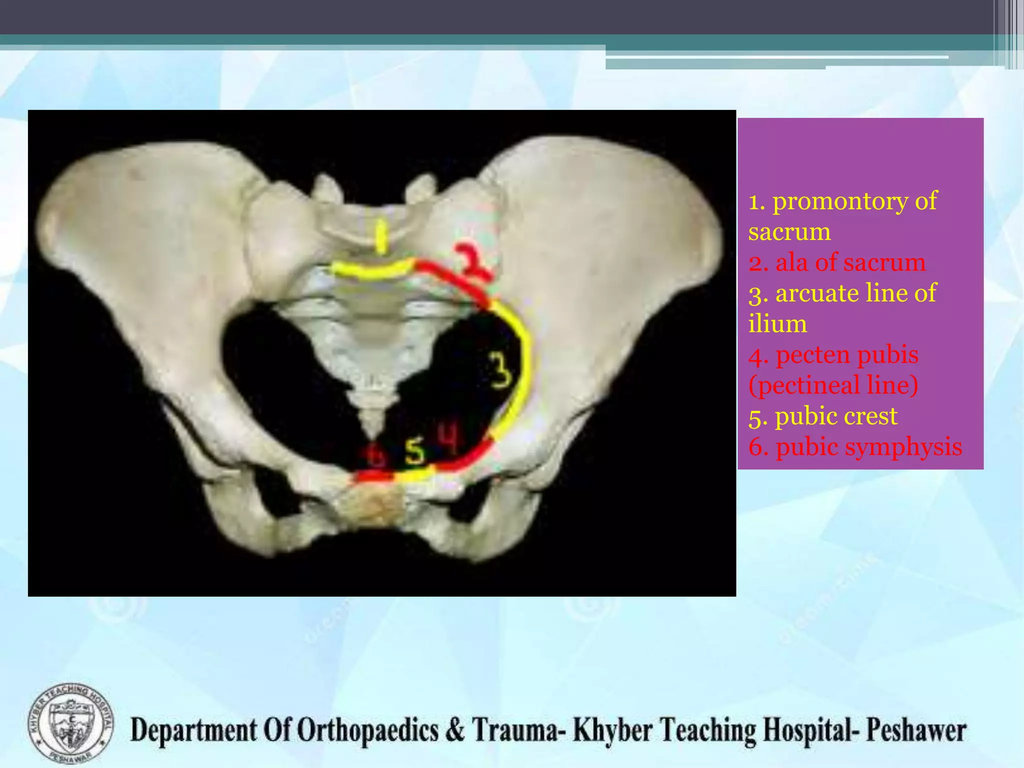
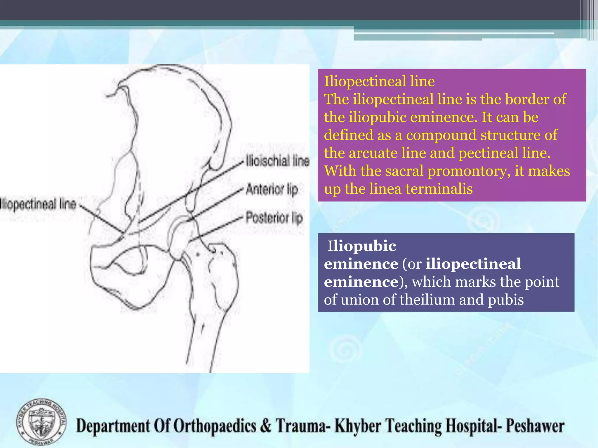
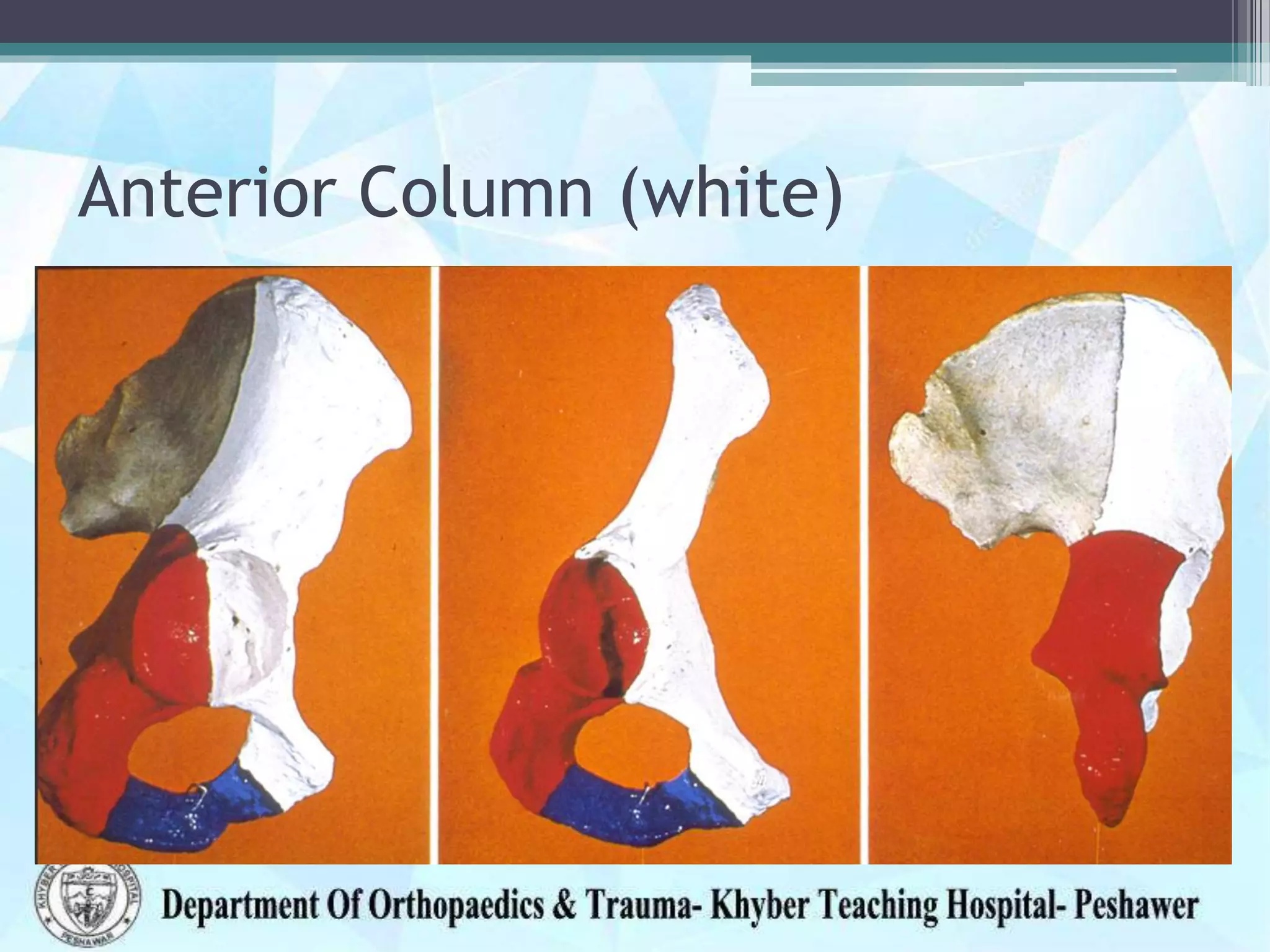
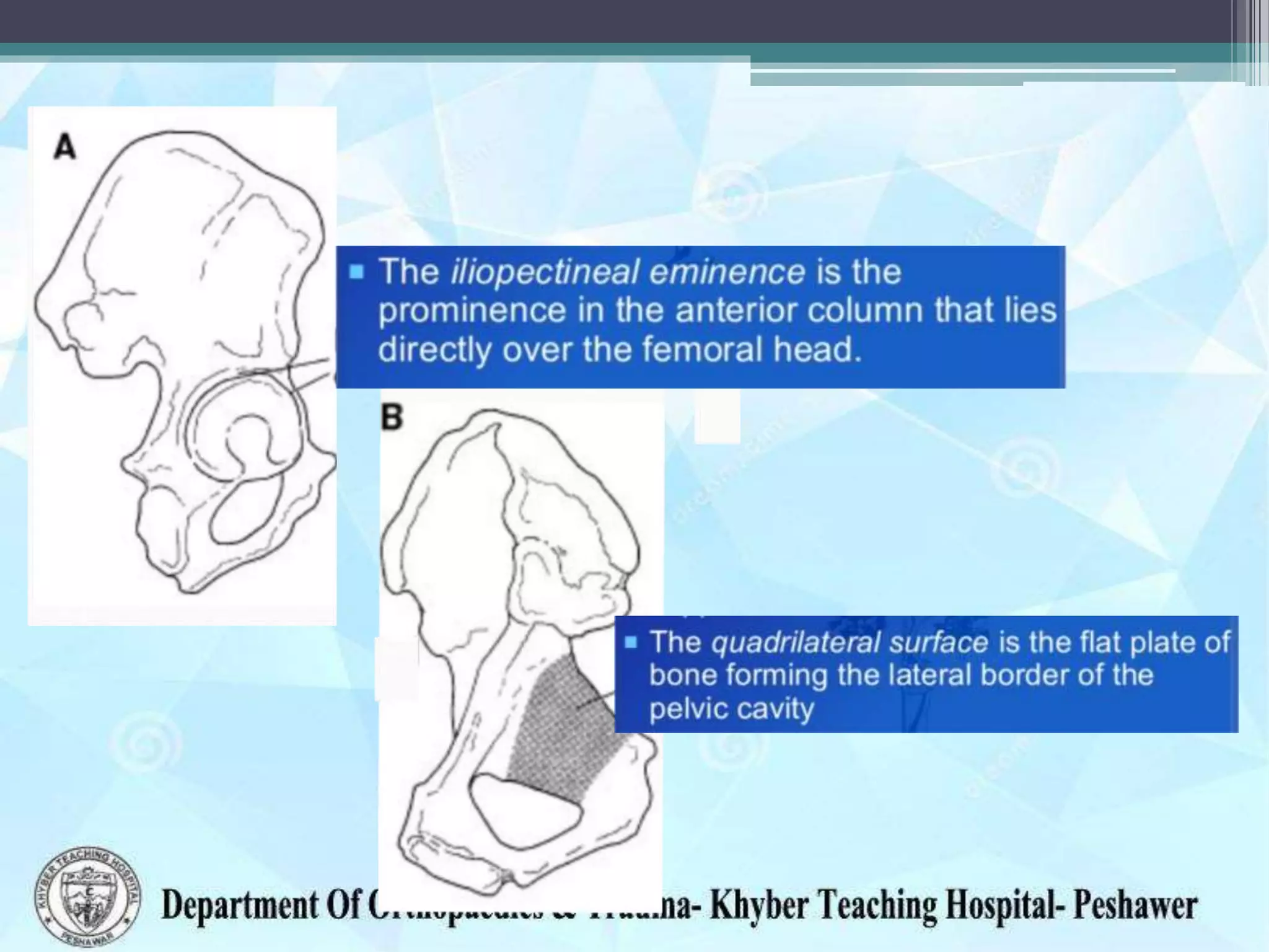
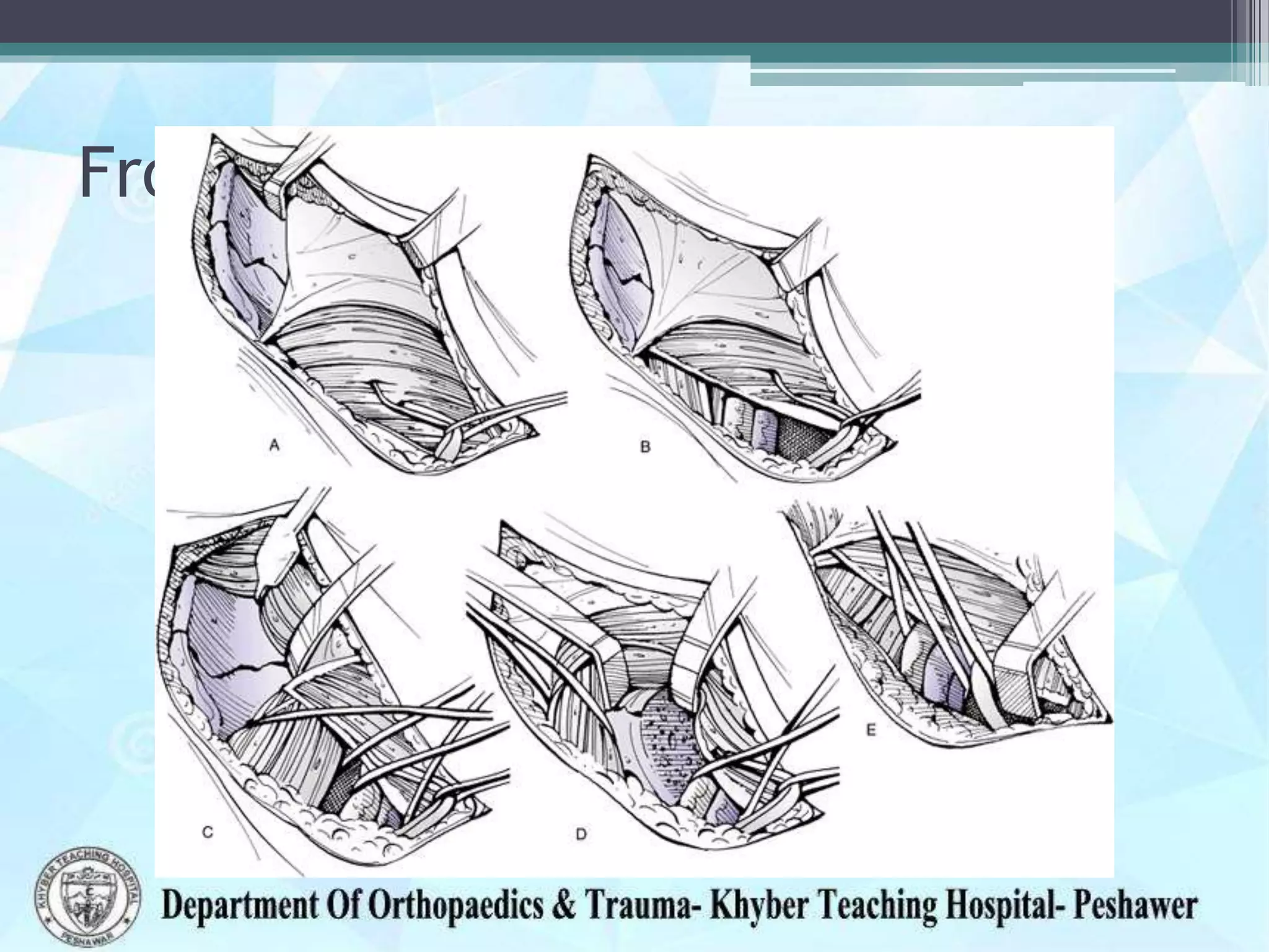
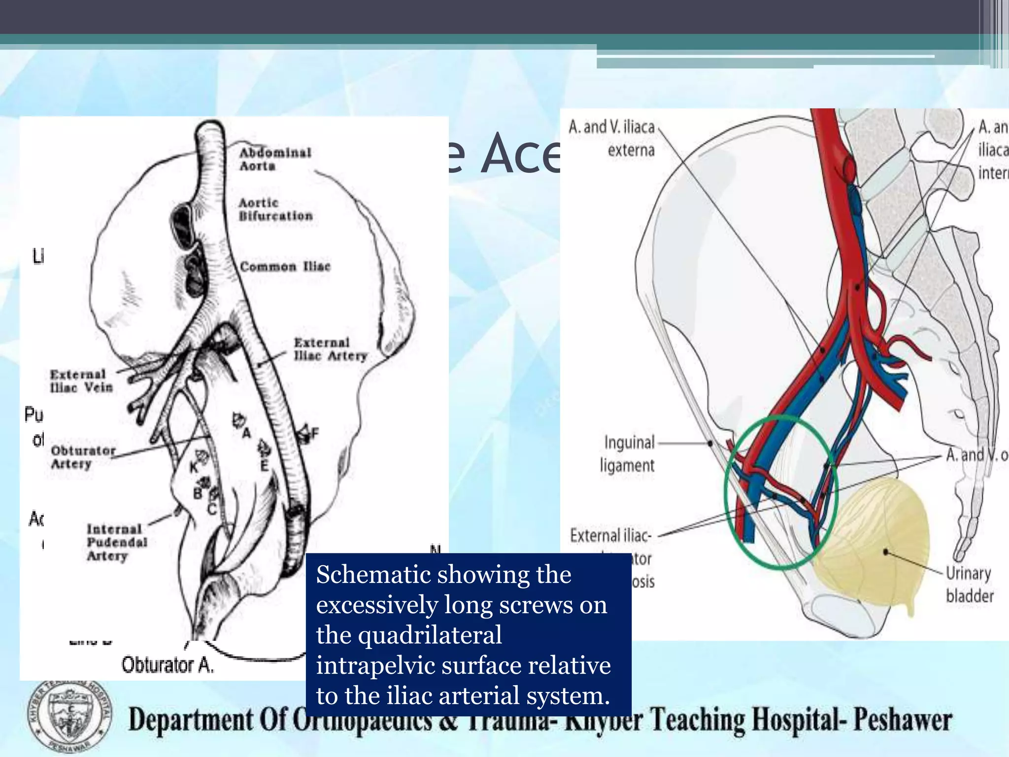
The document provides information on the anatomy of the acetabulum. It discusses:
1) The components that make up the acetabulum where the three bones of the pelvis meet, including the articular surface and acetabular fossa.
2) The embryological development of the hip joint from 4-11 weeks of gestation and continued growth after birth.
3) Structures around the acetabulum including nerves, vessels, muscles and bony landmarks.
4) Forces across the hip joint during various activities from walking to running.
Total Hip replacement for Ankylosing Spondylitis: Planning & Execution Vaibhav Bagaria
Performing Total Hip replacement in Ankylosing Spondylitis requires a well thought of strategy. Preoperative planning, Inventory ordering, positioning, cup and stem orientation all play a role.
Total Hip replacement for Ankylosing Spondylitis: Planning & Execution Vaibhav Bagaria
Performing Total Hip replacement in Ankylosing Spondylitis requires a well thought of strategy. Preoperative planning, Inventory ordering, positioning, cup and stem orientation all play a role.
Patella dislocation is a common problem in the young. Recurrence of dislocation can be significant problem causing pain and discomfort. The assessment and guidelines towards non-surgical and surgical treatment options are discussed here.
Evolution of tunnel placement in ACL reconstructionDhananjaya Sabat
One of my talks at Delhi Arthroscopy Club....... this presentation provides a insight regarding the conceptual evolution in tunnel placement during ACL reconstruction.
Marketiing Proposal to Verizon Wirelessjaimegarciacb
This is an example of a marketing proposal presented to Verizon Wireless. They soon joined the Chamber and we worked with them on their Latino Small Business marketing campaign
«Консультирование «жертв» деструктивных культов и их родственников»Michel Vershinin
Авторский семинар Вершинина Михаила
«Консультирование «жертв» деструктивных культов и их родственников»
Специализированный семинар для психологов, религиоведов, социальных работников и специалистов по работе с жертвами психологического и физического насилия.
Одесса, 31 Мая 2013
"Продвижение и мониторинг в социальных сетях и Твиттере: анализ трафика, раб...Michel Vershinin
Анонс практическогого семинара "Продвижение и мониторинг в социальных сетях и Твиттере: анализ трафика, работа с обращениями, создание контента" для корпоративного клиента
Anatomy of the pelvis, understand the clinical relevance and key landmarks,parts and function,blood and nerve supply and disorders associated with the pelvis.
1. Anatomy of the Acetabulum
Dr Zahid Askar
MBBS, FCPS(Ortho),FRCS(Tr & Ortho)
Prof. of Orthopaedics
Khyber Medical College
Peshawer
2. • Acetabulum (Gr: ὀξίς, ὀξύβαφον, ὀξυβάφιον)
was a vinegar-cup, used by
Greeks and Romans placed on the dining-table .
3. • The acetabulum is the point where the three
components of the pelvis (ilium, ischium, and
pubis) meet.
4. The articular Lunate Surface/DOME
The nonarticular Acetabular
Fossa
The inferior Acetabular
Notch.
Anatomical restoration of the
dome with concentric
reduction of the femoral head
beneath this dome is the goal
of both operative and
nonoperative treatment.
5. Embryology
• At 4 to 6 gestational weeks, the hip joint
develops from the cartilaginous analogue -
• By 7 weeks a cleft appears
• By 11 weeks, hip joint formation is mostly
complete
6. • At late gestation, femoral head grows more
rapidly
• At birth the femoral head is < 50% covered
• Acetabulum continues to grow till 7 years with
maximum growth is in the first 4 years.
7. ASIS
AIIS
Ischial Tuberosity
Pubis
Ischial Spine
tendon of obturator internus m.
internal pudendal a.
internal pudendal v.
pudendal n.
nerve to obturator internus
- 7 nerves:
- Sciatic Nerve
- Superior Gluteal Nerve
- Inferior Gluteal Nerve
- Internal Pudendal Nerve:
- Posterior Femoral Cutaneous Nerve
- Nerve to Quadratus Femoris
- Nerve to Obturator Externus
- 3 Vessel Sets:
- Superior Gluteal Artery & Vein
- Inferior Gluteal Artery & vein
- Internal Pudendal Artery & vein
- 1 Muscle:
- Piriformis
8. Forces Across The Hip Joint
• Walking upto 4 times BW
• Running upto 7 times BW
• Supported gait (w/ a walker or a cane) 2-3 BW
Wednesday,
December 17,
2014
8
Supine straight leg raising 2 times BW.
Getting on & off bed pan 4 times BW
Jogging and very fast walking both raised the forces to about 5.5 BW.
Stumbling on one occasion caused magnitudes of 7.20 BW
9. 1. promontory of
sacrum
2. ala of sacrum
3. arcuate line of
ilium
4. pecten pubis
(pectineal line)
5. pubic crest
6. pubic symphysis
10. Iliopectineal line
The iliopectineal line is the border of
the iliopubic eminence. It can be
defined as a compound structure of
the arcuate line and pectineal line.
With the sacral promontory, it makes
up the linea terminalis
Iliopubic
eminence (or iliopectineal
eminence), which marks the point
of union of theilium and pubis
11. Ant. Column
The two Columns
Post. Column
Acetabulum
These two columns serve as
struts, mechanically
representing the coalescence of
bony trabeculae along lines of
stress
12. Anterior Column (white)
Consists of
• Anterior half of the Iliac crest,
• The Iliac spines,
• Anterior half of the acetabulum, and the pubis. ;
13. Posterior Column(red)
• Consists of:
• Ischium,
• the ischial spine,
• the posterior half of the acetabulum,
• the dense bone forming the sciatic notch.
14. • The posterior column ends at its intersection
with the anterior column at the top of the sciatic
notch
20. Sciatic Nerve
• The largest and longest nerve in the body
• Arises from the lumber and sacral plexus
• Comes out of the pelvis through the Sciatic
Notch
• Runs anterior (deep) to Piriformis and lies
posterior (superficial) to the short external
rotators
21.
22. Leaves pelvis through the
greater sciatic notch along-
with superior gluteal artery
and vein
It runs over the piriformis
between the gluteus medius
and minimus supplying
these muscles
Superior Gluteal Nerve
Do not split gluteus medius
more than 5-7 cm proximal
to greater trochanter due to
risk of denervating the
muscle at risk during the
lateral (Hardinge)
approach to the hip
23. Leaves the pelvis through
the greater sciatic foramen
Runs underneath the
piriformis
Divides into muscular
branches to supply
the Gluteus Maximus
Inferior Gluteal Nerve
25. Vessels near the Acetabulum
Schematic showing the
excessively long screws on
the quadrilateral
intrapelvic surface relative
to the iliac arterial system.
26. corona mortis
"corona mortis" or crown of death
Chances of significant hemorrhage
An anastomosis between the
obturator and the external iliac or
inferior epigastric arteries or veins.
Located behind the superior pubic
ramus at a variable distance from
the symphysis pubis (range 40-96
mm).
60% had a large diameter (>3 mm)
27. “We carried out forty cadaver dissections (80 hemi-pelvises) through the
ilioinguinal approach. A vascular anastomosis was found in 83% of specimens.
Of these, 60% had a large diameter (>3 mm) channel along the posterior
aspect of the superior pubic ramus”
“In clinical practice, however, 492 anterior approaches (to the best
of our knowledge the largest series described) have been carried
out over the last 15 years by the senior author (MB) and only five of
these problematic vessels were discovered, and in only two cases
was there troublesome bleeding. “
Darmanis S, Lewis A, Mansoor A, Bircher M
Corona mortis: an anatomical study with clinical implications in
approaches to the pelvis and acetabulum.
Clin Anat. 2007 May;20(4):433-9.