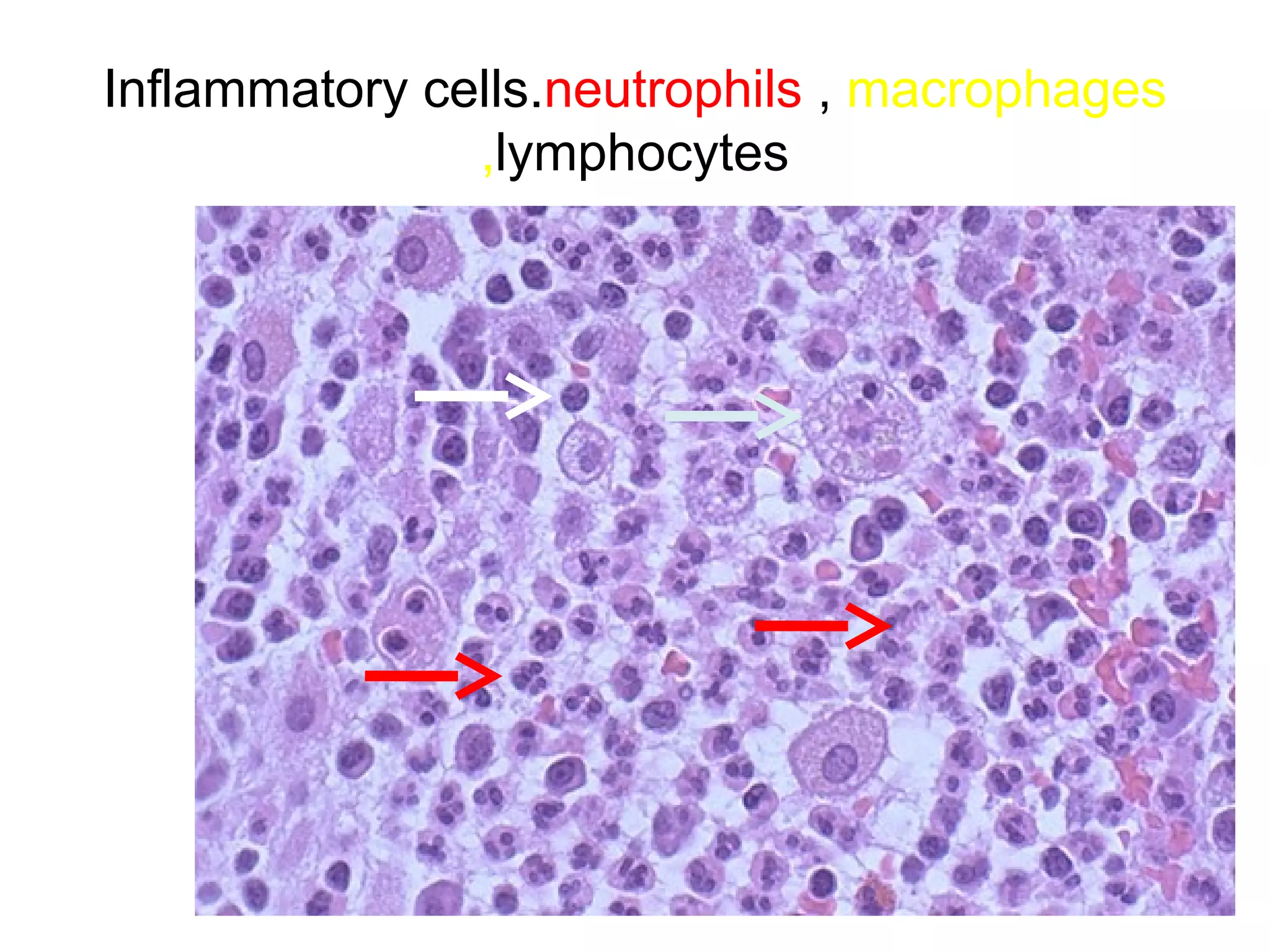
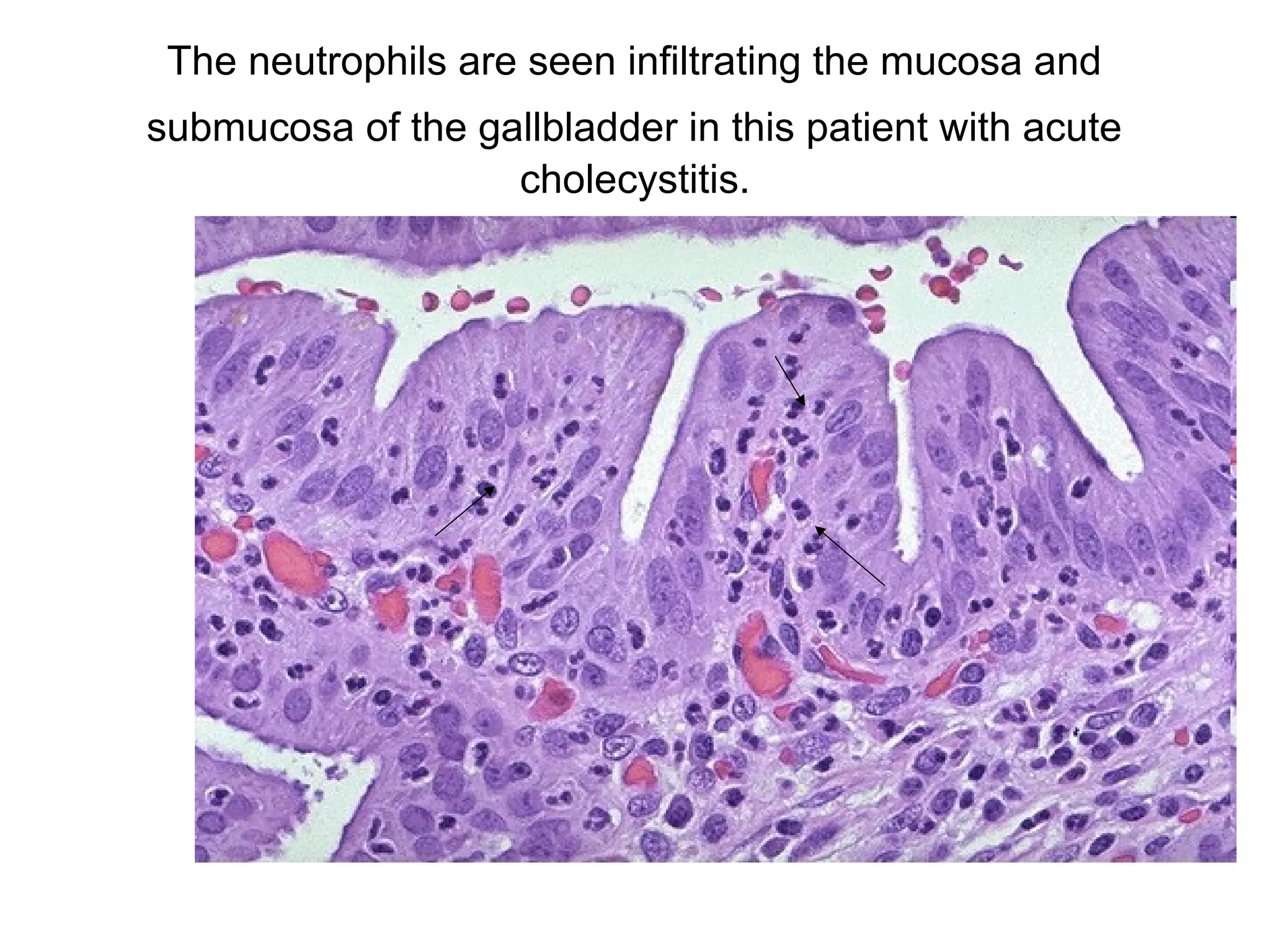
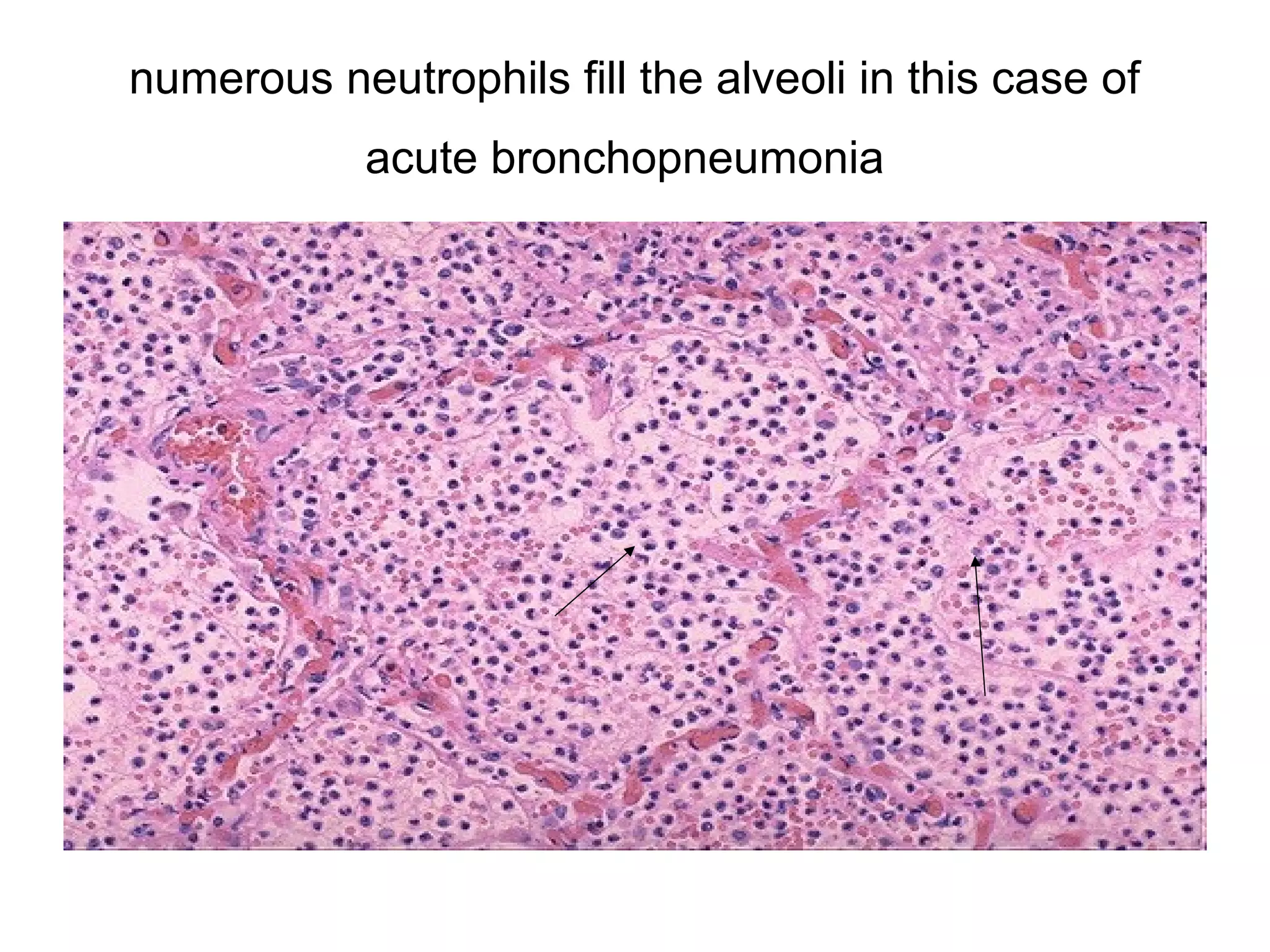
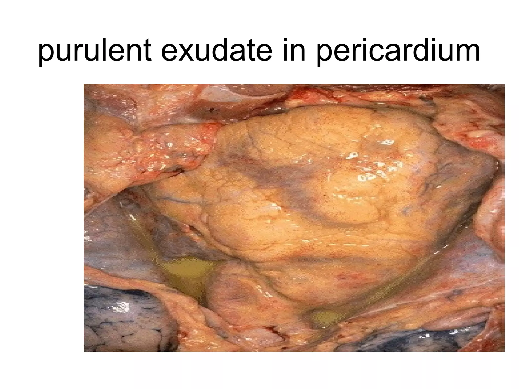
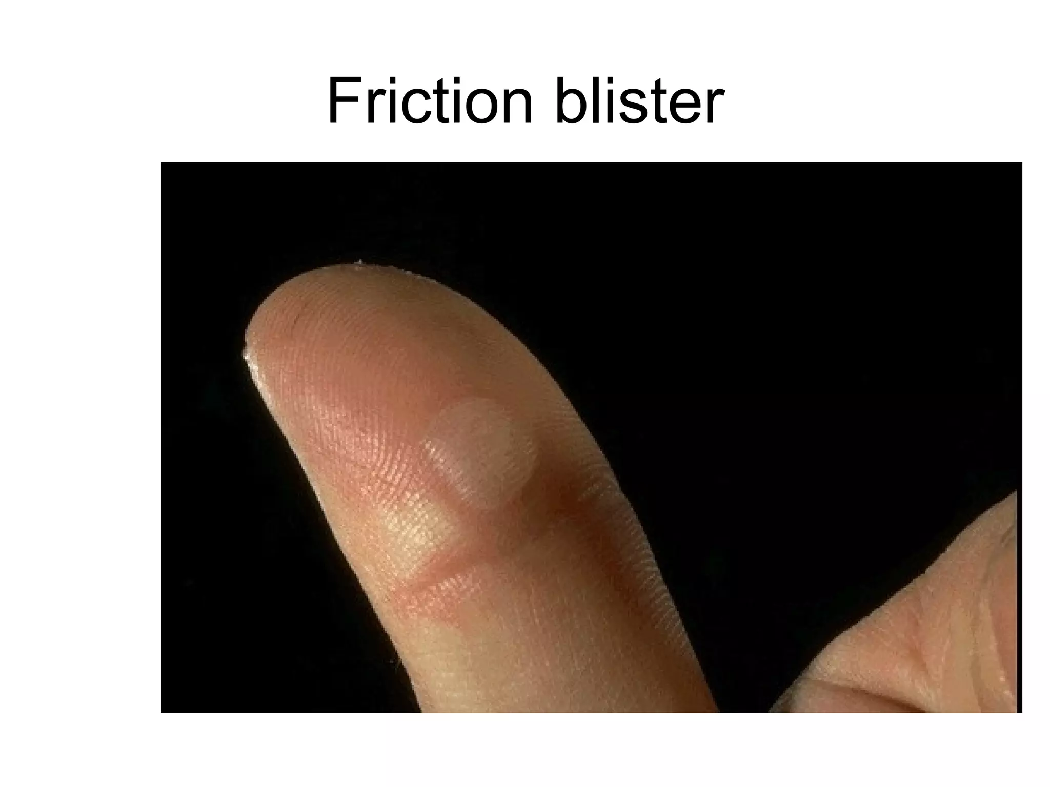
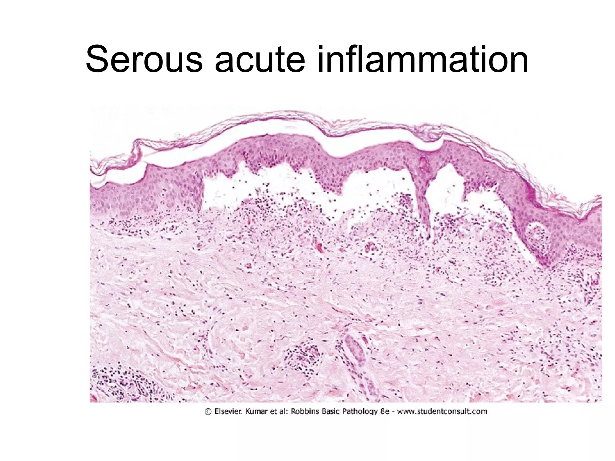
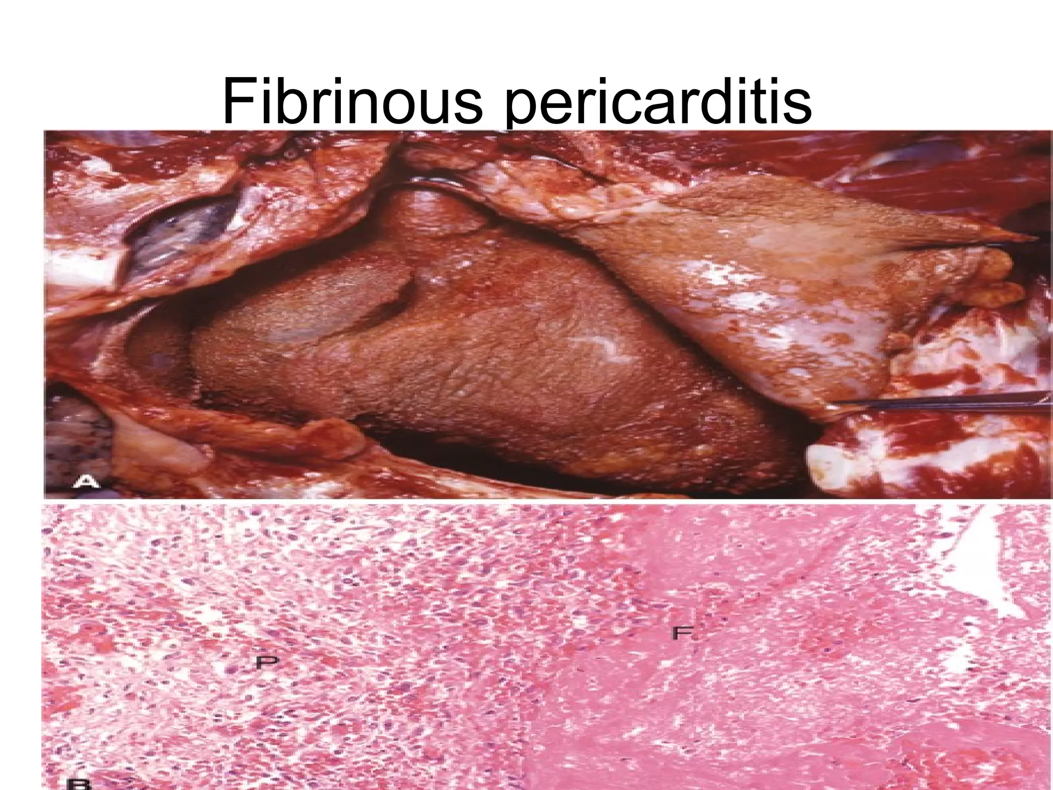
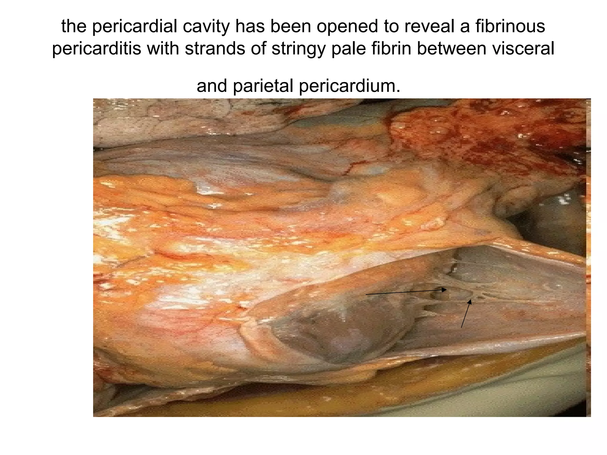
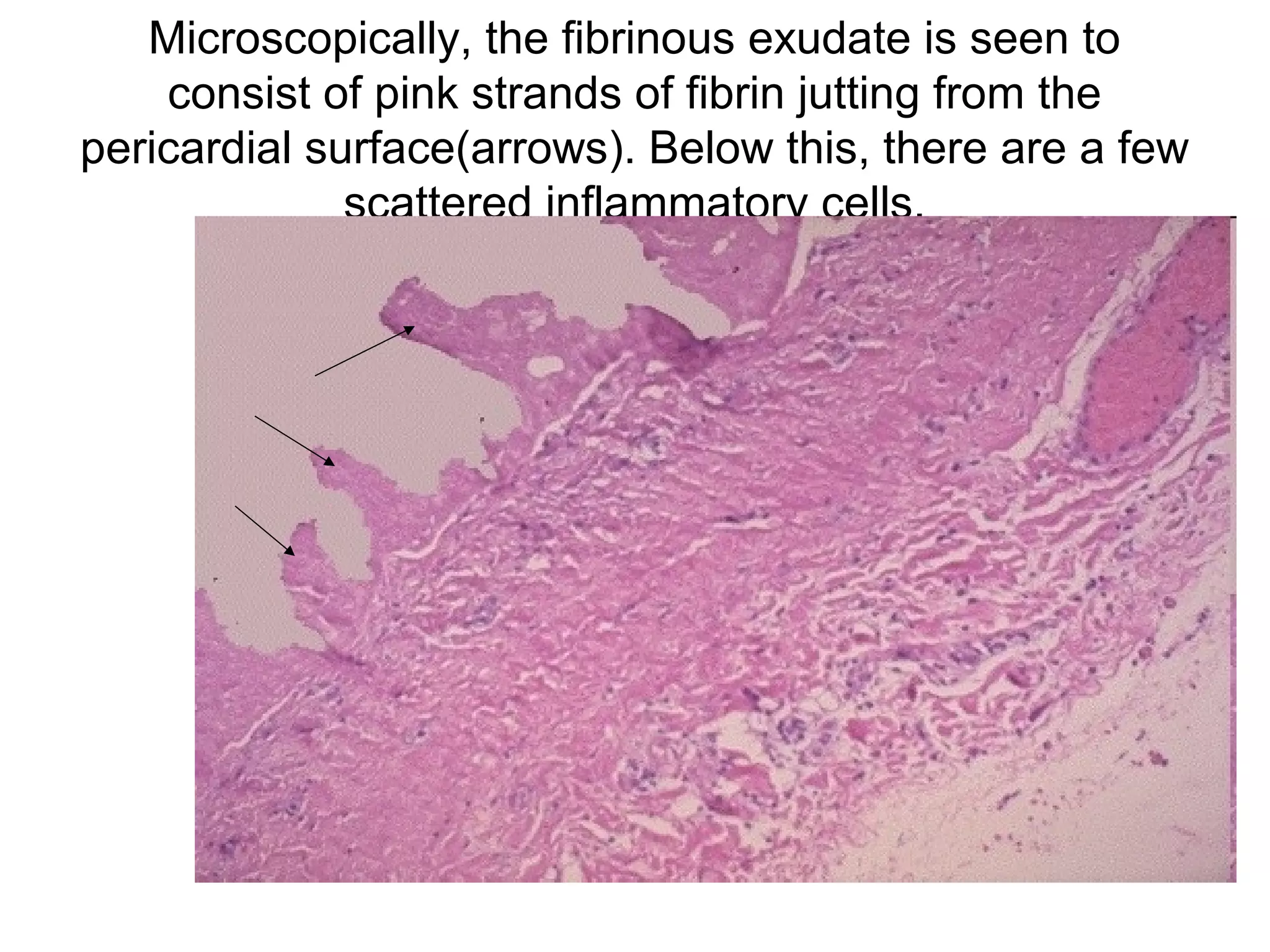
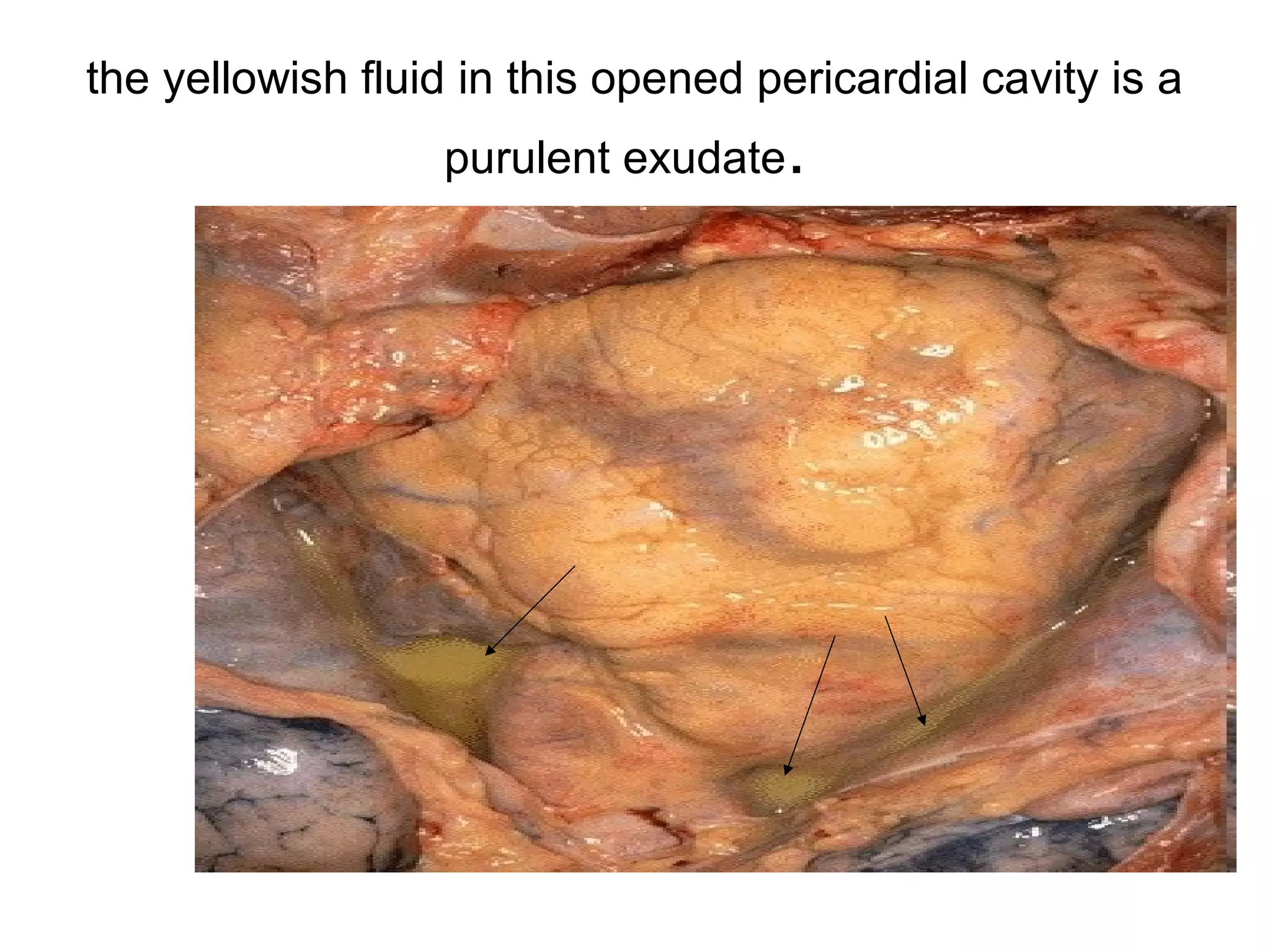
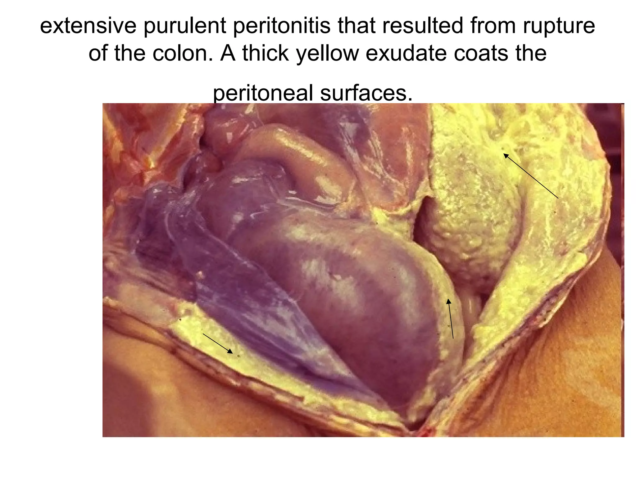
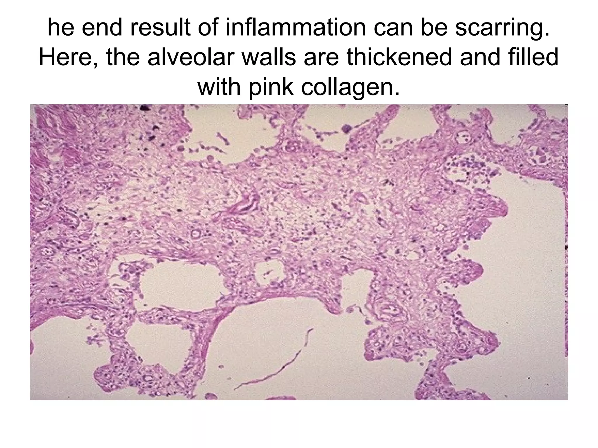
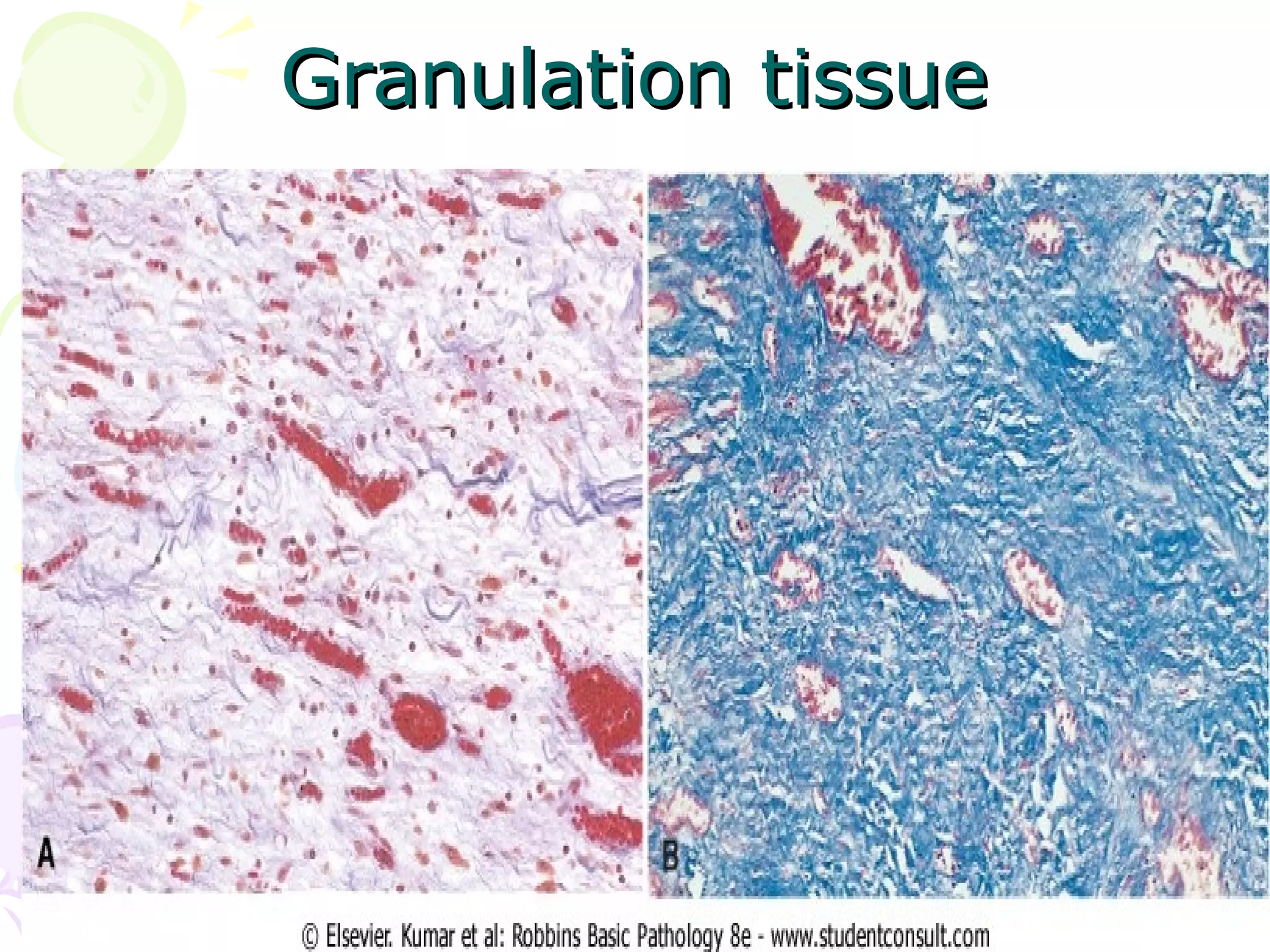
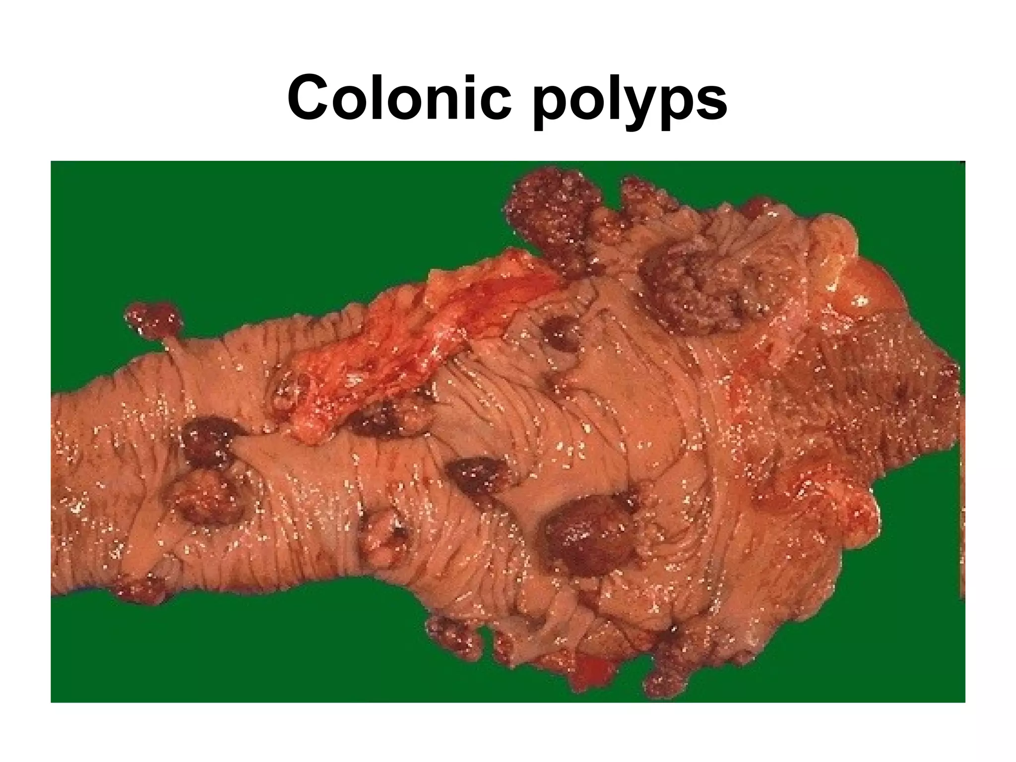
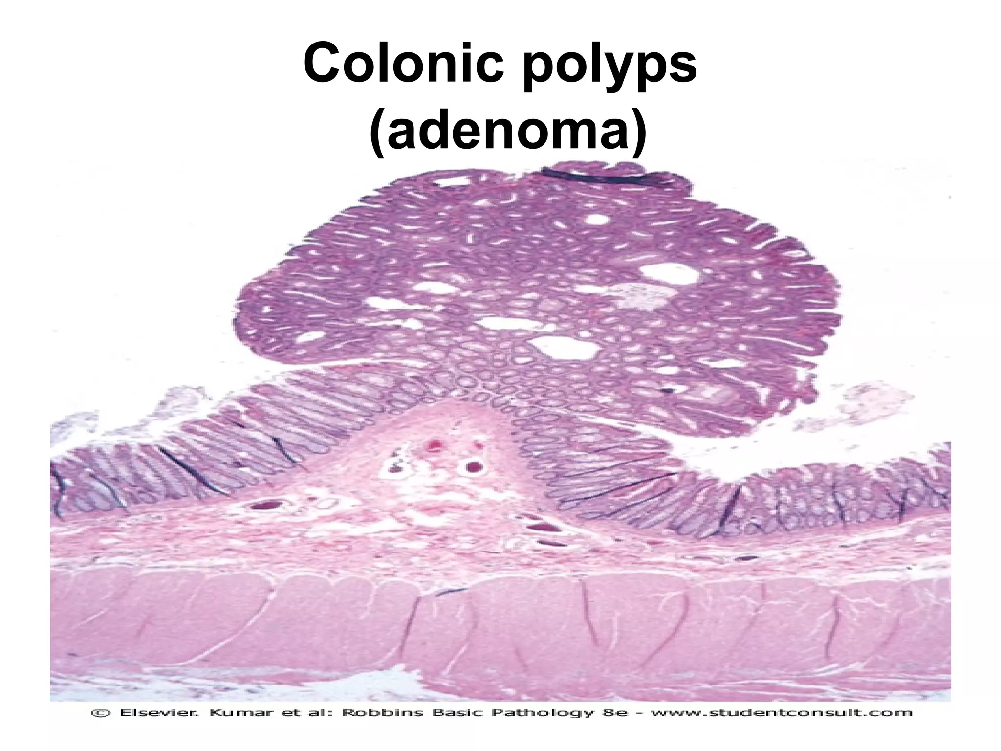
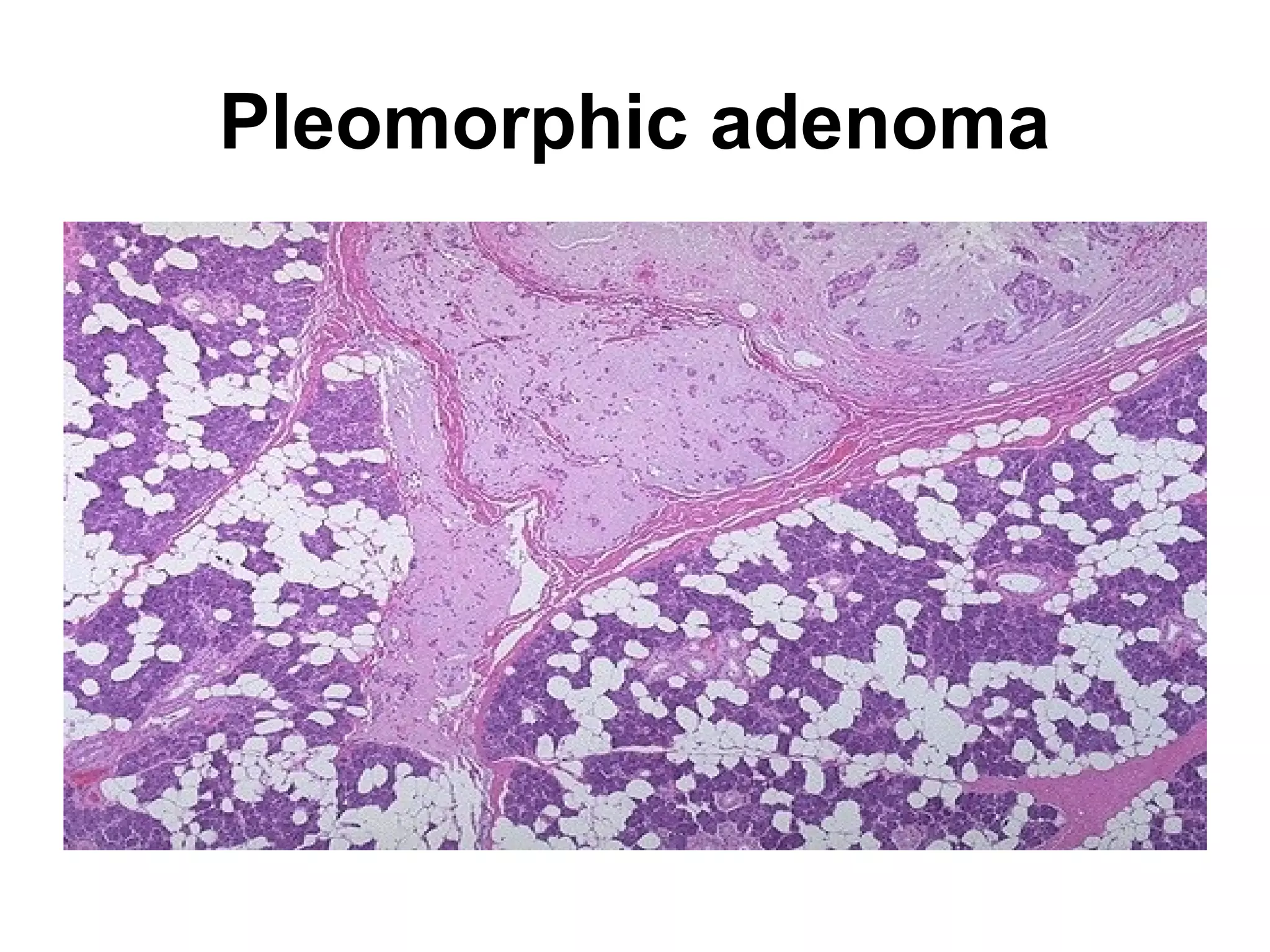
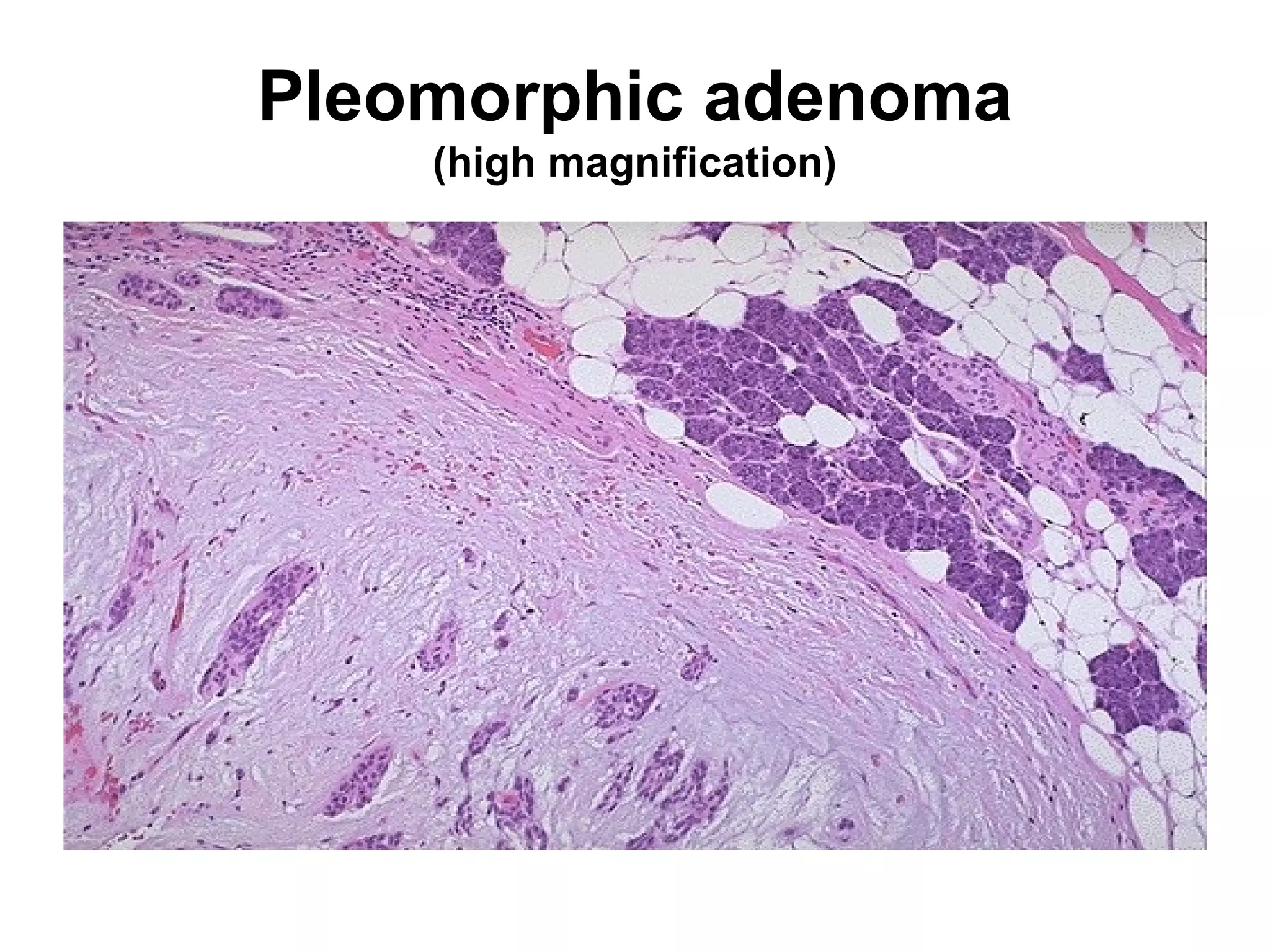
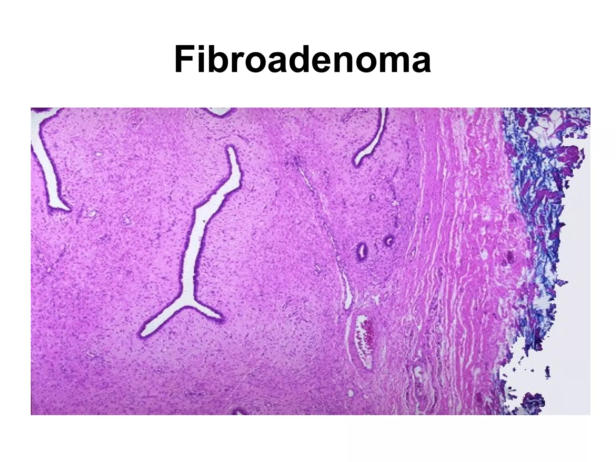
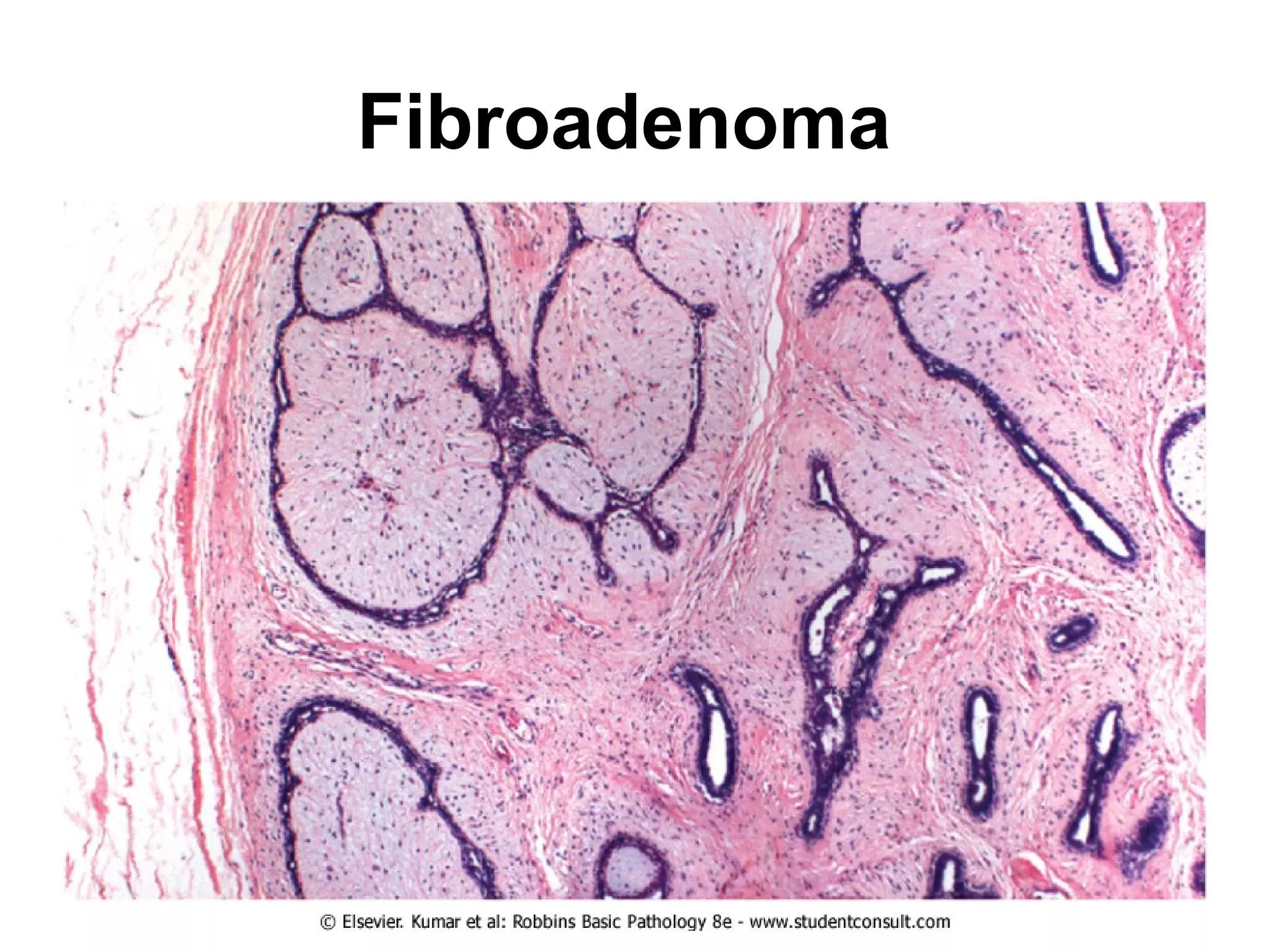
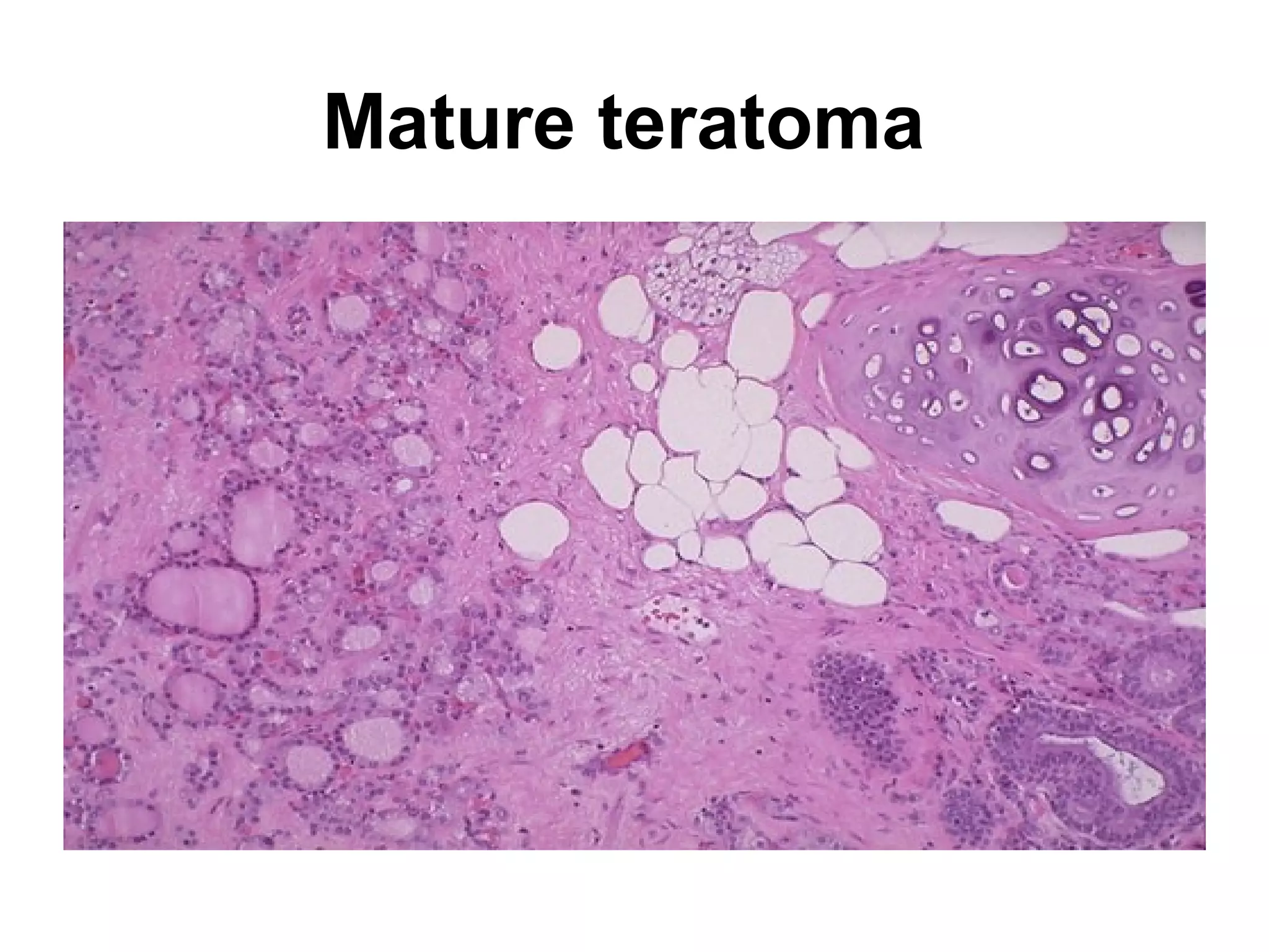
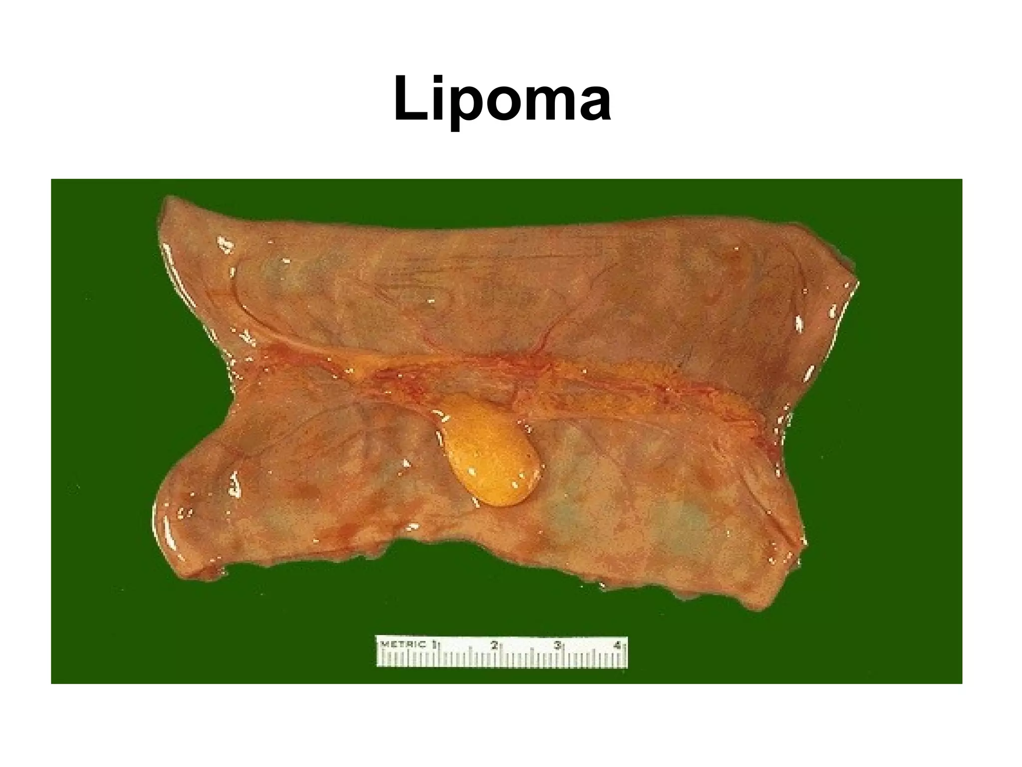
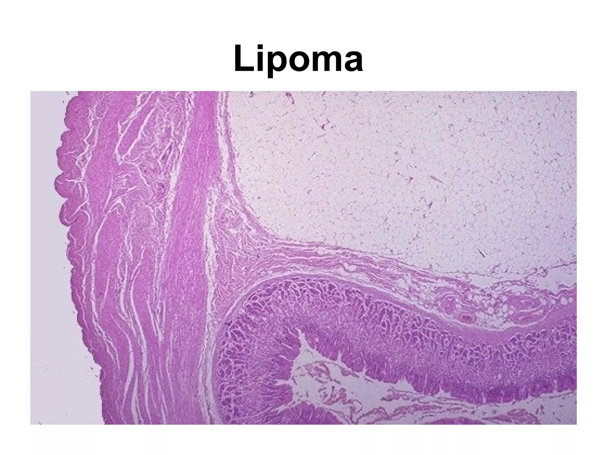
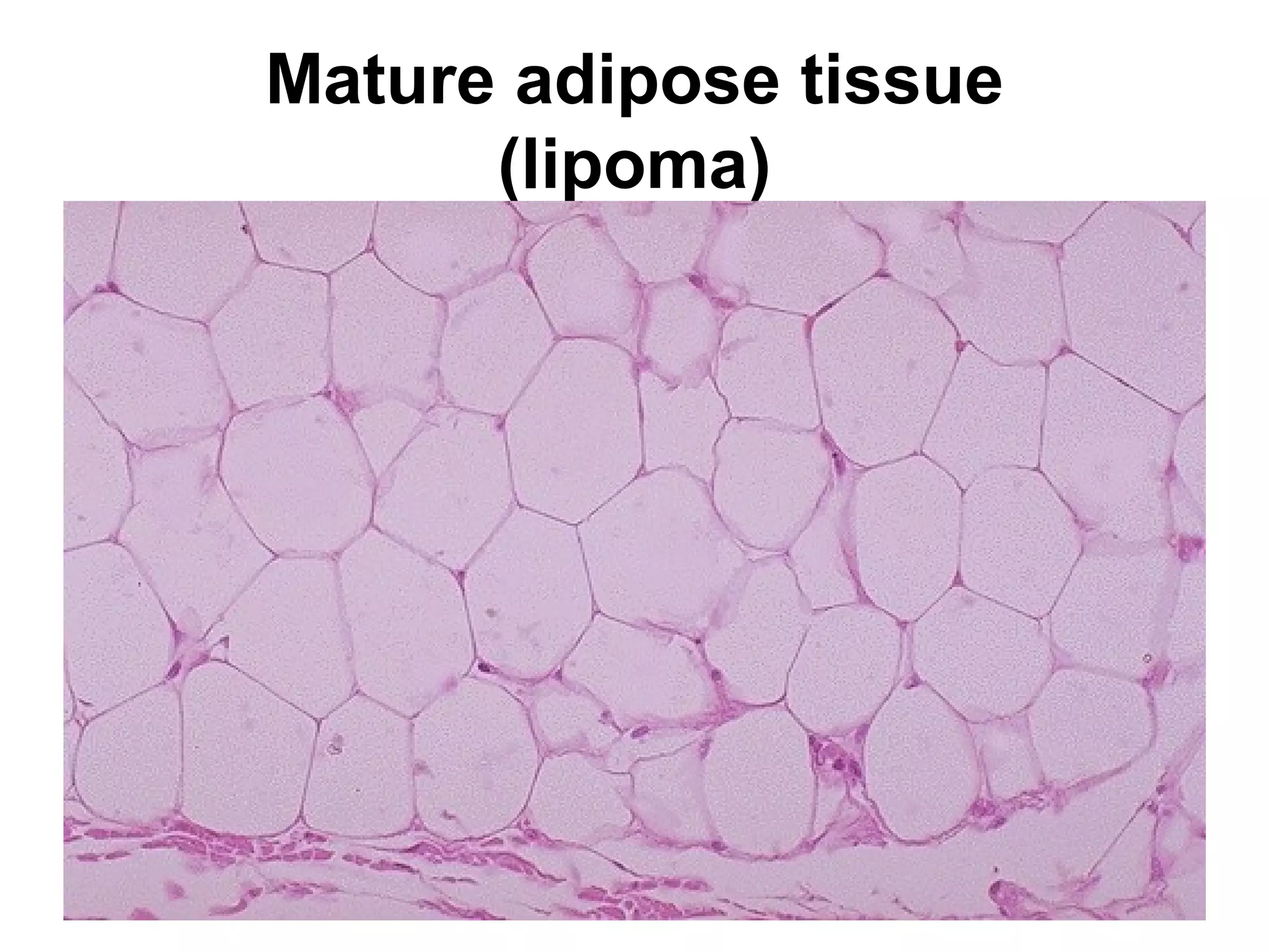
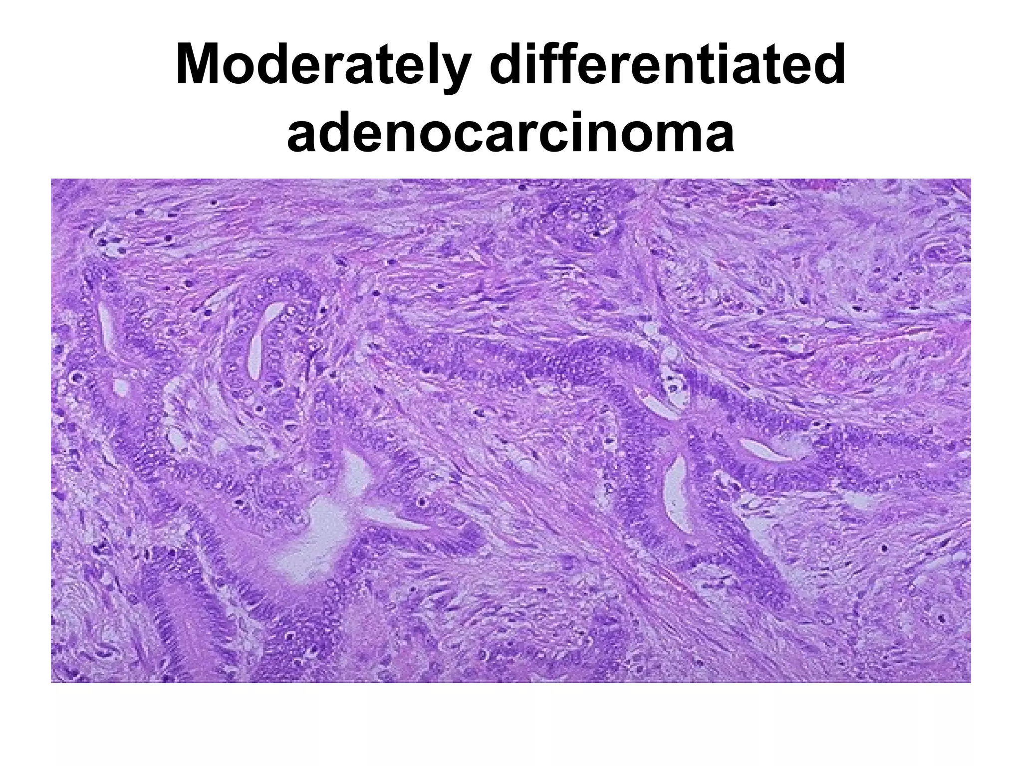
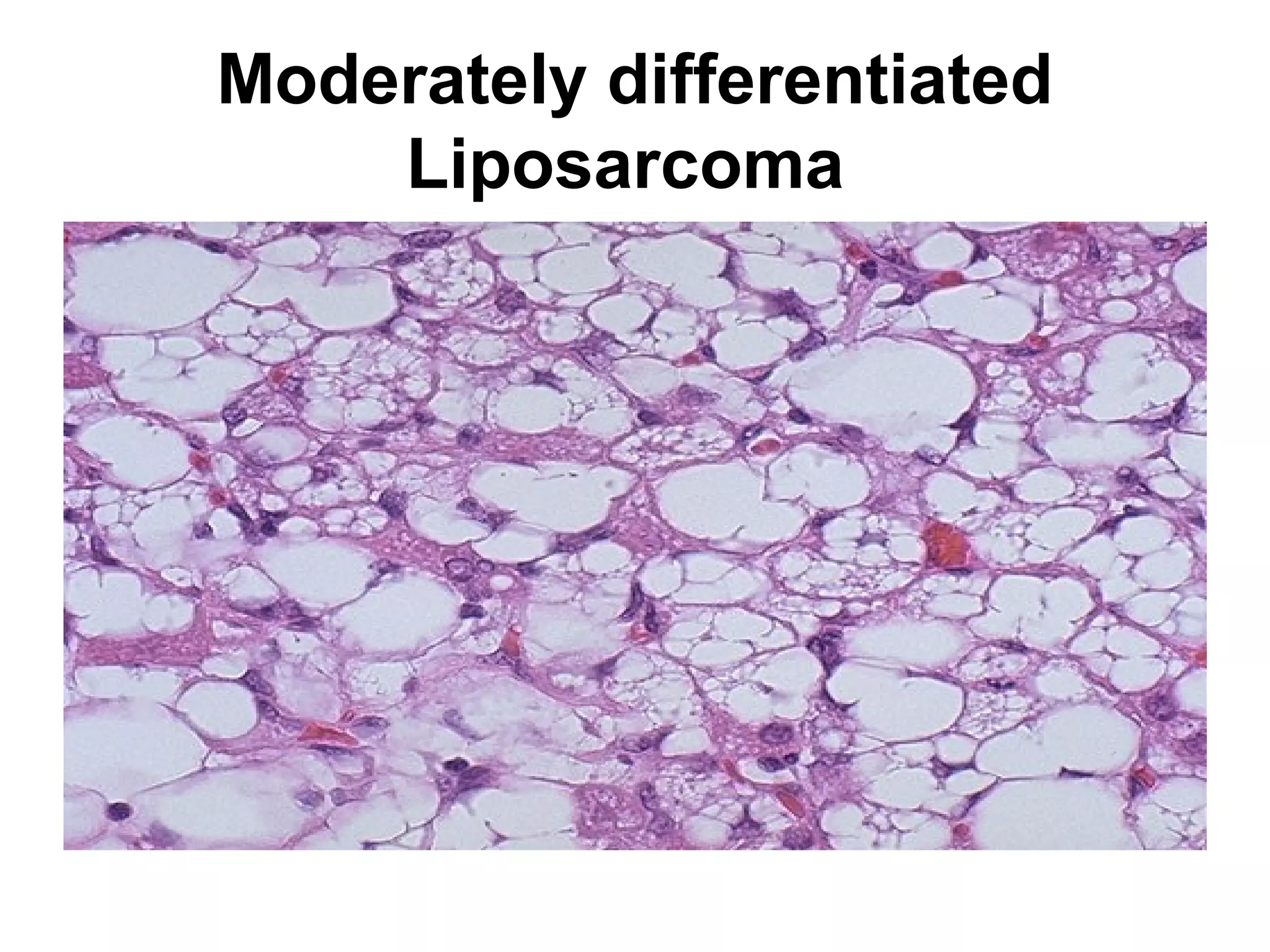
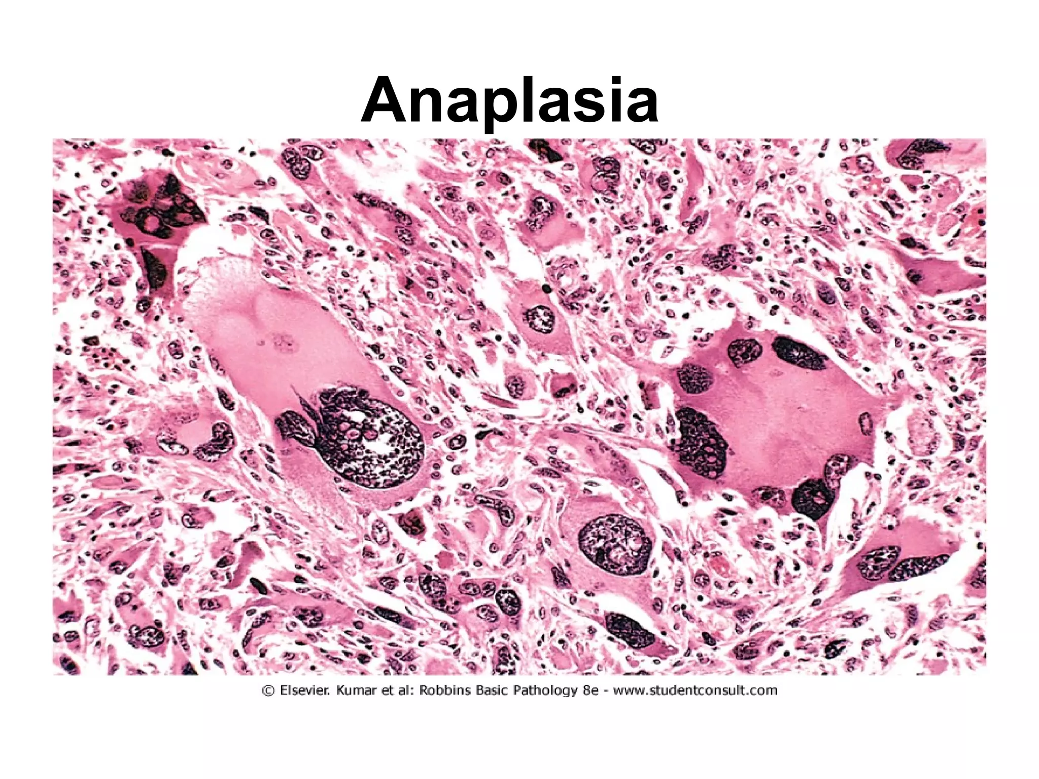
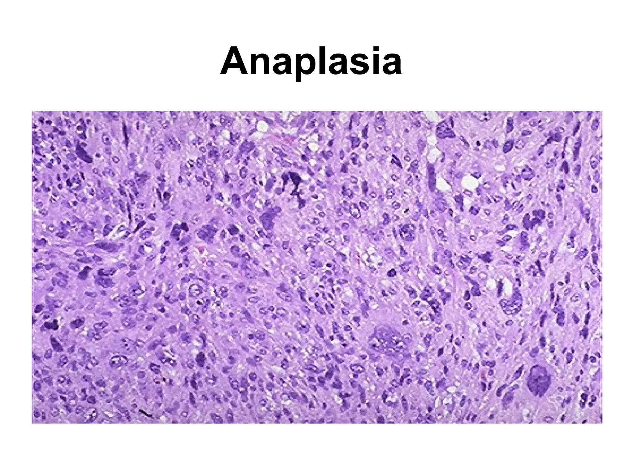
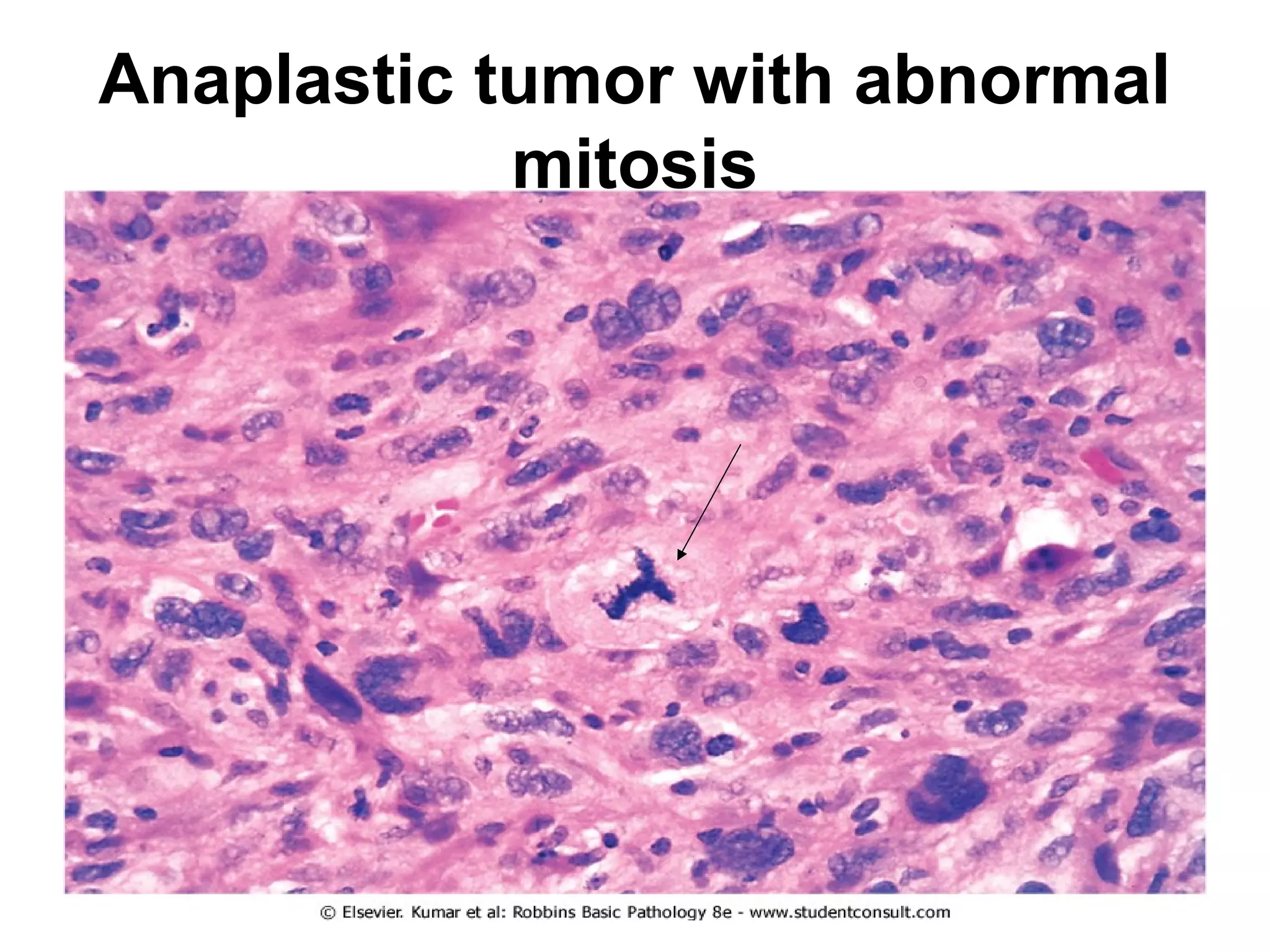
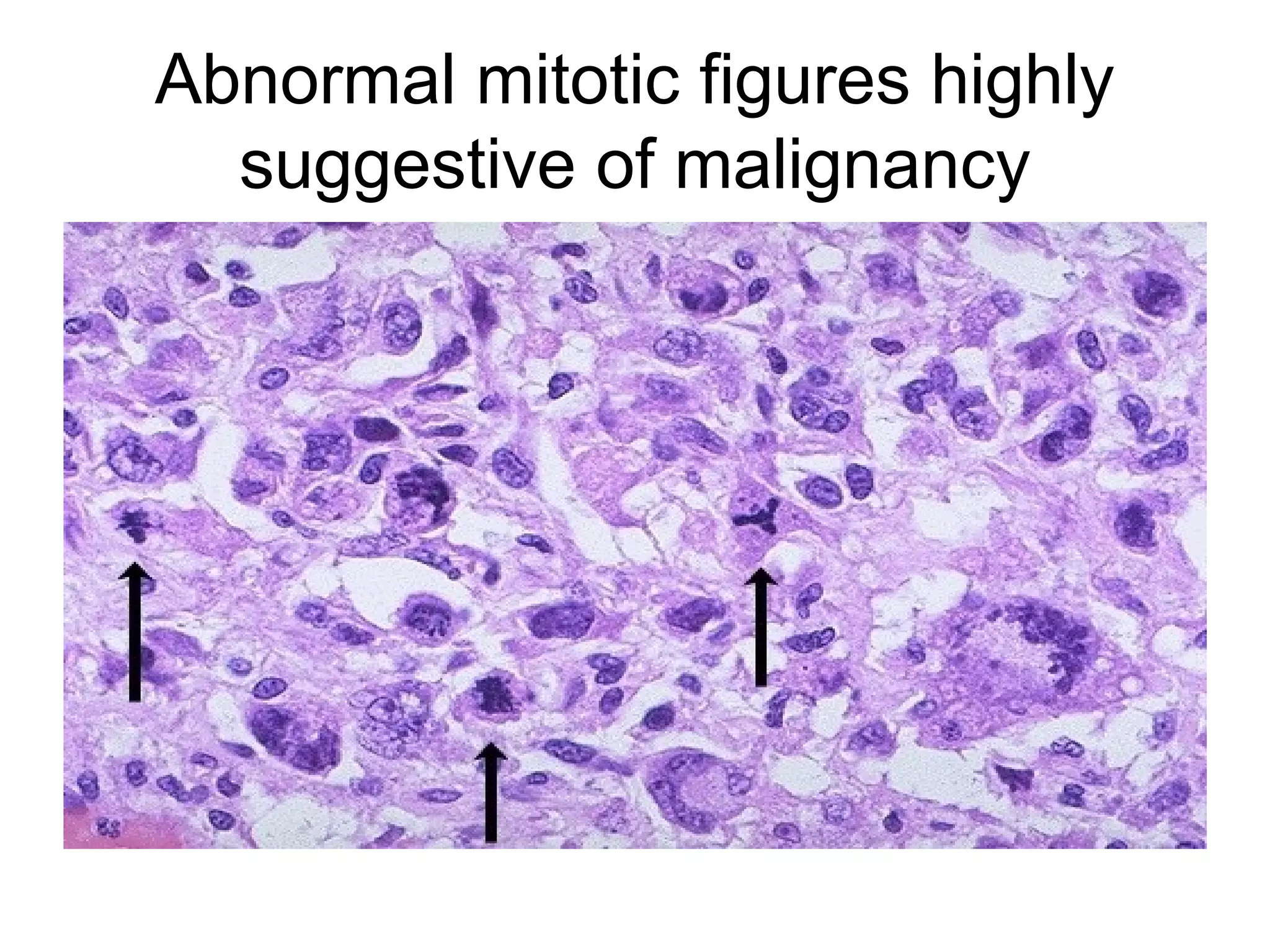
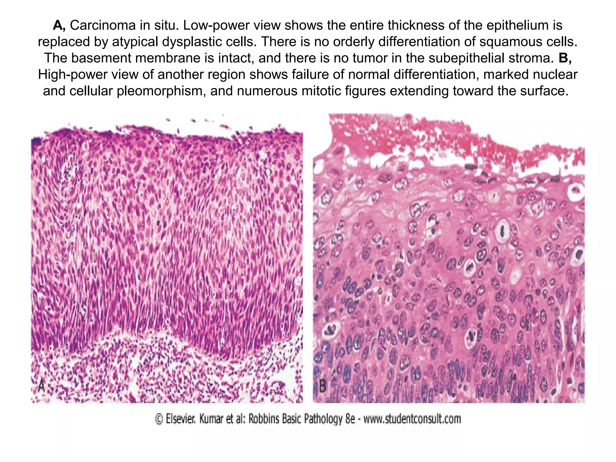
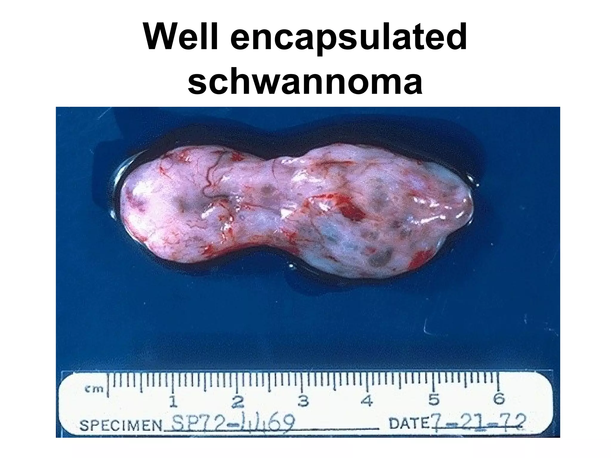
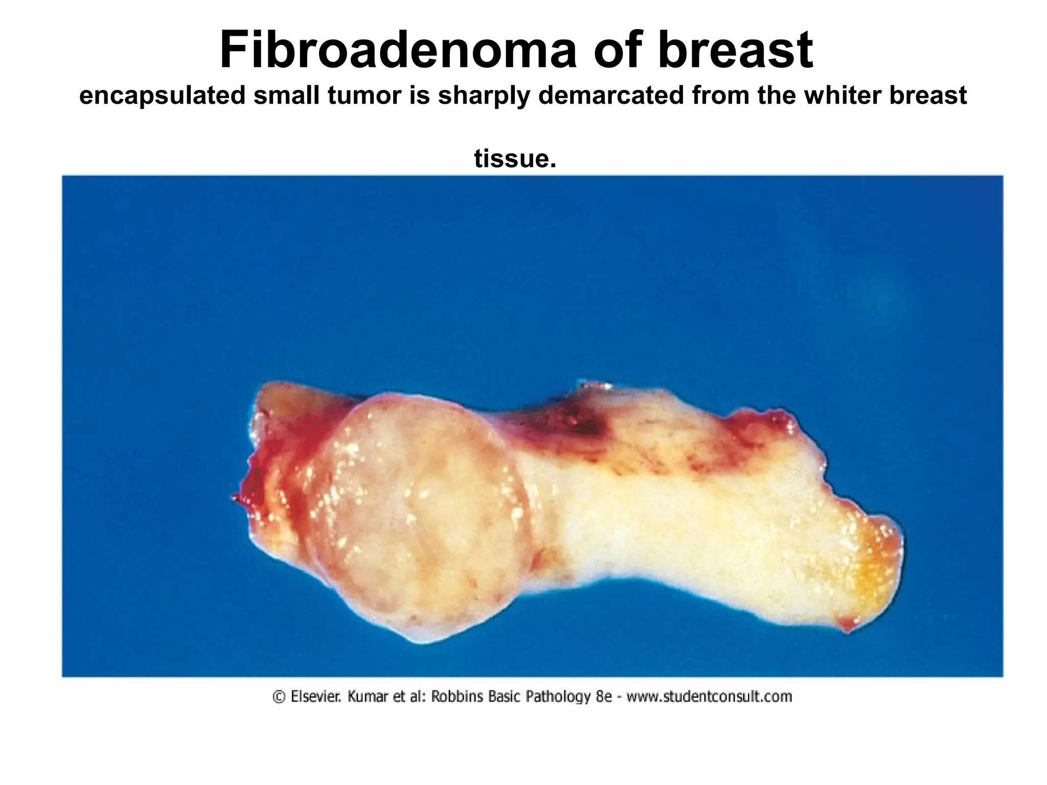
This document contains numerous pathology slides and descriptions of different types of cell and tissue changes seen microscopically. Some key points summarized:
1) Slides show examples of necrosis, inflammation, repair processes like granulation tissue and scarring, and various neoplastic conditions including benign tumors like lipomas and malignant tumors with areas of necrosis, abnormal mitosis, and anaplasia.
2) Specific examples include liver necrosis showing pale hepatocytes without nuclei, acute bronchopneumonia with neutrophils filling alveoli, and granulation tissue forming during repair.
3) Benign tumors discussed include pleomorphic adenomas, fibroadenomas, and mature teratomas, while malignant tumors shown
PMU third/fourth year Clinical pathonanatomy Part 1BenjiH
Part one of the full clinical anatomy micro and macroscopic slides.
this part covers the first 32 microscopic slide topics. For medical students Plovdiv medical university.
This slide is about secondary lymphoid organs. Majorly focusing on lymphnode, spleen and splenic circulation, tonsils, mucosal associated lymphoid tissue, appendix and their medical applications.
Indian Dental Academy: will be one of the most relevant and exciting training center with best faculty and flexible training programs for dental professionals who wish to advance in their dental practice,Offers certified courses in Dental implants,Orthodontics,Endodontics,Cosmetic Dentistry, Prosthetic Dentistry, Periodontics and General Dentistry.
Prix Galien International 2024 Forum ProgramLevi Shapiro
June 20, 2024, Prix Galien International and Jerusalem Ethics Forum in ROME. Detailed agenda including panels:
- ADVANCES IN CARDIOLOGY: A NEW PARADIGM IS COMING
- WOMEN’S HEALTH: FERTILITY PRESERVATION
- WHAT’S NEW IN THE TREATMENT OF INFECTIOUS,
ONCOLOGICAL AND INFLAMMATORY SKIN DISEASES?
- ARTIFICIAL INTELLIGENCE AND ETHICS
- GENE THERAPY
- BEYOND BORDERS: GLOBAL INITIATIVES FOR DEMOCRATIZING LIFE SCIENCE TECHNOLOGIES AND PROMOTING ACCESS TO HEALTHCARE
- ETHICAL CHALLENGES IN LIFE SCIENCES
- Prix Galien International Awards Ceremony
Factory Supply Best Quality Pmk Oil CAS 28578–16–7 PMK Powder in Stockrebeccabio
Factory Supply Best Quality Pmk Oil CAS 28578–16–7 PMK Powder in Stock
Telegram: bmksupplier
signal: +85264872720
threema: TUD4A6YC
You can contact me on Telegram or Threema
Communicate promptly and reply
Free of customs clearance, Double Clearance 100% pass delivery to USA, Canada, Spain, Germany, Netherland, Poland, Italy, Sweden, UK, Czech Republic, Australia, Mexico, Russia, Ukraine, Kazakhstan.Door to door service
Hot Selling Organic intermediates
Lung Cancer: Artificial Intelligence, Synergetics, Complex System Analysis, S...Oleg Kshivets
RESULTS: Overall life span (LS) was 2252.1±1742.5 days and cumulative 5-year survival (5YS) reached 73.2%, 10 years – 64.8%, 20 years – 42.5%. 513 LCP lived more than 5 years (LS=3124.6±1525.6 days), 148 LCP – more than 10 years (LS=5054.4±1504.1 days).199 LCP died because of LC (LS=562.7±374.5 days). 5YS of LCP after bi/lobectomies was significantly superior in comparison with LCP after pneumonectomies (78.1% vs.63.7%, P=0.00001 by log-rank test). AT significantly improved 5YS (66.3% vs. 34.8%) (P=0.00000 by log-rank test) only for LCP with N1-2. Cox modeling displayed that 5YS of LCP significantly depended on: phase transition (PT) early-invasive LC in terms of synergetics, PT N0—N12, cell ratio factors (ratio between cancer cells- CC and blood cells subpopulations), G1-3, histology, glucose, AT, blood cell circuit, prothrombin index, heparin tolerance, recalcification time (P=0.000-0.038). Neural networks, genetic algorithm selection and bootstrap simulation revealed relationships between 5YS and PT early-invasive LC (rank=1), PT N0—N12 (rank=2), thrombocytes/CC (3), erythrocytes/CC (4), eosinophils/CC (5), healthy cells/CC (6), lymphocytes/CC (7), segmented neutrophils/CC (8), stick neutrophils/CC (9), monocytes/CC (10); leucocytes/CC (11). Correct prediction of 5YS was 100% by neural networks computing (area under ROC curve=1.0; error=0.0).
CONCLUSIONS: 5YS of LCP after radical procedures significantly depended on: 1) PT early-invasive cancer; 2) PT N0--N12; 3) cell ratio factors; 4) blood cell circuit; 5) biochemical factors; 6) hemostasis system; 7) AT; 8) LC characteristics; 9) LC cell dynamics; 10) surgery type: lobectomy/pneumonectomy; 11) anthropometric data. Optimal diagnosis and treatment strategies for LC are: 1) screening and early detection of LC; 2) availability of experienced thoracic surgeons because of complexity of radical procedures; 3) aggressive en block surgery and adequate lymph node dissection for completeness; 4) precise prediction; 5) adjuvant chemoimmunoradiotherapy for LCP with unfavorable prognosis.
Couples presenting to the infertility clinic- Do they really have infertility...Sujoy Dasgupta
Dr Sujoy Dasgupta presented the study on "Couples presenting to the infertility clinic- Do they really have infertility? – The unexplored stories of non-consummation" in the 13th Congress of the Asia Pacific Initiative on Reproduction (ASPIRE 2024) at Manila on 24 May, 2024.
Explore natural remedies for syphilis treatment in Singapore. Discover alternative therapies, herbal remedies, and lifestyle changes that may complement conventional treatments. Learn about holistic approaches to managing syphilis symptoms and supporting overall health.
Anti ulcer drugs and their Advance pharmacology ||
Anti-ulcer drugs are medications used to prevent and treat ulcers in the stomach and upper part of the small intestine (duodenal ulcers). These ulcers are often caused by an imbalance between stomach acid and the mucosal lining, which protects the stomach lining.
||Scope: Overview of various classes of anti-ulcer drugs, their mechanisms of action, indications, side effects, and clinical considerations.
Title: Sense of Taste
Presenter: Dr. Faiza, Assistant Professor of Physiology
Qualifications:
MBBS (Best Graduate, AIMC Lahore)
FCPS Physiology
ICMT, CHPE, DHPE (STMU)
MPH (GC University, Faisalabad)
MBA (Virtual University of Pakistan)
Learning Objectives:
Describe the structure and function of taste buds.
Describe the relationship between the taste threshold and taste index of common substances.
Explain the chemical basis and signal transduction of taste perception for each type of primary taste sensation.
Recognize different abnormalities of taste perception and their causes.
Key Topics:
Significance of Taste Sensation:
Differentiation between pleasant and harmful food
Influence on behavior
Selection of food based on metabolic needs
Receptors of Taste:
Taste buds on the tongue
Influence of sense of smell, texture of food, and pain stimulation (e.g., by pepper)
Primary and Secondary Taste Sensations:
Primary taste sensations: Sweet, Sour, Salty, Bitter, Umami
Chemical basis and signal transduction mechanisms for each taste
Taste Threshold and Index:
Taste threshold values for Sweet (sucrose), Salty (NaCl), Sour (HCl), and Bitter (Quinine)
Taste index relationship: Inversely proportional to taste threshold
Taste Blindness:
Inability to taste certain substances, particularly thiourea compounds
Example: Phenylthiocarbamide
Structure and Function of Taste Buds:
Composition: Epithelial cells, Sustentacular/Supporting cells, Taste cells, Basal cells
Features: Taste pores, Taste hairs/microvilli, and Taste nerve fibers
Location of Taste Buds:
Found in papillae of the tongue (Fungiform, Circumvallate, Foliate)
Also present on the palate, tonsillar pillars, epiglottis, and proximal esophagus
Mechanism of Taste Stimulation:
Interaction of taste substances with receptors on microvilli
Signal transduction pathways for Umami, Sweet, Bitter, Sour, and Salty tastes
Taste Sensitivity and Adaptation:
Decrease in sensitivity with age
Rapid adaptation of taste sensation
Role of Saliva in Taste:
Dissolution of tastants to reach receptors
Washing away the stimulus
Taste Preferences and Aversions:
Mechanisms behind taste preference and aversion
Influence of receptors and neural pathways
Impact of Sensory Nerve Damage:
Degeneration of taste buds if the sensory nerve fiber is cut
Abnormalities of Taste Detection:
Conditions: Ageusia, Hypogeusia, Dysgeusia (parageusia)
Causes: Nerve damage, neurological disorders, infections, poor oral hygiene, adverse drug effects, deficiencies, aging, tobacco use, altered neurotransmitter levels
Neurotransmitters and Taste Threshold:
Effects of serotonin (5-HT) and norepinephrine (NE) on taste sensitivity
Supertasters:
25% of the population with heightened sensitivity to taste, especially bitterness
Increased number of fungiform papillae
micro teaching on communication m.sc nursing.pdfAnurag Sharma
Microteaching is a unique model of practice teaching. It is a viable instrument for the. desired change in the teaching behavior or the behavior potential which, in specified types of real. classroom situations, tends to facilitate the achievement of specified types of objectives.
Title: Sense of Smell
Presenter: Dr. Faiza, Assistant Professor of Physiology
Qualifications:
MBBS (Best Graduate, AIMC Lahore)
FCPS Physiology
ICMT, CHPE, DHPE (STMU)
MPH (GC University, Faisalabad)
MBA (Virtual University of Pakistan)
Learning Objectives:
Describe the primary categories of smells and the concept of odor blindness.
Explain the structure and location of the olfactory membrane and mucosa, including the types and roles of cells involved in olfaction.
Describe the pathway and mechanisms of olfactory signal transmission from the olfactory receptors to the brain.
Illustrate the biochemical cascade triggered by odorant binding to olfactory receptors, including the role of G-proteins and second messengers in generating an action potential.
Identify different types of olfactory disorders such as anosmia, hyposmia, hyperosmia, and dysosmia, including their potential causes.
Key Topics:
Olfactory Genes:
3% of the human genome accounts for olfactory genes.
400 genes for odorant receptors.
Olfactory Membrane:
Located in the superior part of the nasal cavity.
Medially: Folds downward along the superior septum.
Laterally: Folds over the superior turbinate and upper surface of the middle turbinate.
Total surface area: 5-10 square centimeters.
Olfactory Mucosa:
Olfactory Cells: Bipolar nerve cells derived from the CNS (100 million), with 4-25 olfactory cilia per cell.
Sustentacular Cells: Produce mucus and maintain ionic and molecular environment.
Basal Cells: Replace worn-out olfactory cells with an average lifespan of 1-2 months.
Bowman’s Gland: Secretes mucus.
Stimulation of Olfactory Cells:
Odorant dissolves in mucus and attaches to receptors on olfactory cilia.
Involves a cascade effect through G-proteins and second messengers, leading to depolarization and action potential generation in the olfactory nerve.
Quality of a Good Odorant:
Small (3-20 Carbon atoms), volatile, water-soluble, and lipid-soluble.
Facilitated by odorant-binding proteins in mucus.
Membrane Potential and Action Potential:
Resting membrane potential: -55mV.
Action potential frequency in the olfactory nerve increases with odorant strength.
Adaptation Towards the Sense of Smell:
Rapid adaptation within the first second, with further slow adaptation.
Psychological adaptation greater than receptor adaptation, involving feedback inhibition from the central nervous system.
Primary Sensations of Smell:
Camphoraceous, Musky, Floral, Pepperminty, Ethereal, Pungent, Putrid.
Odor Detection Threshold:
Examples: Hydrogen sulfide (0.0005 ppm), Methyl-mercaptan (0.002 ppm).
Some toxic substances are odorless at lethal concentrations.
Characteristics of Smell:
Odor blindness for single substances due to lack of appropriate receptor protein.
Behavioral and emotional influences of smell.
Transmission of Olfactory Signals:
From olfactory cells to glomeruli in the olfactory bulb, involving lateral inhibition.
Primitive, less old, and new olfactory systems with different path
3. Many cells have died that
the tissue is not
recognizable. Many nuclei
have become pyknotic
(shrunken and dark) and
have then undergone
karorrhexis
(fragmentation) and
karyolysis (dissolution).
The cytoplasm and cell
borders are no longer
recognizable. In this case,
loss of the blood supply
from a major coronary
artery led to ischemia and
cell death.
5. In these images you can see this preservation of architecture: this is quite recognizably liver,
despite the absence of nuclei in the hepatocytes in the affected area. Notice the thread-like
bacteria present in the sinusoids: these are the organism causing the lesion! Darker staining
viable hepatocytes are visible around the portal regions (right) and pale, necrotic hepatocytes are
visible in the central lobular zones. Hepatocytes near the blood supply have more physiologic
reserve and will be more able to fend off the infection than those in the oxygen and nutrient-poor
central region: but in this case the "advantage" is moot.
7. A typical gross pathology finding in a case like this would look similar to those
you see here. At left is an example of liquefactive necrosis in the brain, as seen
at autopsy in a coronal section. The area of the cerebral infarct is grossly
visible at the upper left, and can be highlighted by scrolling the cursor over the
image. Notice also the formation of a "clear" area (arrow) visible in the gross
specimen. Liquefactive necrosis of brain tissue in the infarcted area has
occurred. At right you see the clear area at low magnification in an H&E slide.
The region marked by the line is that which has been destroyed by cell death.
The rest of the field is normal brain tissue.
8. Figure 7.11 Fat necrosis, ordinary type. Multinucleated histiocytic giant cells surround a
large lipid vacuole formed by fusion of destroyed adipocytes. Scattered lymphocytes and
monocytes occupy expanded spaces between cells at top.
9. Intracellular accumulations of a variety of materials can occur in response to cellular
injury. Here is fatty metamorphosis (fatty change) of the liver in which deranged
lipoprotein transport from injury (most often alcoholism) leads to accumulation of lipid in
the cytoplasm of hepatocytes.
10. • 26. Lungs:
anthracotic pigment in
a patient with "black
lung" disease
• Note the heavy
deposition of black
pigment in this lung (see
arrow) from a patient
with coal worker s
pneumoconiosis (dust-
related pulmonary
disease). The large
spaces in the apex of
this lung represent
emphysema secondary
to damage associated to
the respiratory
bronchioles from the
excess anthracotic
pigment. "Black lung"
disease is a crippling
disease and is associated
with an increased
incidence of TB.
13. A Prussian blue reaction is seen in this iron stain of the liver to demonstrate large
amounts of hemosiderin that are present within the cytoplasm of the hepatocytes and
Kupffer cells. Ordinarily, only a small amount of hemosiderin would be present in the
fixed macrophage-like cells in liver, the Kupffer cells, as part of iron recycling.
14. Some of these skeletal muscle fibers here show atrophy, compared to normal fibers. The
number of cells is the same as before the atrophy occurred, but the size of some fibers
is reduced. This is a response to injury by "downsizing" to conserve the cell. In this case,
innervation to the small, atrophic fibers was lost. (This is a trichrome stain.)
22. there is marked laryngeal edema such that the airway is narrowed. This is life-
threatening. Thus, fluid collections can be serious depending upon their location.
24. The arm at the botton is swollen(edematous) &
reddened(erythematous)
25. fibrin mesh in fluid with PMN's that has formed in the
area of acute inflammation.
26. PMN's that are marginated along the dilated venule wall (arrow)
are squeezing through the basement membrane (the process of
diapedesis) and spilling out into extravascular space.
33. the pericardial cavity has been opened to reveal a fibrinous
pericarditis with strands of stringy pale fibrin between visceral
and parietal pericardium.
34. Microscopically, the fibrinous exudate is seen to
consist of pink strands of fibrin jutting from the
pericardial surface(arrows). Below this, there are a few
scattered inflammatory cells.
35. the yellowish fluid in this opened pericardial cavity is a
purulent exudate.
36. A purulent exudate is seen beneath the meninges in the brain of
this patient with acute meningitis from Streptococcus
pneumoniae infection. The exudate obscures the sulci.
37. extensive purulent peritonitis that resulted from rupture
of the colon. A thick yellow exudate coats the
peritoneal surfaces.
38. The white arrows mark areas of abscess formation in the upper
lobe of this lung. The liquefactive necrosis of an abscess is
apparent, because the purulent contents are draining out to
leave a cavity.
39. focal abscess in the lung. The
alveoli in that area have been
destroyed.
41. One consequence of acute inflammation is ulceration.
This occurs on epithelial surfaces. Here the gastric
mucosa has been lost, or ulcerated.
42. Below the vocal cords in this larynx are large ulcerations. Such
subglottic ulcers are produced with prolonged endotracheal
intubation in which the cuff of the endotracheal tube fits too tight
70. A, Carcinoma in situ. Low-power view shows the entire thickness of the epithelium is
replaced by atypical dysplastic cells. There is no orderly differentiation of squamous cells.
The basement membrane is intact, and there is no tumor in the subepithelial stroma. B,
High-power view of another region shows failure of normal differentiation, marked nuclear
and cellular pleomorphism, and numerous mitotic figures extending toward the surface.