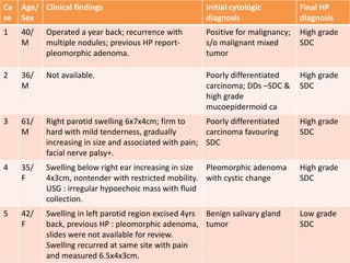
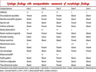
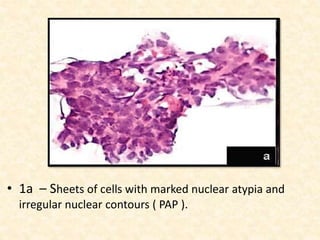
Bangalore Call Girls Hebbal Kempapura Number 7001035870 Meetin With Bangalor...
MY JC.pptx
1. Salivary Duct Carcinoma :
Correlation of morphologic
features by fine needle aspiration
cytology and histopathology
Moderator : DR. K. PUSHPALATHA PAI
Senior professor and hod
Presenter : Dr. Anu Sumi
Issac
2. Indian Journal of Pathology and Microbiology –
54 (1), January-March 2011.
Authors :
Rajesh N.G , Aruna K. Prayaga , Sundaram C.
Department of Pathology, Nizam’s Institute of
Medical Science, Hyderabad, India.
3. To review cytomorphological features
of histology proven SDC and evaluate
potential pitfalls in cytologic diagnosis
4. • An unusual but very aggressive type of salivary
gland carcinoma with a poor prognosis.
• Early diagnosis and treatment are important as
this tumor is known to metastasize early.
• SDC is histologically similar to intraductal and
infiltrating carcinoma of the breast.
5. • Comedonecrosis - common feature.
• Aggressive biologic behaviour with a high
incidence of lymph node metastasis, local
recurrence and significant mortality justify
categorisation of SDC as a high grade
malignancy in the current classification of
salivary gland neoplasms.
6. • This study was conducted by the Department of
Pathology, Nizam’s Institute of Medical Science,
Hyderabad.
• Analysis of the cytomorphological features in fine
needle aspiration cytology was done.
• The difficulties related to specific diagnosis and
differential diagnosis in five patients with
histologically proven SDC was studied.
7. • Five histologically confirmed cases of parotid
gland SDCs were identified from histopathology
files of the institute.
• FNAC slides and clinical details of the five patients
were retrieved from cytopathology files.
• FNAC was performed using 23- or 24- gauge
needles mounted on 10ml syringe.
8. • All the smears were air dried for May Grunwald
Giemsa stain and alcohol fixed for Papanicalaou
( PAP ) stain.
• All subsequent surgical specimens were fixed in
10% buffered formalin, embedded in paraffin and
sectioned at 5μm and stained with H&E and alcian
blue PAS stains.
9. • FNAC slides were quantified for the following : cellularity, fine
cytoplasmic vacuolation, abundant eosinophilic cytoplasm,
apocrine cells, indistinct cell borders, nuclear pleomorphism,
nuclear membrane irregularity, chromatin clumping, nuclear
eccentricity, metachromatic stroma, necrosis, cyst
macrophages, mitotic activity, papillary pattern, cribriform
configuration and three-dimensional clusters.
• Among these, cellularity, nuclear pleomorphism,
metachromatic stroma and mitotic activity were
semiquantitively assessed and the other parameters were
quantified for their presence or absence.
10. Semiquantitative scoring system
A . Cellularity :
Hypocellular smears +
Moderate cellularity ++
High cellularity +++
B. Nuclear pleomorphism :
Absent -
Minimal +
Marked pleomorphism ++
C. Metachromatic stroma :
Absent -
<2 low power field +
2-5 LPF ++
>5 LPF +++
D. Mitotic activity/HPF :
Absent -
Occasional +
Numerous ++
11. • Table 1 : Summary of the clinical findings,
initial cytologic and final histopathologic
diagnosis
Ca
se
Age/
Sex
Clinical findings Initial cytologic
diagnosis
Final HP
diagnosis
1 40/
M
Operated a year back; recurrence with
multiple nodules; previous HP report-
pleomorphic adenoma.
Positive for malignancy;
s/o malignant mixed
tumor
High grade
SDC
2 36/
M
Not available. Poorly differentiated
carcinoma; DDs –SDC &
high grade
mucoepidermoid ca
High grade
SDC
3 61/
M
Right parotid swelling 6x7x4cm; firm to
hard with mild tenderness, gradually
increasing in size and associated with pain;
facial nerve palsy+.
Poorly differentiated
carcinoma favouring
SDC
High grade
SDC
4 35/
F
Swelling below right ear increasing in size
4x3cm, nontender with restricted mobility.
USG : irregular hypoechoic mass with fluid
collection.
Pleomorphic adenoma
with cystic change
High grade
SDC
5 42/
F
Swelling in left parotid region excised 4yrs
back, previous HP : pleomorphic adenoma,
slides were not available for review.
Swelling recurred at same site with pain
and measured 6.5x4x3cm.
Benign salivary gland
tumor
Low grade
SDC
12.
13. • Interpreted cytologically as poorly differentiated carcinoma
with a possibility of SDC.
• Case 2 had a DD of high grade mucoepidermoid carcinoma.
• Cells were arranged in sheets and cohesive clusters with
abundant eosinophilic cytoplasm, indistinct cytoplasmic
boundaries, round to oval pleomorphic nuclei with coarsely
clumped chromatin, inconspicous nucleoli, and nuclear
membrane irregularity.
14. • The DD of mucoepidermoid carcinoma was
suggested in view of the younger age of the patient
( 36 yrs ) coupled with the presence of scattered
clusters of tumor cells with vacuolated cytoplasm
and rare intracytoplasmic mucin like droplets.
• Both the cases had bright metachromatic stroma
prominently seen in MGG stain.
15. • 1a – Sheets of cells with marked nuclear atypia and
irregular nuclear contours ( PAP ).
17. • Diagnosed as suggestive of malignant mixed tumor.
• 40yr/M who had undergone superficial parotidectomy
one year back at an outside hospital.
• HP report of the earlier surgical specimen revealed
pleomorphic adenoma. The original slides and blocks
were not available for review.
• There was a recurrence of multiple nodules at the
surgical site.
18. • FNAC showed cellular smears with sheets and three-
dimensional clusters of polygonal cells ( cribriform
pattern ). Nuclei were round to oval with coarsely
clumped chromatin, irregular nuclear membrane and
inconspicuous nucleoli. The cytoplasm showed fine
vacuolation in MGG stain. The cells were intimately
admixed with prominent granular metachromatic stroma.
21. • 35yr/F with swelling below the right ear and it was slowly
increasing in size. The swelling was nontender with
restricted mobility. There was no facial palsy.
• FNAC smears showed moderate cellularity with clusters
of round to oval eccentric nuclei admixed with numerous
cyst macrophages. MGG stained smears showed
prominent metachromatic stroma. Nuclear pleomorphism
was minimal.
22. • (b) cells in pseudopapillary clusters, bland nuclear morphology and myxoid matrix (MGG);
• (c) numerous cyst macrophages in a background of mucin (MGG);
• (d) apocrine cells arranged in cribriform pattern with mild nuclear pleomorphism (PAP)
23. • 42yr/F with recurrent swelling in the parotid region. The
patient had mild pain since few days. There was a history
of previous excision 4 years back at an outside hospital.
• The histopathology slides of the previous excision biopsy
were not available for review.
• FNAC done on the recurrent swelling showed moderately
cellular smears with sheets of cyst macrophages and
occasional clusters of cells with round to oval nuclei and
minimal nuclear pleomorphism. Many cells also showed
apocrine change. The background showed bright
metachromatic stroma in MGG stain.
24. • First four tumors had features of high grade SDC with
prominent comedonecrosis and high mitotic activity. Two
of the tumors revealed lymphatic and perineural invasion.
• Fifth case was low grade SDC, was cystic on gross
examination and showed intraductal proliferation
exhibiting cystic ducts with micropapillary, tufted and
plaque like intraluminal proliferation. Some of the ducts
were distended by solid or pseudocribriform proliferation
with varied cystic dilatation and exhibited mild nuclear
atypia. There were no goblet cells and mucin stains were
negative.
25. • SDC is an uncommon but distinctive type of salivary gland tumor
that mimics the histologic patterns of intraductal and infiltrating
ductal carcinoma of the breast.
• One of the most aggressive types of salivary gland carcinoma with a
marked tendency toward distant metastases and local recurrence.
• Kleinsasser et al.first described SDC in 1968. Patients commonly
present with rapidly growing mass, often with facial nerve
involvement, localized pain and cervical adenopathy.
• Approximately, 76% of the patients reported with this condition
have been men.
• The age range at presentation is 22-91 years with a peak incidence
in the sixth and seventh decades.
26. • Young age at diagnosis, primary tumor >3.0 cm and origin in
submandibular gland, all worsen the prognosis. Even with
aggressive therapy, prognosis is poor with a mortality rate of 60-
70%.
• On gross examination, the tumors are usually poorly circumscribed
and often multinodular, sometimes with scattered cysts and focal
areas of necrosis. They often induce fibrotic reaction.
• Mucicarmine and alcian blue PAS stains occasionally demonstrate
luminal or interstitial reactivity, but the tumor cells generally are
negative although Hui et al reported occasional intracellular mucin.
Perineural infiltration and lymphatic infiltration are frequently
present.
27. • Twenty five cases describing the cytomorphology of SDC
by FNAC have appeared in the literature.
• The cytomorphologic features described in these reports
include broad flat sheets of cells, tightly cohesive three-
dimensional clusters, papillary and cribriform
configurations, large polygonal or low columnar cells with
abundant, granular cytoplasm and eccentric,
hyperchromatic nuclei.
• Conversely, polygonal cells with round to oval
hypochromatic nuclei having prominent nucleoli have also
been described.
28. • Nuclear atypia has ranged from mild to moderate to
"malignant". A necrotic background has been observed in
some, but not all cases.
• A necrotic background has been observed in some cases.
• These authors included high grade mucoepidermoid
carcinoma, acinic cell carcinoma, oncocytic neoplasms,
papillary cystadenocarcinoma and polymorphous low
grade adenocarcinoma in the differential diagnosis.
29. • The authors concluded in retrospect that the presence of cribriform areas and a
background of necrosis should have prompted a consideration of SDC in the first
case. Oncocytic appearing areas in SDC are generally apocrine rather than being
truly oncocytic.
• The most useful feature for distinction of SDC from an oncocytic neoplasm by
FNAC is the presence of cribriform groups. Absence of cribriform and papillary
groups, however, leads to inconclusive diagnosis and, as noted in previous
reports, this feature is also more apparent on cell block material than in
histologic smears.
• High grade mucoepidermoid carcinoma was described by Elsheikh et al. as the
most difficult neoplasm to be distinguished cytologically from SDC. In the
absence of papillary groups or cribriform areas, one may not be able to
distinguish such an aspirate from high grade mucoepidermoid carcinoma or
squamous carcinoma.
30. • None of the three subsequent reports describing the
cytomorphologic findings on SDC confirmed the presence
of intranuclear inclusions or emphasized the importance
of nuclear eccentricity.
• In general, FNAC of parotid masses have a high accuracy
with over 99% specificity and 85% sensitivity.
• Immunohistochemistry (IHC) with AR, GCDFP-15 and p63,
as well as morphometric analysis of these tumors can be
very useful adjuncts in cytologic diagnosis of SDCs.
31. • Mucin rich variant of SDC with large pools of extracellular mucin has
been described and this variant carries poor prognosis.
• IHC with carcinoembryonic antigen (CEA), cytokeratins, S-100
protein, estrogen receptor (ER), progesterone receptor (PR) and
HER 2/neu have low sensitivity and specificity, while mucin antigen
profile MUC2, MUC5B, and MUC6 have shown positivity in mucin
rich variant.
• IHC studies have revealed a consistent expression of androgen
receptor (AR) and GCDFP-15. Negative expression of p63 is another
sensitive test in the diagnosis of SDC.
32. • Gal et al. described intranuclear inclusions and nuclear eccentricity
as a characteristic cytologic finding in SDC. He also described naked
nuclei with anisokaryosis, chromatin clumps and clear vacuolar
zones in his case report.
• None of our five cases demonstrated the presence of intranuclear
inclusions though nuclear eccentricity was observed in one of the
five cases.
• SDC is more common in men with a peak incidence in sixth and
seventh decades.
33. • In the current study, we observed that four of the five cases
were patients presenting at a younger age group and two were
females.
• The relative morphologic and clinical heterogeneity of SDC may
preclude specific cytologic diagnosis.
• Although adequate sampling may ensure diagnosis of a high
grade carcinoma with a possibility of SDC, the distinction of SDC
with mild nuclear atypia or low grade histology may be difficult
or impossible on the basis of cytomorphology alone.
• However, ancillary studies such as IHC with a panel of
antibodies, AR, GCDFP-15 and p63, are helpful in the definitive
diagnosis of SDC by FNAC.