Downloaded 200 times

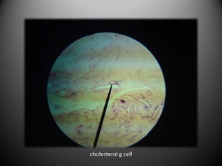
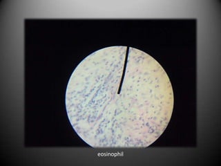
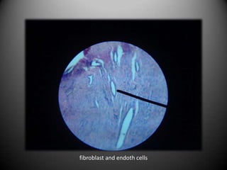
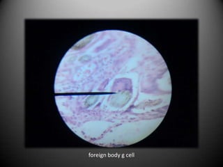
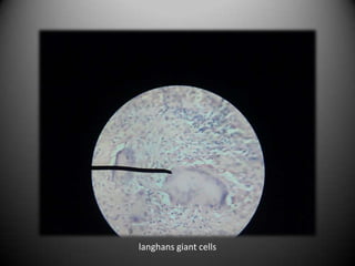
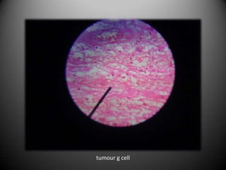
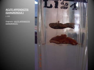
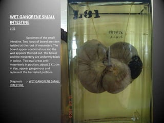
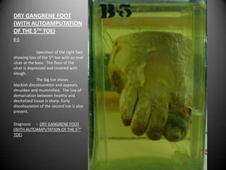
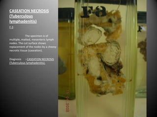
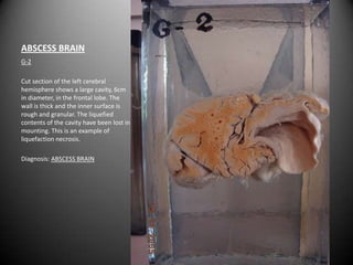
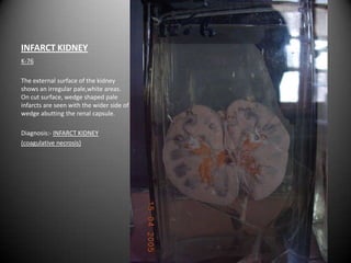
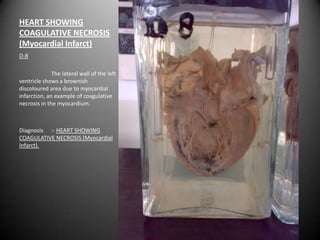
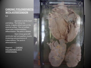
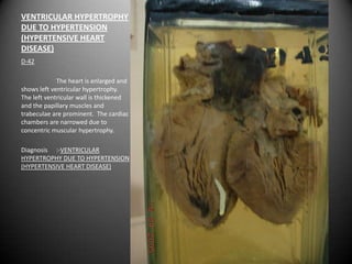
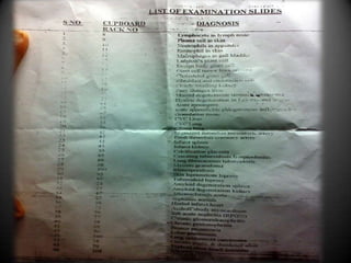
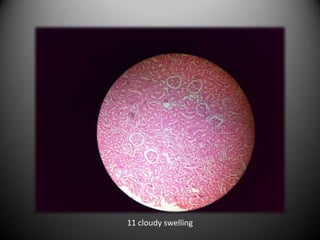
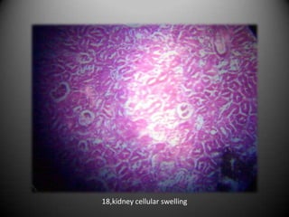
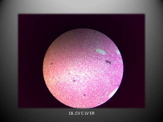
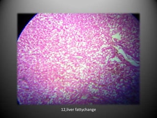
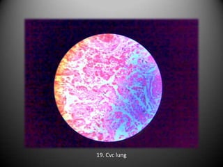
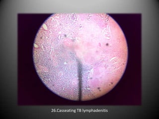
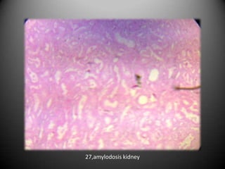
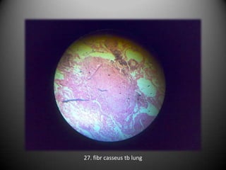
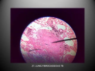
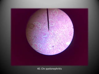

The document provides detailed descriptions of various pathological specimens and diagnoses, including cases of acute appendicitis, gangrene, caseation necrosis in tuberculous lymphadenitis, brain abscess, myocardial infarct, atherosclerosis, chronic venous congestion in the liver, tuberculosis in lungs, and chronic pyelonephritis with hypertension. Each described specimen includes observations and pathological findings relevant to the diagnoses. The document serves as a comprehensive overview of specific pathology examples, illustrating different forms of tissue necrosis and disease manifestations.