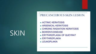
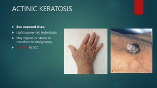
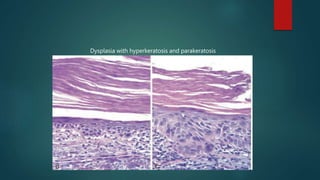
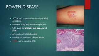
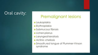
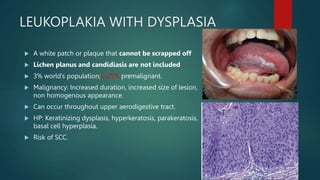
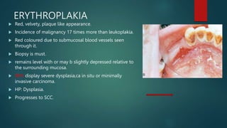
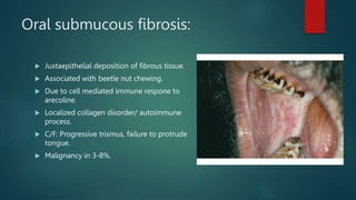
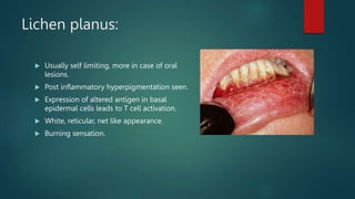
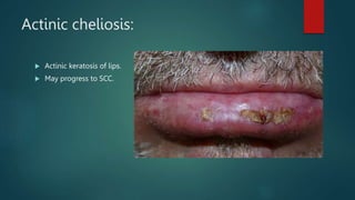
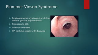
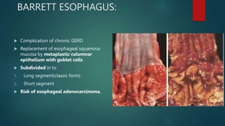
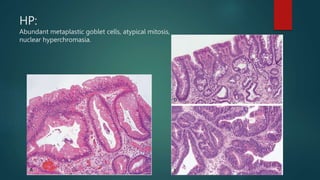
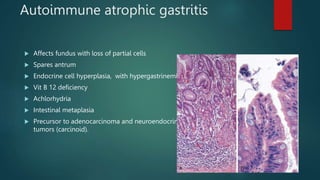
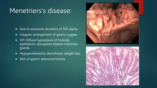
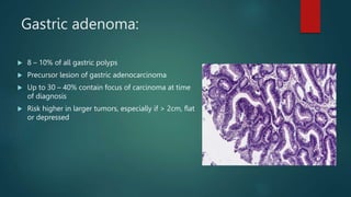
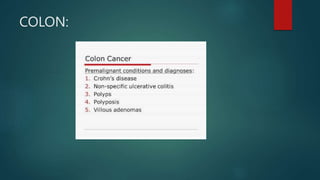
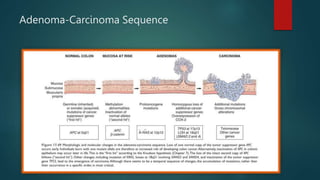
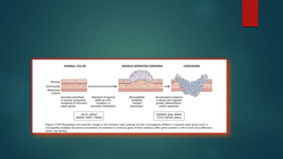
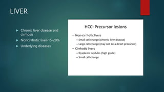
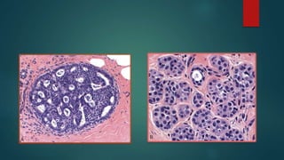
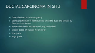
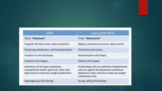
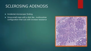
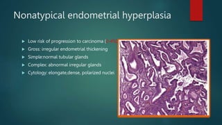
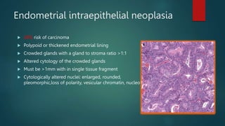
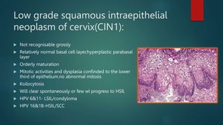
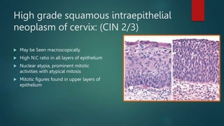
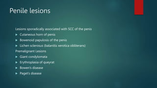
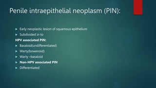
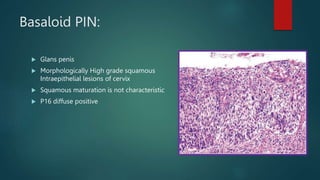
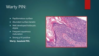
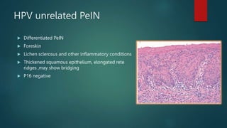
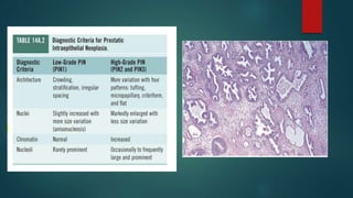
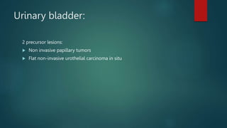
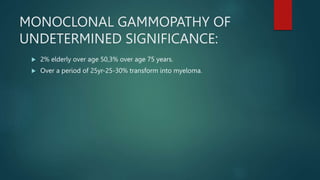
PRENEOPLASTIC
AND NEOPLASTIC
LESIONS.
DR. MOHAN MOHANTY
MODERATOR- DR.ROHAN AGRAWAL
Preneoplastic lesions:
A lesion involving abnormal cells , which are associated with an increased risk of developing into
cancer.
SKIN
ACTINIC KERATOSIS
 Sun exposed sites.
 Light pigmented individuals.
 May regress or stable or
transform to malignancy.
 0.1-10% to SCC
Dysplasia with hyperkeratosis and parakeratosis
BOWEN DISEASE:
 SCC in situ or squamous intraepithelial
neoplasia.
 Indolent scaly, erythematous plaques
 Site- non chronically sun exposured
skin.
 Atypical epithelial changes.
 Involve full thickness of epithelium.
 3-5% risk to develop SCC.
Arsenic keratosis:
 Cutaneous manifestation of chronic
arsenic toxicity.
 Corn like yellowish hyperkeratotic plaques
or papules.
 Affects palms and soles
 May progress to SCC.
 HP: Hyperkeratosis, parakeratosis,
acanthosis.
Chronic radiation keratosis:
 Change in skin appearance: Hypo and
hyperpigmentation, skin atrophy, loss of hair
appendages.
 HP: Hyperkeratosis.
 Occurs due to imbalance of proinflammatory
and profibrotic cytokines.
 Etiology: Radiotherapy inappropriate dose,
genetic factor, CT disorders.
 May occur after years of treatment.
GASTROINTESTINAL TRACT:
Oral cavity:
LEUKOPLAKIA WITH DYSPLASIA
 A white patch or plaque that cannot be scrapped off
 Lichen planus and candidiasis are not included
 3% world’s population;5-25% premalignant.
 Malignancy: Increased duration, increased size of lesion,
non homogenous appearance.
 Can occur throughout upper aerodigestive tract.
 HP: Keratinizing dysplasis, hyperkeratosis, parakeratosis,
basal cell hyperplasia.
 Risk of SCC.
ERYTHROPLAKIA
 Red, velvety, plaque like appearance.
 Incidence of malignancy 17 times more than leukoplakia.
 Red coloured due to submucosal blood vessels seen
through it.
 Biopsy is must.
 remains level with or may b slightly depressed relative to
the surrounding mucosa.
 90% display severe dysplasia,ca in situ or minimally
invasive carcinoma.
 HP: Dysplasia.
 Progresses to SCC.
Oral submucous fibrosis:
 Juxtaepithelial deposition of fibrous tissue.
 Associated with beetle nut chewing.
 Due to cell mediated immune respone to
arecoline.
 Localized collagen disorder/ autoimmune
process.
 C/F: Progressive trismus, failure to protrude
tongue.
 Malignancy in 3-8%.
Lichen planus:
 Usually self limiting, more in case of oral
lesions.
 Post inflammatory hyperpigmentation seen.
 Expression of altered antigen in basal
epidermal cells leads to T cell activation.
 White, reticular, net like appearance.
 Burning sensation.
Actinic cheliosis:
 Actinic keratosis of lips.
 May progress to SCC.
Plummer Vinson Syndrome:
 Esophageal webs , dysphagia, iron deficiency
anemia, glossitis, angular chelitis.
 Progresses to SCC.
 Common in females.
 HP: epithelial atrophy with dysplasia.
ESOPHAGUS AND STOMACH:
BARRETT ESOPHAGUS:
 Complication of chronic GERD
 Replacement of esophageal squamous
mucosa by metaplastic columnar
epithelium with goblet cells
 Subdivided in to
1. Long segment(classic form)
2. Short segment
 Risk of esophageal adenocarcinoma.
HP:
Abundant metaplastic goblet cells, atypical mitosis,
nuclear hyperchromasia.
Autoimmune atrophic gastritis
 Affects fundus with loss of partial cells
 Spares antrum
 Endocrine cell hyperplasia, with hypergastrinemia
 Vit B 12 deficiency
 Achlorhydria
 Intestinal metaplasia
 Precursor to adenocarcinoma and neuroendocrine
tumors (carcinoid).
Menetriers’s disease:
 Due to excessive secretion of TGF-alpha.
 Irregular arrangement of gastric ruggae.
 HP: Diffuse hyperplasia of foveolar
epithelium, elongated dilated corkscrew
glands.
 Hypoproteinemia, diarrohoea, weight loss.
 Risk of gastric adenocarcinoma.
Gastric adenoma:
 8 – 10% of all gastric polyps
 Precursor lesion of gastric adenocarcinoma
 Up to 30 – 40% contain focus of carcinoma at time
of diagnosis
 Risk higher in larger tumors, especially if > 2cm, flat
or depressed
COLON:
Adenoma-Carcinoma Sequence
Neoplastic polyps:
 Colorectal adenomas are characterized by presence of epithelial dysplasia.
 Hallmark-nuclear hyperchromasia, elongation and startifiaction.
 Reduction in number of goblet cells.
 Tubular,tubulovillous or villous.
 Sessile serrated lesions-lack typical cytologic features of dysplasia.
 Size is the most imp risk of malignancy.
Cont….
HNPCC (Lynch Syndrome):
 Type of cancer syndrome.
 Germline mutation in MMR gene
 80% of patients develop colorectal carcinoma
 Also risk for: endometrial cancer(33%),ovarian carcinoma(5%), upper
urinary tract, other GI cancers.
 More often right colon and arises from adenomas
FAP
 AD, defect in APC gene.
 Numerous colorectal adenomas as
teenagers.
 Atleast 100 polyps are necessary for
diagnosis –Classic FAP
 Colorectal adenocarcinoma
develops in 100% in untreated
cases,
Colitis associated neoplasia:
 Long term Ulcerative colitis and colonic crohn disease.
 Risk of dysplasia and subsequent cancer related to
1. Duration of the disease
2. Extent of the disease
3. Nature of inflammatory response
Liver:
PRECANCEROUS LESION
LIVER
 Chronic liver disease and
cirrhosis
 Noncirrhotic liver-15-20%
 Underlying diseases
Small cell changes
Large cell changes
Dysplastic
changes
Lungs & Pleura:
PRECANCEROUS LESIONS
Asbestos related diseases
Family of pro inflammatory crystalline hydrated silicates
Asso with pulmonary fibrosis and various cancers
Lung carcinoma
Malignant mesothelioma
Laryngeal, ovarian , other extrapulmonary neoplasm including colon
carcinoma
90%mesothelioma are asbestos related
Life time risk of developing mesothelioma 7-10%
Breast:
PRECANCEROUS LESION
BREAST
USUAL DUCTAL HYPERPLASIA
 Benign epithelial proliferation
 Polyclonal
 Cohesive proliferation with haphazard architecture,
irregular ,slit like lumina ,often peripherally located
streaming,syncytial pattern
 Variable sized cells with indistinct borders
 Overlapping nuclei
ATYPICAL DUCTAL HYPERPLASIA
(ADH)
 Resembling DCIS but less developed in architecture and extend
 Size- <2mm and <2duct spaces
 Cells-evenly spaced monotonous cells,round nuclei with dense chromatin
DUCTAL CARCINOMA IN SITU
 Often detected on mammography
 Clonal proliferation of epithelial cells limited to ducts and lobules by
basement membrane.
 Myoepithelial cells are preserved ,may diminished
 Graded based on nucleus morphology
 Low grade
 High grade
SCLEROSING ADENOSIS
 Incidental microscopic finding
 Gross:small mass with a disk like , multinodular
configuration that cuts with increase resistance
Female genital tract
PRECANCEROUS LESION
Nonatypical endometrial hyperplasia
 Low risk of progression to carcinoma (2-4%)
 Gross: irregular endometrial thickening
 Simple:normal tubular glands
 Complex: abnormal irregular glands
 Cytology: elongate,dense, polarized nuclei
Endometrial intraepithelial neoplasia
 38% risk of carcinoma
 Polypoid or thickened endometrial lining
 Crowded glands with a gland to stroma ratio >1:1
 Altered cytology of the crowded glands
 Must be >1mm with in single tissue fragment
 Cytologically altered nuclei: enlarged, rounded,
pleomorphic,loss of polarity, vesicular chromatin, nucleoli
Low grade squamous intraepithelial
neoplasm of cervix(CIN1):
 Not recognisable grossly
 Relatively normal basal cell layer,hyperplastic parabasal
layer
 Orderly maturation
 Mitotic activities and dysplasia confinded to the lower
third of epithelium.no abnormal mitosis
 Koilocytosis
 Will clear spontaneously or few wl progress to HSIL
 HPV 6&11- LSIL/condyloma
 HPV 16&18-HSIL/SCC
High grade squamous intraepithelial
neoplasm of cervix: (CIN 2/3)
 May be Seen macroscopically
 High N:C ratio in all layers of epithelium
 Nuclear atypia, prominent mitotic
activities with atypical mitosis
 Mitotic figures found in upper layers of
epithelium
Vulva
Penis
PRECANCEROUS LESION
Penile lesions
Lesions sporadically associated with SCC of the penis
 Cutaneous horn of penis
 Bowenoid papulosis of the penis
 Lichen sclerosus (balanitis xerotica obliterans)
Premalignant Lesions
 Giant condylomata
 Erythroplasia of queyrat
 Bowen’s disease
 Paget’s disease
Penile intraepithelial neoplasm (PIN):
 Early neoplastic lesion of squamous epithelium
 Subdivided in to
HPV associated PIN:
 Basaloid(undifferentiated)
 Warty(bowenoid)
 Warty –basaloid
 Non-HPV associated PIN
 Differentiated
Basaloid PIN:
 Glans penis
 Morphologically High grade squamous
Intraepithelial lesions of cervix
 Squamous maturation is not characteristic
 P16 diffuse positive
Warty PIN:
 Papillomatous surface
 Abundant surface keratin
 Well developed koilocytic
atypia
 Frequent squamous
maturation
 Strong p16 positive
Warty- basaloid PIN.
HPV unrelated PeIN
 Differentiated PeIN
 Foreskin
 Lichen sclerosus and other inflammatory conditions
 Thickened squamous epithelium, elongated rete
ridges ,may show bridging
 P16 negative
Germ cell neoplasia in situ(GCNIS):
 80%-Residual testis in organs harbouring an invasive germ cell malignancy
 5%-C/L testis.
 Restricted to seminiferous tubules,also rete testies-pagetoid appearance
 Atypical germ cell at base of seminiferous tubules.
 Spermatogenesis absent.
Prostate:
2 premalignant Lesions:
 Prostatic intraepithelial Neoplasia (PIN)
(Intraductal dysplasia, primary atypia hyperplasia, large acinar dysplasia
,acinar- ductal dysplasia)
 Atypical adenomatous hyperplasia (AAH)
(adenosis, small gland hyperplasia)
Urinary bladder:
2 precursor lesions:
 Non invasive papillary tumors
 Flat non-invasive urothelial carcinoma in situ
BONE:
 Genetic syndromes.
 Chronic osteomyelitis.
 Paget disease.
 Giant cell tumor
 Irradiation
 Use of metal orthopaedic devices.
Hematogenous:
MONOCLONAL GAMMOPATHY OF
UNDETERMINED SIGNIFICANCE:
 2% elderly over age 50,3% over age 75 years.
 Over a period of 25yr-25-30% transform into myeloma.
MYELODYSPLASTIC SYNDROMES:
 Clonal hematopoietic stem cell disorders characterized by cytopenias,
dysplasia in one or more of the major myeloid cell lines, ineffective
erythropoiesis with cellular marrow .
 Transformation to acute leukemia :30-35%
premalignant lesions new.pptx

premalignant lesions new.pptx

  • 1.
    PRENEOPLASTIC AND NEOPLASTIC LESIONS. DR. MOHANMOHANTY MODERATOR- DR.ROHAN AGRAWAL
  • 2.
    Preneoplastic lesions: A lesioninvolving abnormal cells , which are associated with an increased risk of developing into cancer.
  • 3.
  • 4.
    ACTINIC KERATOSIS  Sunexposed sites.  Light pigmented individuals.  May regress or stable or transform to malignancy.  0.1-10% to SCC
  • 5.
  • 6.
    BOWEN DISEASE:  SCCin situ or squamous intraepithelial neoplasia.  Indolent scaly, erythematous plaques  Site- non chronically sun exposured skin.  Atypical epithelial changes.  Involve full thickness of epithelium.  3-5% risk to develop SCC.
  • 7.
    Arsenic keratosis:  Cutaneousmanifestation of chronic arsenic toxicity.  Corn like yellowish hyperkeratotic plaques or papules.  Affects palms and soles  May progress to SCC.  HP: Hyperkeratosis, parakeratosis, acanthosis.
  • 8.
    Chronic radiation keratosis: Change in skin appearance: Hypo and hyperpigmentation, skin atrophy, loss of hair appendages.  HP: Hyperkeratosis.  Occurs due to imbalance of proinflammatory and profibrotic cytokines.  Etiology: Radiotherapy inappropriate dose, genetic factor, CT disorders.  May occur after years of treatment.
  • 9.
  • 10.
  • 11.
    LEUKOPLAKIA WITH DYSPLASIA A white patch or plaque that cannot be scrapped off  Lichen planus and candidiasis are not included  3% world’s population;5-25% premalignant.  Malignancy: Increased duration, increased size of lesion, non homogenous appearance.  Can occur throughout upper aerodigestive tract.  HP: Keratinizing dysplasis, hyperkeratosis, parakeratosis, basal cell hyperplasia.  Risk of SCC.
  • 12.
    ERYTHROPLAKIA  Red, velvety,plaque like appearance.  Incidence of malignancy 17 times more than leukoplakia.  Red coloured due to submucosal blood vessels seen through it.  Biopsy is must.  remains level with or may b slightly depressed relative to the surrounding mucosa.  90% display severe dysplasia,ca in situ or minimally invasive carcinoma.  HP: Dysplasia.  Progresses to SCC.
  • 13.
    Oral submucous fibrosis: Juxtaepithelial deposition of fibrous tissue.  Associated with beetle nut chewing.  Due to cell mediated immune respone to arecoline.  Localized collagen disorder/ autoimmune process.  C/F: Progressive trismus, failure to protrude tongue.  Malignancy in 3-8%.
  • 14.
    Lichen planus:  Usuallyself limiting, more in case of oral lesions.  Post inflammatory hyperpigmentation seen.  Expression of altered antigen in basal epidermal cells leads to T cell activation.  White, reticular, net like appearance.  Burning sensation.
  • 15.
    Actinic cheliosis:  Actinickeratosis of lips.  May progress to SCC.
  • 16.
    Plummer Vinson Syndrome: Esophageal webs , dysphagia, iron deficiency anemia, glossitis, angular chelitis.  Progresses to SCC.  Common in females.  HP: epithelial atrophy with dysplasia.
  • 17.
  • 18.
    BARRETT ESOPHAGUS:  Complicationof chronic GERD  Replacement of esophageal squamous mucosa by metaplastic columnar epithelium with goblet cells  Subdivided in to 1. Long segment(classic form) 2. Short segment  Risk of esophageal adenocarcinoma.
  • 19.
    HP: Abundant metaplastic gobletcells, atypical mitosis, nuclear hyperchromasia.
  • 20.
    Autoimmune atrophic gastritis Affects fundus with loss of partial cells  Spares antrum  Endocrine cell hyperplasia, with hypergastrinemia  Vit B 12 deficiency  Achlorhydria  Intestinal metaplasia  Precursor to adenocarcinoma and neuroendocrine tumors (carcinoid).
  • 21.
    Menetriers’s disease:  Dueto excessive secretion of TGF-alpha.  Irregular arrangement of gastric ruggae.  HP: Diffuse hyperplasia of foveolar epithelium, elongated dilated corkscrew glands.  Hypoproteinemia, diarrohoea, weight loss.  Risk of gastric adenocarcinoma.
  • 22.
    Gastric adenoma:  8– 10% of all gastric polyps  Precursor lesion of gastric adenocarcinoma  Up to 30 – 40% contain focus of carcinoma at time of diagnosis  Risk higher in larger tumors, especially if > 2cm, flat or depressed
  • 23.
  • 24.
  • 26.
    Neoplastic polyps:  Colorectaladenomas are characterized by presence of epithelial dysplasia.  Hallmark-nuclear hyperchromasia, elongation and startifiaction.  Reduction in number of goblet cells.  Tubular,tubulovillous or villous.  Sessile serrated lesions-lack typical cytologic features of dysplasia.  Size is the most imp risk of malignancy.
  • 27.
  • 28.
    HNPCC (Lynch Syndrome): Type of cancer syndrome.  Germline mutation in MMR gene  80% of patients develop colorectal carcinoma  Also risk for: endometrial cancer(33%),ovarian carcinoma(5%), upper urinary tract, other GI cancers.  More often right colon and arises from adenomas
  • 29.
    FAP  AD, defectin APC gene.  Numerous colorectal adenomas as teenagers.  Atleast 100 polyps are necessary for diagnosis –Classic FAP  Colorectal adenocarcinoma develops in 100% in untreated cases,
  • 30.
    Colitis associated neoplasia: Long term Ulcerative colitis and colonic crohn disease.  Risk of dysplasia and subsequent cancer related to 1. Duration of the disease 2. Extent of the disease 3. Nature of inflammatory response
  • 31.
  • 32.
    LIVER  Chronic liverdisease and cirrhosis  Noncirrhotic liver-15-20%  Underlying diseases
  • 33.
    Small cell changes Largecell changes Dysplastic changes
  • 34.
  • 35.
    Asbestos related diseases Familyof pro inflammatory crystalline hydrated silicates Asso with pulmonary fibrosis and various cancers Lung carcinoma Malignant mesothelioma Laryngeal, ovarian , other extrapulmonary neoplasm including colon carcinoma 90%mesothelioma are asbestos related Life time risk of developing mesothelioma 7-10%
  • 36.
  • 37.
  • 38.
    USUAL DUCTAL HYPERPLASIA Benign epithelial proliferation  Polyclonal  Cohesive proliferation with haphazard architecture, irregular ,slit like lumina ,often peripherally located streaming,syncytial pattern  Variable sized cells with indistinct borders  Overlapping nuclei
  • 39.
    ATYPICAL DUCTAL HYPERPLASIA (ADH) Resembling DCIS but less developed in architecture and extend  Size- <2mm and <2duct spaces  Cells-evenly spaced monotonous cells,round nuclei with dense chromatin
  • 41.
    DUCTAL CARCINOMA INSITU  Often detected on mammography  Clonal proliferation of epithelial cells limited to ducts and lobules by basement membrane.  Myoepithelial cells are preserved ,may diminished  Graded based on nucleus morphology  Low grade  High grade
  • 43.
    SCLEROSING ADENOSIS  Incidentalmicroscopic finding  Gross:small mass with a disk like , multinodular configuration that cuts with increase resistance
  • 44.
  • 45.
    Nonatypical endometrial hyperplasia Low risk of progression to carcinoma (2-4%)  Gross: irregular endometrial thickening  Simple:normal tubular glands  Complex: abnormal irregular glands  Cytology: elongate,dense, polarized nuclei
  • 46.
    Endometrial intraepithelial neoplasia 38% risk of carcinoma  Polypoid or thickened endometrial lining  Crowded glands with a gland to stroma ratio >1:1  Altered cytology of the crowded glands  Must be >1mm with in single tissue fragment  Cytologically altered nuclei: enlarged, rounded, pleomorphic,loss of polarity, vesicular chromatin, nucleoli
  • 47.
    Low grade squamousintraepithelial neoplasm of cervix(CIN1):  Not recognisable grossly  Relatively normal basal cell layer,hyperplastic parabasal layer  Orderly maturation  Mitotic activities and dysplasia confinded to the lower third of epithelium.no abnormal mitosis  Koilocytosis  Will clear spontaneously or few wl progress to HSIL  HPV 6&11- LSIL/condyloma  HPV 16&18-HSIL/SCC
  • 48.
    High grade squamousintraepithelial neoplasm of cervix: (CIN 2/3)  May be Seen macroscopically  High N:C ratio in all layers of epithelium  Nuclear atypia, prominent mitotic activities with atypical mitosis  Mitotic figures found in upper layers of epithelium
  • 49.
  • 50.
  • 51.
    Penile lesions Lesions sporadicallyassociated with SCC of the penis  Cutaneous horn of penis  Bowenoid papulosis of the penis  Lichen sclerosus (balanitis xerotica obliterans) Premalignant Lesions  Giant condylomata  Erythroplasia of queyrat  Bowen’s disease  Paget’s disease
  • 52.
    Penile intraepithelial neoplasm(PIN):  Early neoplastic lesion of squamous epithelium  Subdivided in to HPV associated PIN:  Basaloid(undifferentiated)  Warty(bowenoid)  Warty –basaloid  Non-HPV associated PIN  Differentiated
  • 53.
    Basaloid PIN:  Glanspenis  Morphologically High grade squamous Intraepithelial lesions of cervix  Squamous maturation is not characteristic  P16 diffuse positive
  • 54.
    Warty PIN:  Papillomatoussurface  Abundant surface keratin  Well developed koilocytic atypia  Frequent squamous maturation  Strong p16 positive Warty- basaloid PIN.
  • 55.
    HPV unrelated PeIN Differentiated PeIN  Foreskin  Lichen sclerosus and other inflammatory conditions  Thickened squamous epithelium, elongated rete ridges ,may show bridging  P16 negative
  • 56.
    Germ cell neoplasiain situ(GCNIS):  80%-Residual testis in organs harbouring an invasive germ cell malignancy  5%-C/L testis.  Restricted to seminiferous tubules,also rete testies-pagetoid appearance  Atypical germ cell at base of seminiferous tubules.  Spermatogenesis absent.
  • 57.
    Prostate: 2 premalignant Lesions: Prostatic intraepithelial Neoplasia (PIN) (Intraductal dysplasia, primary atypia hyperplasia, large acinar dysplasia ,acinar- ductal dysplasia)  Atypical adenomatous hyperplasia (AAH) (adenosis, small gland hyperplasia)
  • 59.
    Urinary bladder: 2 precursorlesions:  Non invasive papillary tumors  Flat non-invasive urothelial carcinoma in situ
  • 62.
    BONE:  Genetic syndromes. Chronic osteomyelitis.  Paget disease.  Giant cell tumor  Irradiation  Use of metal orthopaedic devices.
  • 63.
  • 64.
    MONOCLONAL GAMMOPATHY OF UNDETERMINEDSIGNIFICANCE:  2% elderly over age 50,3% over age 75 years.  Over a period of 25yr-25-30% transform into myeloma.
  • 65.
    MYELODYSPLASTIC SYNDROMES:  Clonalhematopoietic stem cell disorders characterized by cytopenias, dysplasia in one or more of the major myeloid cell lines, ineffective erythropoiesis with cellular marrow .  Transformation to acute leukemia :30-35%

Editor's Notes

  • #31 1.Risk increases upto 8-10yr after duration onset. 2.Pancolitis patients are at greater risk 3.Severity of active inflammation increases the risk of cancer 4.On the basis of dysplasia
  • #34 1.Dysplastic nodule are commonly a/w small cell change 2.Dysplastic nodule differs from cirrhotic nodule in size,colour and vascularisation
  • #41 Monomorphic proliferation of the regularly spaced cells. More columnar cells in the periphery and rounded cells in the centre.
  • #55 MORE COMPLEX ARCHITECH AS THE NAME IMPLIES
  • #56 ASSO WITH LICHEN SCLEROSUS AND OTHER CHRONIC INFLAMMATORY CONDITIONS
  • #57 By who 2016CURRENT CLASSIFICATION OF TESTICULAR NEOPLASIA IS BASED ON their relationship with gcnis. IN SITU STAGE OF GERM CELL NEOPLASIA AKA TESTICULAR INTRAEPITHELIAL NEOPLASIA.pic a row of atypical germ cells with clear cytoplasm is seen against the thickend basement memb.
  • #58 Not proven to be a malignant lesion. Archit atypia,small gland proliferation without cytologic atypia
  • #59 Complex architech,epithelial proliferation and nuclear stratification