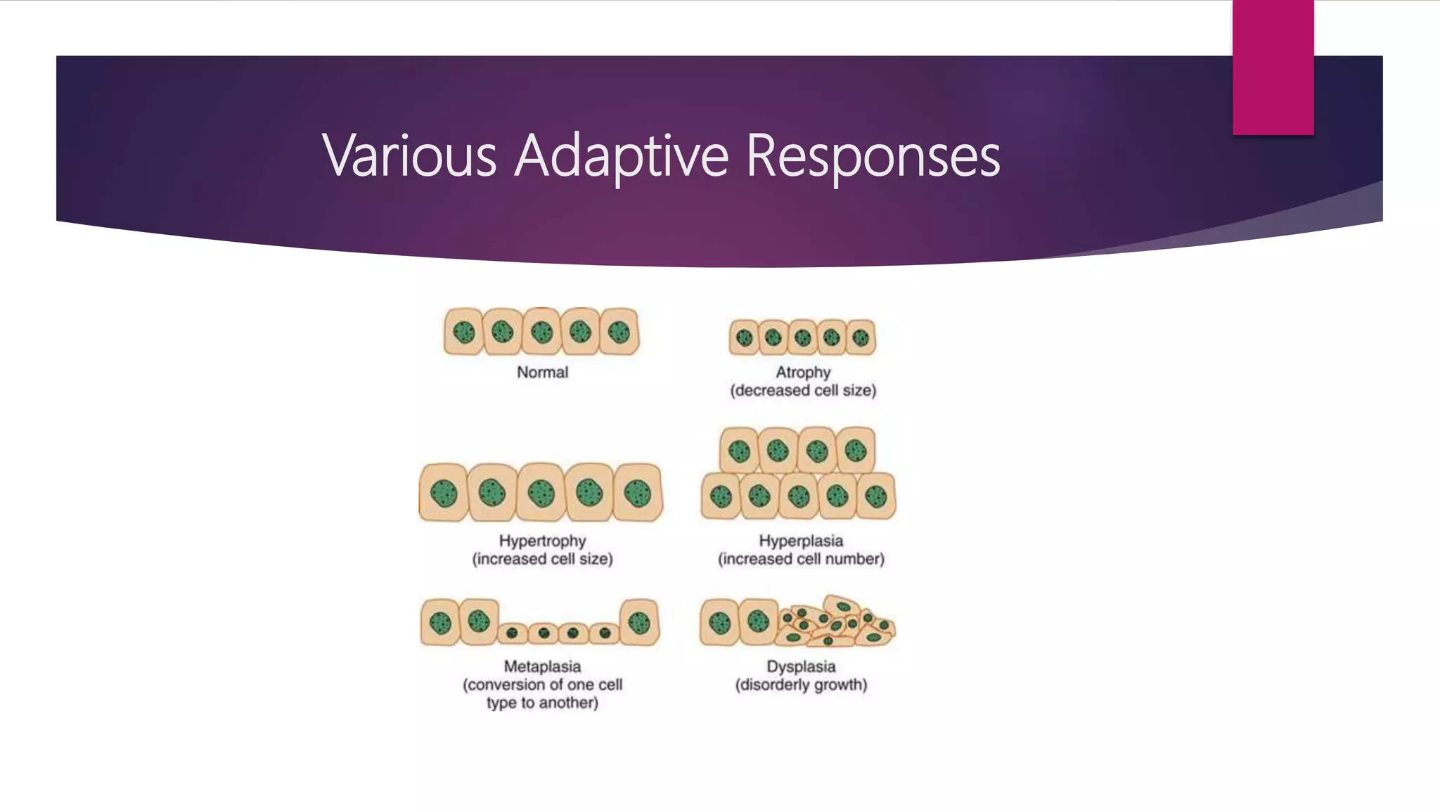
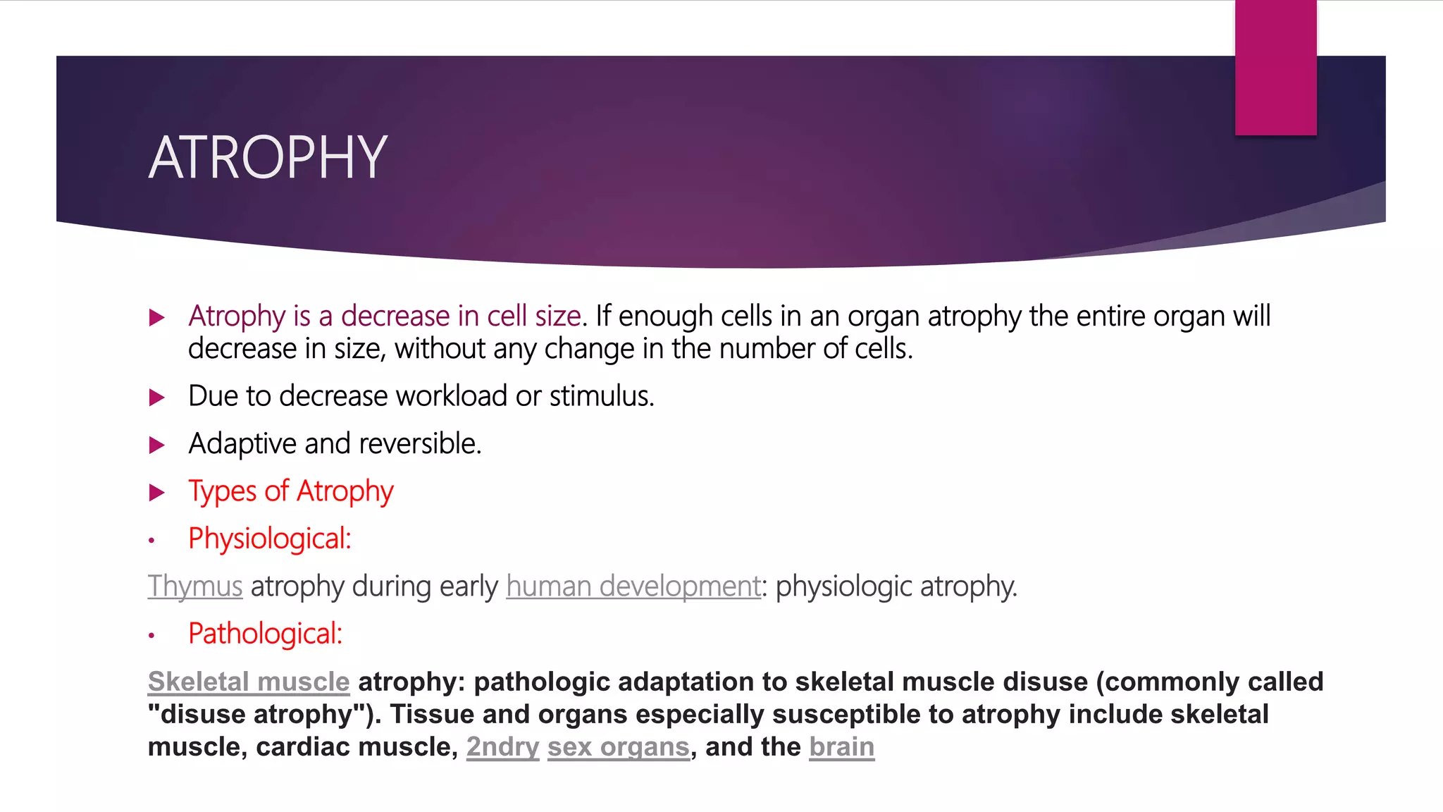
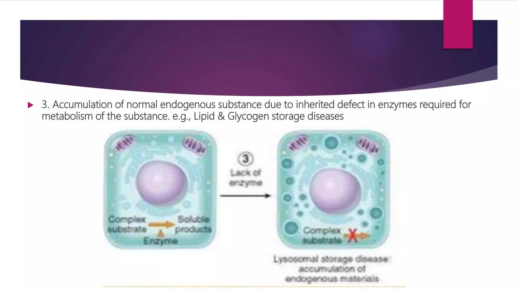
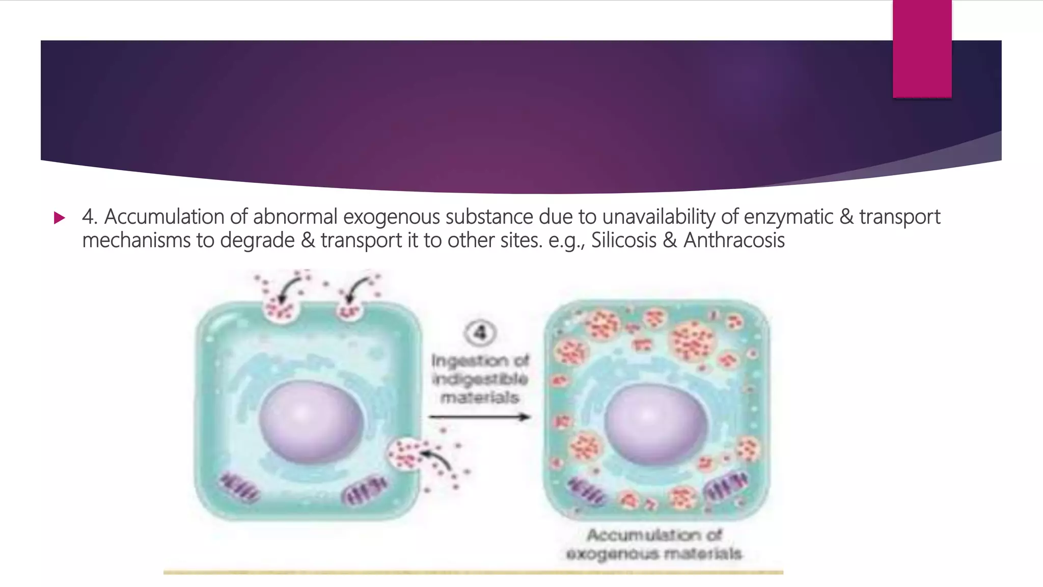
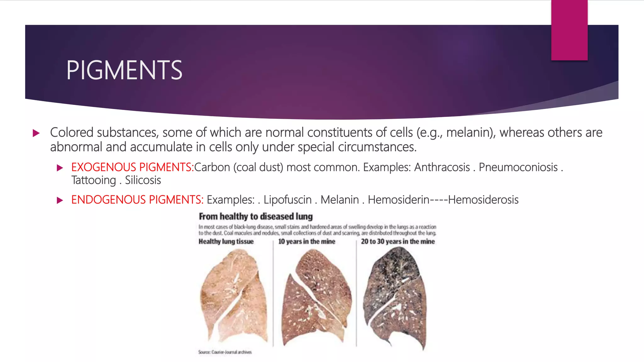
Cellular Adaptations
Cellular adaptation refers to changes made by a cell in response to
adverse environmental changes.
Various Adaptive Responses
ATROPHY
 Atrophy is a decrease in cell size. If enough cells in an organ atrophy the entire organ will
decrease in size, without any change in the number of cells.
 Due to decrease workload or stimulus.
 Adaptive and reversible.
 Types of Atrophy
• Physiological:
Thymus atrophy during early human development: physiologic atrophy.
• Pathological:
Skeletal muscle atrophy: pathologic adaptation to skeletal muscle disuse (commonly called
"disuse atrophy"). Tissue and organs especially susceptible to atrophy include skeletal
muscle, cardiac muscle, 2ndry sex organs, and the brain
Examples
 Disuse atrophy (paralysis)
 Degeneration atrophy (MS)
 Ischemic atrophy (kidney, heart)
 Malnutrition atrophy (starvation)
 Loss of endocrine stimulation (uterine, breast)
 Atrophy can be unilateral or bilateral
HYPERTROPHY
 Hypertrophy is an increase in cell size, resulting in the increase in overall size of organ, without any
change in the number of cells.
 Physiological:
 Uterine growth during pregnancy
Gross: increased myometrial thickness.
Microscopic ID:
1. increased smooth muscle size
2. increased number of feeding blood vessel
 Skeletal muscle bulk increase by exercise
 Pathological:
• Cardiac hypertrophy in hypertension.
Examples
Hyperplasia
 Hyperplasia is an increase in number of cells, resulting in the increase in overall
size of organ, it is a reversible adaptive response, if stimulus is withdrawn, organs
comes back to normal size.
 Restricted to cells capable of mitosis (epidermis, intestinal epithelium, and
glandular tissue)
 Physiological: breast & uterus in pregnancy
 Pathological: Benign Prostatic Hyperplasia (BPH), Endometrial Hyperplasia due
to hormonal stimulation, Thyroid Goitre, warts.
Examples
Metaplasia
 One cell type is replaced by another.
 Involves reprogramming of undifferentiated stem cells.
 Allows cells to survive better in hostile environment.
 Is reversible and in response to chronic irritation and inflammation.
 May predispose to cancer.(if stimulus is prolonged).
 Cells that are normally columnar or stratified may changed to squamous.
 Ciliated columnar Stratified squamous
Dysplasia
 Deranged cell growth resulting in cells of varying size, shape, and appearance
 May be associated with chronic irritation or inflammation
 May be reversible if offending agent is removed
 Dysplasia is considered A STRONG PRECURSOR OF CANCER!!!
 Example: Cervical dysplasia
 However, dysplasia is an adaptive process – may or may not lead to cancer
 Decrease risk if irritation is removed or inflammation treated.
Anaplasia
 Cells differentiate to a more IMMATURE or embryonic form.
 Malignant tumors are characterized by anaplastic cell growth.
Summary
INTRACELLULAR ACCUMULATIONS
 DEFINITION: Accumulation of abnormal amounts of various substances due to
manifestations of metabolic derangements in the cell.
 CATEGORIES:
 1. Normal cellular constituents e.g., water, lipids, CHO
 2. Abnormal substances
 a) Exogenous e.g., mineral or products of infectious agents
 b) Endogenous e.g., products of abnormal synthesis or metabolism
 SITES: a) Cytoplasm (phagolysosomes) b) Nucleus
 SOURCE:
 Produced by the affected cell
 Produced elsewhere in the body, but stored in the cell
PROCESSES OF ACCUMULATIONS
 1. Production of a normal endogenous substance at normal or increased
rate, but the rate of metabolism is inadequate to remove it. e.g., fatty liver,
reabsorption protein droplets in tubules of kidney
 2. Accumulation of an abnormal endogenous substance (product of mutated gene) due to
defects in protein folding, transport & inability to degrade abnormal proteins efficiently. e.g.,
accumulation of mutated proteins in liver cells
 3. Accumulation of normal endogenous substance due to inherited defect in enzymes required for
metabolism of the substance. e.g., Lipid & Glycogen storage diseases
 4. Accumulation of abnormal exogenous substance due to unavailability of enzymatic & transport
mechanisms to degrade & transport it to other sites. e.g., Silicosis & Anthracosis
Accumulation Of Lipids
 Triglycerides, cholesterol/cholesterol esters, phospholipids
 STEATOSIS(FATTY CHANGE): Abnormal accumulations of TGs within parenchymal cells.
 SITES: Liver (most common site) . may also occur in heart, skeletal muscle, kidney, and other
organs
 CAUSES:
 Toxins (most importantly: Alcohol abuse)
 Diabetes Mellitus .Protein malnutrition (starvation)
 Obesity
 Anoxia
MECHANISMS OF FATTY CHANGE
MORPHOLOGY OF FATTY CHANGE
 MOST COMMON SITE: • liver •heart. • with increasing accumulation, the
organ enlarges and becomes progressively yellow, soft & greasy
LIGHT MICROSCOPY OF FATTY CHANGE
 Early: small fat vacuoles in the cytoplasm around the nucleus.
 Later stages: the vacuoles coalesce to create cleared spaces that displace the nucleus to the cell
periphery Occasionally contiguous cells rupture (fatty cysts)
ACCUMULATION OF CHOLESTEROL&
CHOLESTEROL ESTERS CONDITIONS
 1. ATHEROSCLEROSIS: . In atherosclerotic plaques, SMCs and macrophages within intimal
layer of aorta & large arteries are filled with lipid vacuoles, most of which are made up of
cholesterol & cholesterol esters. . Have foamy appearance (foamy cells) . produce yellow
cholesterol-laden atheromas
 2. XANTHOMAS: formed by clusters of foamy cells found in the subepithelial connective
tissue of the skin and in tendons
 3.CHOLESTEROLOSIS: focal accumulations of cholesterol-laden macrophages in the lamina
propria of gallbladder
 4.NIEMANN-PICK DISEASE, TYPE C: Lysosomal storage disease caused by mutations affecting
an enzyme involved in cholesterol trafficking, resulting in cholesterol accumulation in
multiple organs
Foam cells
ACCUMULATION OF PROTEINS
 Appear as rounded, eosinophilic droplets, vacuoles or aggregates in cytoplasm.
 On Electron Microscopy they can be amorphous, fibrillar, or crystalline in appearance.
Protein reabsorption droplets in renal tubular epithelium
MODES OF PROTEIN ACCUMULATION
 1. Reabsorption droplets in proximal renal tubules are seen in renal diseases associated with protein
loss in urine (proteinuria).in disorders with heavy protein leakage across the gromerular filter there is
increased reabsorption of protein into vesicles and the protein appears as pink hyaline droplets within
the cytoplasm of the tubular cell
 2. Proteins accumulated may be normal secreted proteins that are produced in excessive amounts as
occurs in certain plasma cells engaged in active synthesis of immunoglobulins.ER becomes hugely
distended producing large, homogenous eosinophilic inclusions called Russell Bodies.
 3. Defective intracellular transport and secretion of critical proteins e.g., α1- antitrypsin deficiency
 4. Accumulation of cytoskeleton proteins e.g. Microtubules .Actin filaments. Myosin filaments
.Intermediate filaments (keratin filaments, neurofilaments, desmin filaments, vimentin filaments & glial
filaments)
 5. Aggregation of abnormal proteins e.g., Amyloidosis (proteinopathies or protein – aggregation
diseases)
HYALINE CHANGE
 Refers to alteration within cells or in the extracellular space that gives a
homogeneous, glassy, pink appearance in routine histologic sections stained
with H&E.
 EXAMPLES: . Reabsorption droplets . Russell bodies . Alcoholic hyaline .
Hyalinization of walls of renal arterioles in long standing HTN & DM
ACCUMULATION OF GLYCOGEN
 Excessive intracellular deposits of glycogen are seen in patients with an
abnormality in either glucose or glycogen metabolism.
 Examples Of Disorders Of Glycogen Metabolism:
 Diabetes Mellitus
 Glycogen storage diseases
PIGMENTS
 Colored substances, some of which are normal constituents of cells (e.g., melanin), whereas others are
abnormal and accumulate in cells only under special circumstances.
 EXOGENOUS PIGMENTS:Carbon (coal dust) most common. Examples: Anthracosis . Pneumoconiosis .
Tattooing . Silicosis
 ENDOGENOUS PIGMENTS: Examples: . Lipofuscin . Melanin . Hemosiderin----Hemosiderosis
Calcification
 Abnormal deposits of calcium salts occur in any tissues except bones and teeth.
 Two distinct types of pathologic calcification: –
 Dystrophic calcification: characterized by deposition of calcium salts in dead or degenerated tissues with
normal calcium metabolism and normal serum calcium levels.
 Metastatic calcification: apparently normal tissues and is associated with deranged calcium metabolism
and hypercalcaemia.
Examples of Dystrophic Calcification
Examples Of Metastatic Calcification
Thanks

Cellular adaptations & accumulations

  • 1.
    Cellular Adaptations Cellular adaptationrefers to changes made by a cell in response to adverse environmental changes.
  • 2.
  • 3.
    ATROPHY  Atrophy isa decrease in cell size. If enough cells in an organ atrophy the entire organ will decrease in size, without any change in the number of cells.  Due to decrease workload or stimulus.  Adaptive and reversible.  Types of Atrophy • Physiological: Thymus atrophy during early human development: physiologic atrophy. • Pathological: Skeletal muscle atrophy: pathologic adaptation to skeletal muscle disuse (commonly called "disuse atrophy"). Tissue and organs especially susceptible to atrophy include skeletal muscle, cardiac muscle, 2ndry sex organs, and the brain
  • 4.
    Examples  Disuse atrophy(paralysis)  Degeneration atrophy (MS)  Ischemic atrophy (kidney, heart)  Malnutrition atrophy (starvation)  Loss of endocrine stimulation (uterine, breast)  Atrophy can be unilateral or bilateral
  • 5.
    HYPERTROPHY  Hypertrophy isan increase in cell size, resulting in the increase in overall size of organ, without any change in the number of cells.  Physiological:  Uterine growth during pregnancy Gross: increased myometrial thickness. Microscopic ID: 1. increased smooth muscle size 2. increased number of feeding blood vessel  Skeletal muscle bulk increase by exercise  Pathological: • Cardiac hypertrophy in hypertension.
  • 6.
  • 8.
    Hyperplasia  Hyperplasia isan increase in number of cells, resulting in the increase in overall size of organ, it is a reversible adaptive response, if stimulus is withdrawn, organs comes back to normal size.  Restricted to cells capable of mitosis (epidermis, intestinal epithelium, and glandular tissue)  Physiological: breast & uterus in pregnancy  Pathological: Benign Prostatic Hyperplasia (BPH), Endometrial Hyperplasia due to hormonal stimulation, Thyroid Goitre, warts.
  • 9.
  • 10.
    Metaplasia  One celltype is replaced by another.  Involves reprogramming of undifferentiated stem cells.  Allows cells to survive better in hostile environment.  Is reversible and in response to chronic irritation and inflammation.  May predispose to cancer.(if stimulus is prolonged).  Cells that are normally columnar or stratified may changed to squamous.  Ciliated columnar Stratified squamous
  • 11.
    Dysplasia  Deranged cellgrowth resulting in cells of varying size, shape, and appearance  May be associated with chronic irritation or inflammation  May be reversible if offending agent is removed  Dysplasia is considered A STRONG PRECURSOR OF CANCER!!!  Example: Cervical dysplasia  However, dysplasia is an adaptive process – may or may not lead to cancer  Decrease risk if irritation is removed or inflammation treated.
  • 12.
    Anaplasia  Cells differentiateto a more IMMATURE or embryonic form.  Malignant tumors are characterized by anaplastic cell growth.
  • 14.
  • 15.
    INTRACELLULAR ACCUMULATIONS  DEFINITION:Accumulation of abnormal amounts of various substances due to manifestations of metabolic derangements in the cell.  CATEGORIES:  1. Normal cellular constituents e.g., water, lipids, CHO  2. Abnormal substances  a) Exogenous e.g., mineral or products of infectious agents  b) Endogenous e.g., products of abnormal synthesis or metabolism  SITES: a) Cytoplasm (phagolysosomes) b) Nucleus  SOURCE:  Produced by the affected cell  Produced elsewhere in the body, but stored in the cell
  • 16.
    PROCESSES OF ACCUMULATIONS 1. Production of a normal endogenous substance at normal or increased rate, but the rate of metabolism is inadequate to remove it. e.g., fatty liver, reabsorption protein droplets in tubules of kidney
  • 17.
     2. Accumulationof an abnormal endogenous substance (product of mutated gene) due to defects in protein folding, transport & inability to degrade abnormal proteins efficiently. e.g., accumulation of mutated proteins in liver cells
  • 18.
     3. Accumulationof normal endogenous substance due to inherited defect in enzymes required for metabolism of the substance. e.g., Lipid & Glycogen storage diseases
  • 19.
     4. Accumulationof abnormal exogenous substance due to unavailability of enzymatic & transport mechanisms to degrade & transport it to other sites. e.g., Silicosis & Anthracosis
  • 20.
    Accumulation Of Lipids Triglycerides, cholesterol/cholesterol esters, phospholipids  STEATOSIS(FATTY CHANGE): Abnormal accumulations of TGs within parenchymal cells.  SITES: Liver (most common site) . may also occur in heart, skeletal muscle, kidney, and other organs  CAUSES:  Toxins (most importantly: Alcohol abuse)  Diabetes Mellitus .Protein malnutrition (starvation)  Obesity  Anoxia
  • 21.
  • 22.
    MORPHOLOGY OF FATTYCHANGE  MOST COMMON SITE: • liver •heart. • with increasing accumulation, the organ enlarges and becomes progressively yellow, soft & greasy
  • 23.
    LIGHT MICROSCOPY OFFATTY CHANGE  Early: small fat vacuoles in the cytoplasm around the nucleus.  Later stages: the vacuoles coalesce to create cleared spaces that displace the nucleus to the cell periphery Occasionally contiguous cells rupture (fatty cysts)
  • 24.
    ACCUMULATION OF CHOLESTEROL& CHOLESTEROLESTERS CONDITIONS  1. ATHEROSCLEROSIS: . In atherosclerotic plaques, SMCs and macrophages within intimal layer of aorta & large arteries are filled with lipid vacuoles, most of which are made up of cholesterol & cholesterol esters. . Have foamy appearance (foamy cells) . produce yellow cholesterol-laden atheromas  2. XANTHOMAS: formed by clusters of foamy cells found in the subepithelial connective tissue of the skin and in tendons  3.CHOLESTEROLOSIS: focal accumulations of cholesterol-laden macrophages in the lamina propria of gallbladder  4.NIEMANN-PICK DISEASE, TYPE C: Lysosomal storage disease caused by mutations affecting an enzyme involved in cholesterol trafficking, resulting in cholesterol accumulation in multiple organs
  • 25.
  • 26.
    ACCUMULATION OF PROTEINS Appear as rounded, eosinophilic droplets, vacuoles or aggregates in cytoplasm.  On Electron Microscopy they can be amorphous, fibrillar, or crystalline in appearance. Protein reabsorption droplets in renal tubular epithelium
  • 27.
    MODES OF PROTEINACCUMULATION  1. Reabsorption droplets in proximal renal tubules are seen in renal diseases associated with protein loss in urine (proteinuria).in disorders with heavy protein leakage across the gromerular filter there is increased reabsorption of protein into vesicles and the protein appears as pink hyaline droplets within the cytoplasm of the tubular cell  2. Proteins accumulated may be normal secreted proteins that are produced in excessive amounts as occurs in certain plasma cells engaged in active synthesis of immunoglobulins.ER becomes hugely distended producing large, homogenous eosinophilic inclusions called Russell Bodies.  3. Defective intracellular transport and secretion of critical proteins e.g., α1- antitrypsin deficiency  4. Accumulation of cytoskeleton proteins e.g. Microtubules .Actin filaments. Myosin filaments .Intermediate filaments (keratin filaments, neurofilaments, desmin filaments, vimentin filaments & glial filaments)  5. Aggregation of abnormal proteins e.g., Amyloidosis (proteinopathies or protein – aggregation diseases)
  • 28.
    HYALINE CHANGE  Refersto alteration within cells or in the extracellular space that gives a homogeneous, glassy, pink appearance in routine histologic sections stained with H&E.  EXAMPLES: . Reabsorption droplets . Russell bodies . Alcoholic hyaline . Hyalinization of walls of renal arterioles in long standing HTN & DM
  • 29.
    ACCUMULATION OF GLYCOGEN Excessive intracellular deposits of glycogen are seen in patients with an abnormality in either glucose or glycogen metabolism.  Examples Of Disorders Of Glycogen Metabolism:  Diabetes Mellitus  Glycogen storage diseases
  • 30.
    PIGMENTS  Colored substances,some of which are normal constituents of cells (e.g., melanin), whereas others are abnormal and accumulate in cells only under special circumstances.  EXOGENOUS PIGMENTS:Carbon (coal dust) most common. Examples: Anthracosis . Pneumoconiosis . Tattooing . Silicosis  ENDOGENOUS PIGMENTS: Examples: . Lipofuscin . Melanin . Hemosiderin----Hemosiderosis
  • 31.
    Calcification  Abnormal depositsof calcium salts occur in any tissues except bones and teeth.  Two distinct types of pathologic calcification: –  Dystrophic calcification: characterized by deposition of calcium salts in dead or degenerated tissues with normal calcium metabolism and normal serum calcium levels.  Metastatic calcification: apparently normal tissues and is associated with deranged calcium metabolism and hypercalcaemia.
  • 33.
  • 34.
  • 35.