Downloaded 473 times


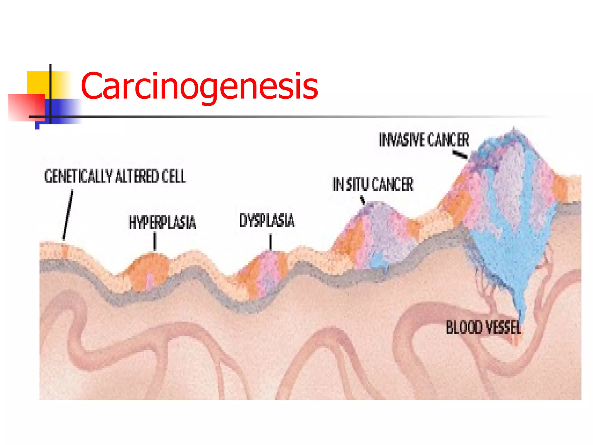


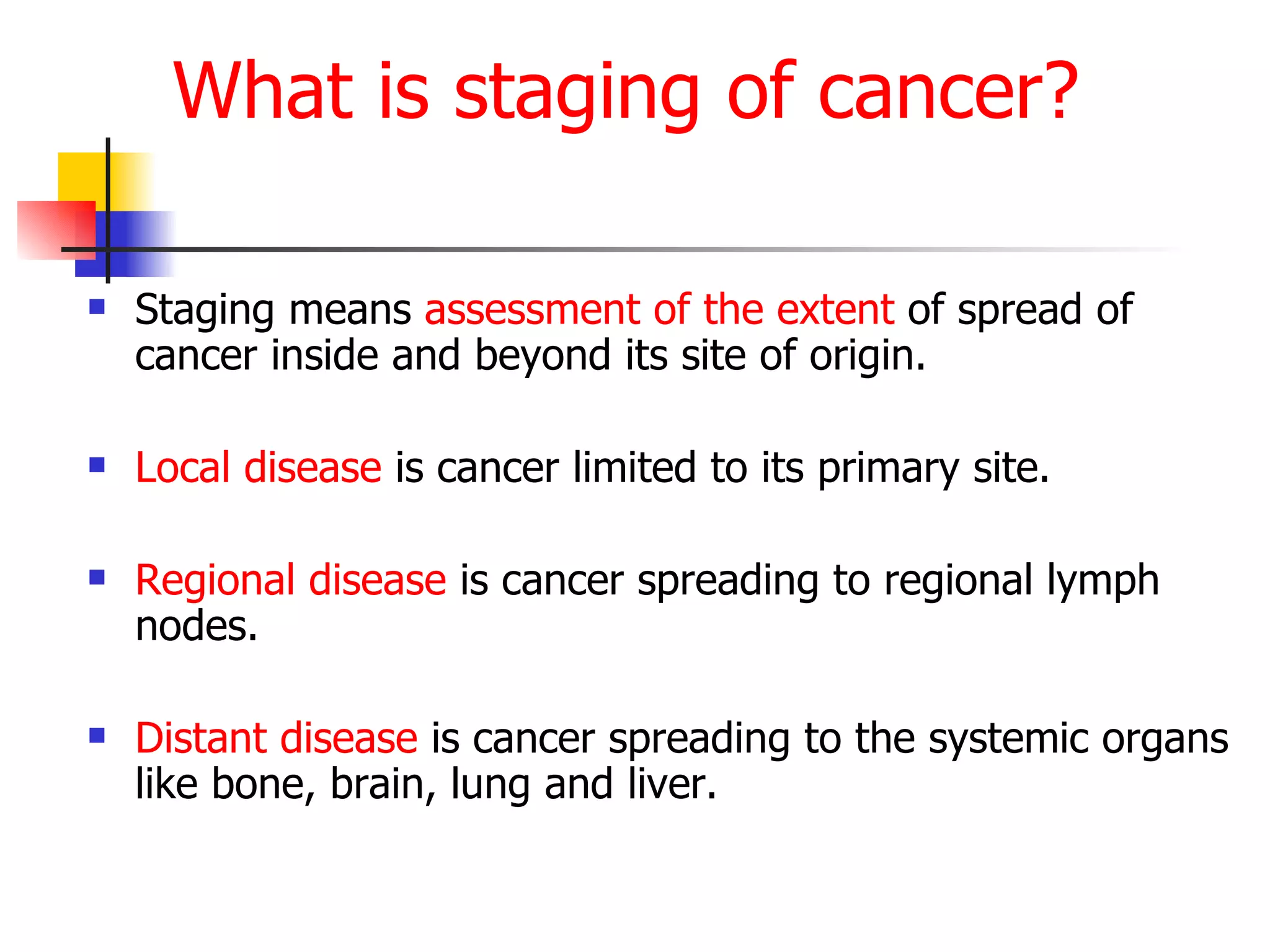
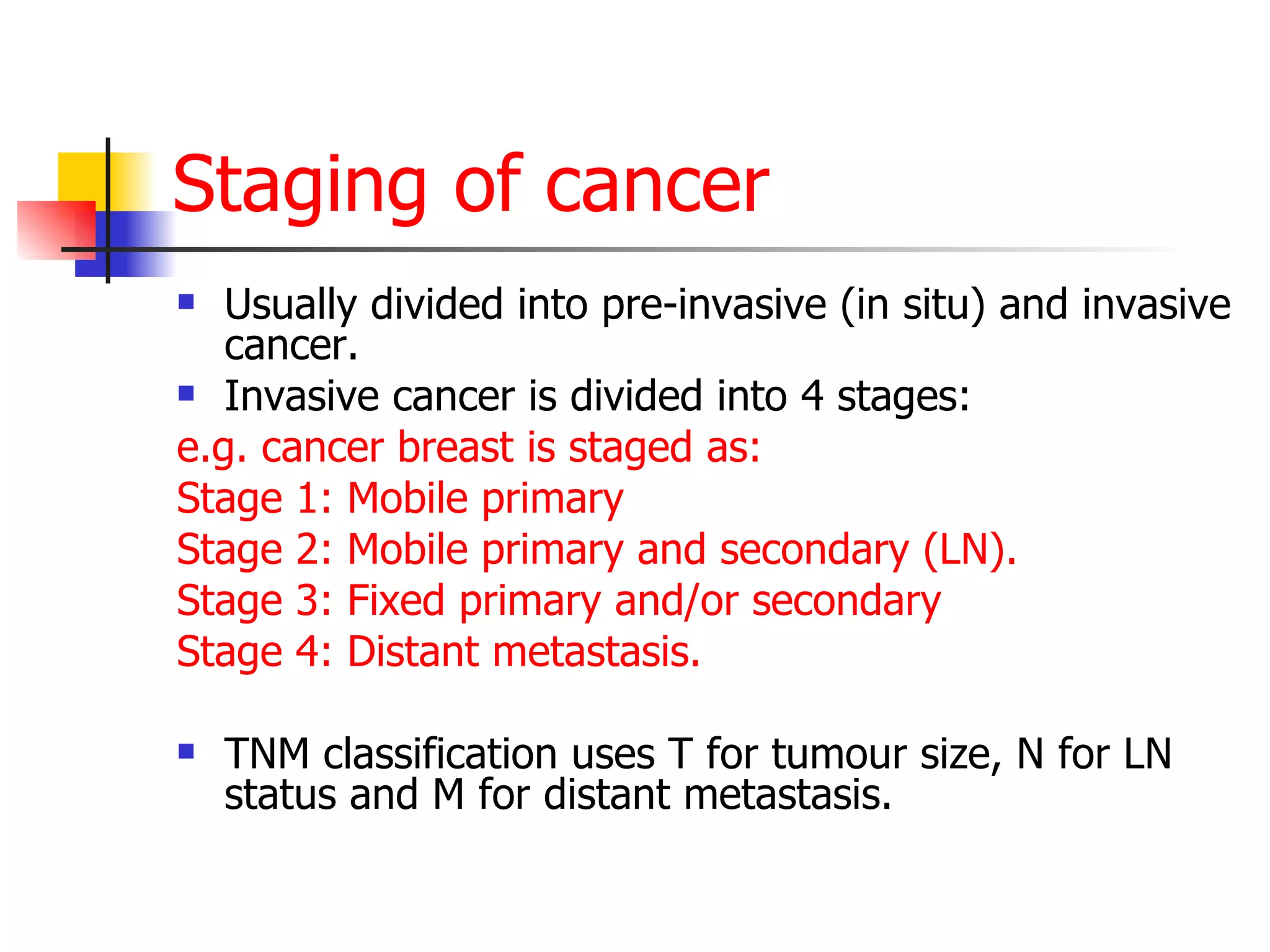
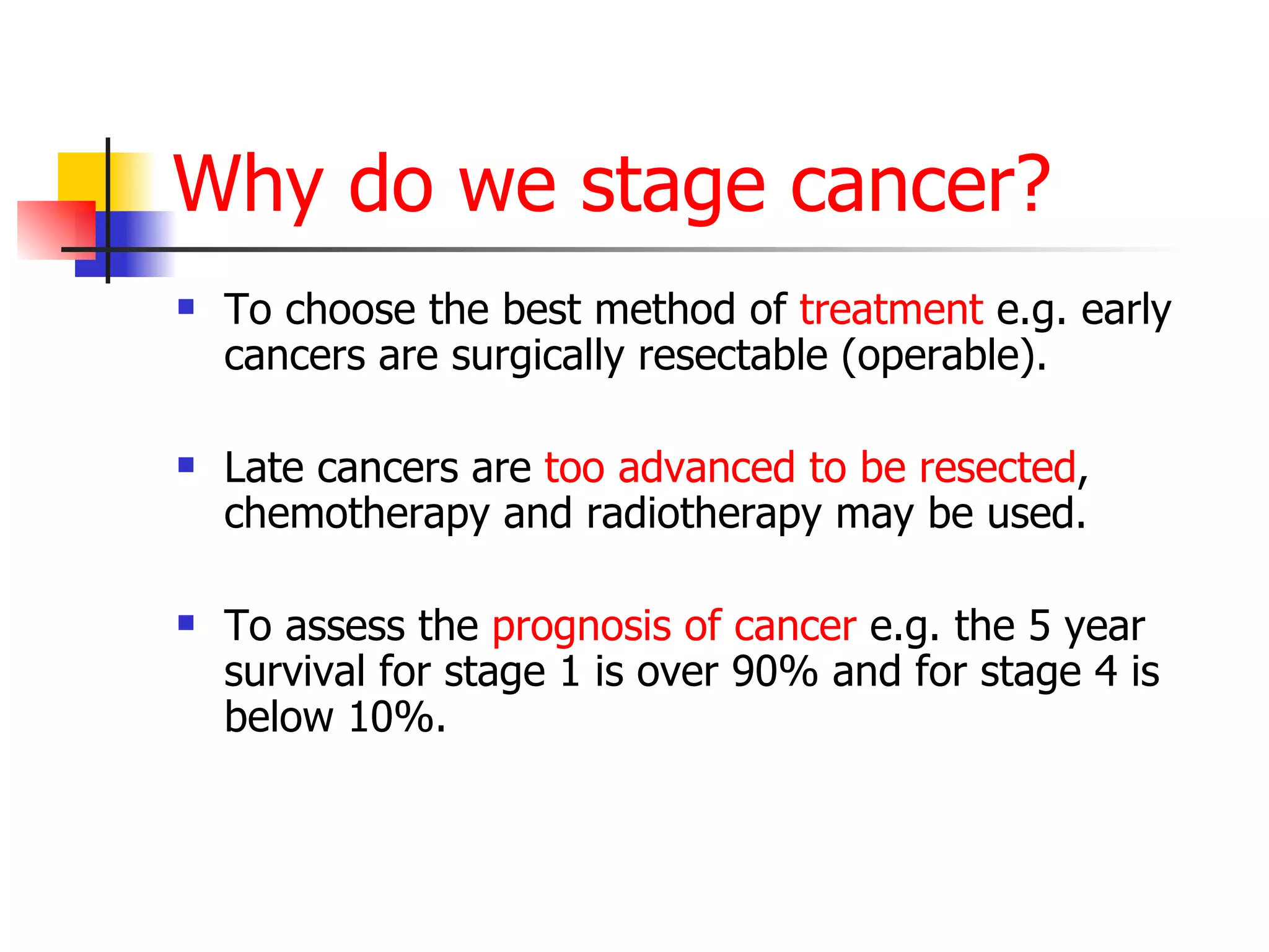
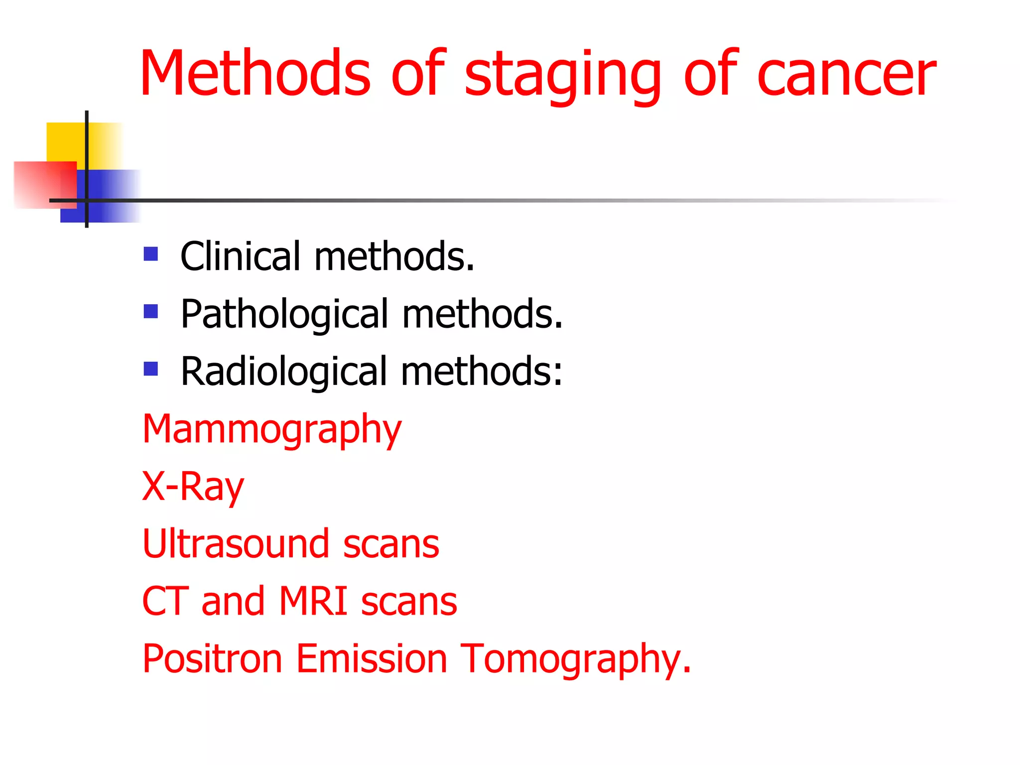

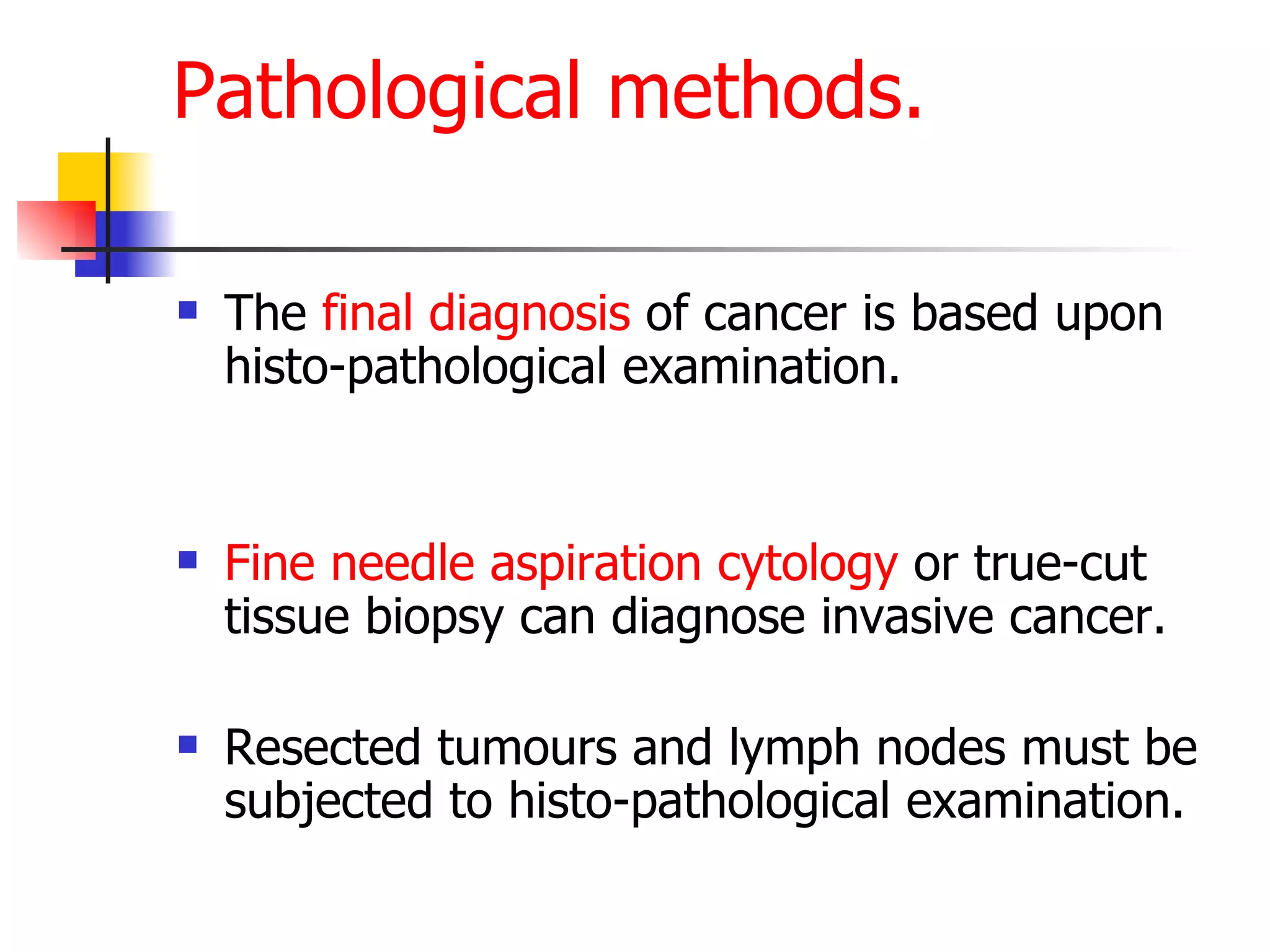
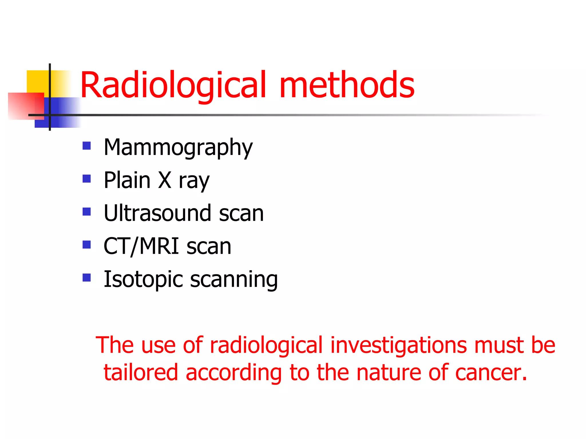


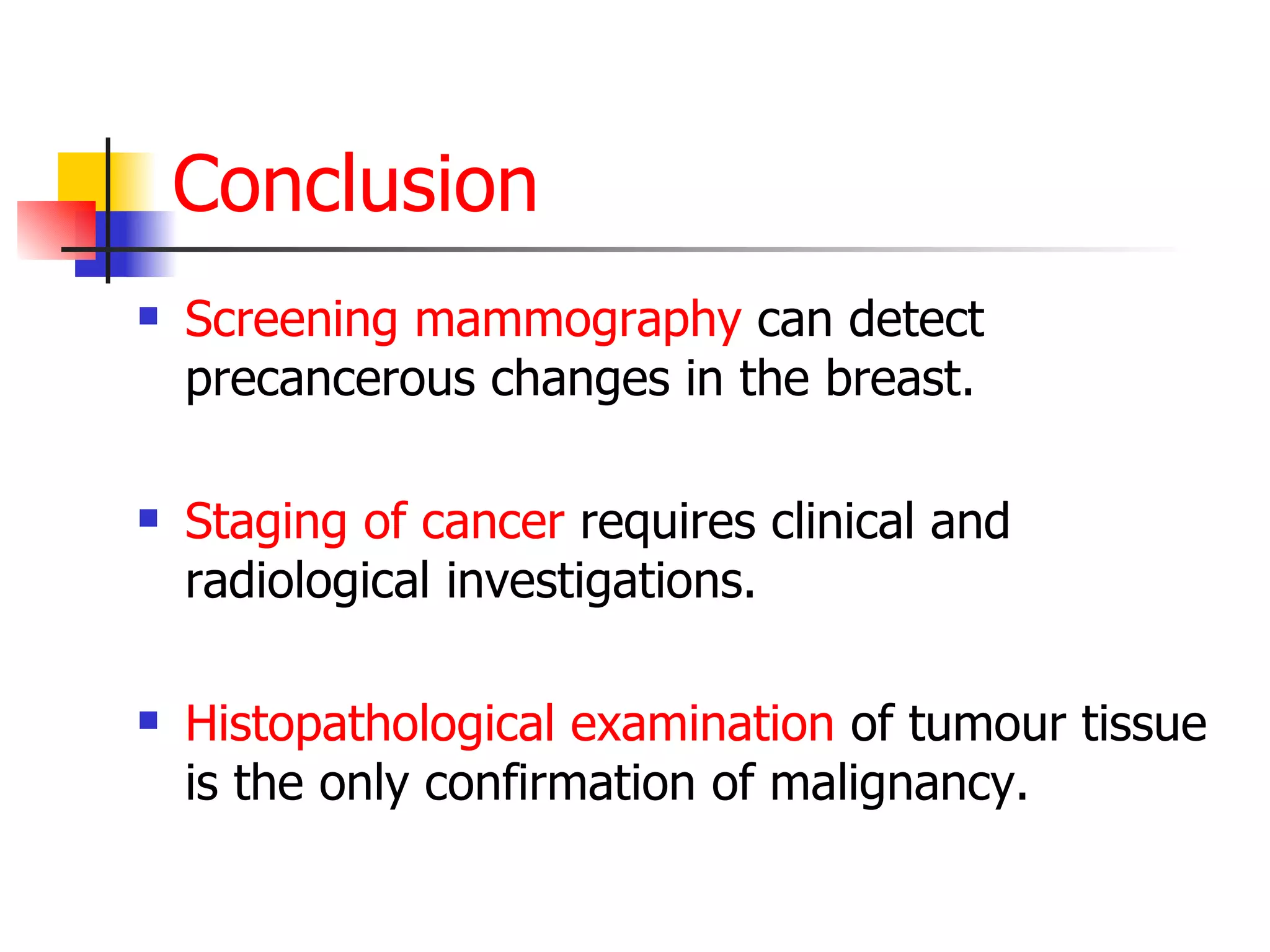


Staging of cancer involves assessing how far a cancer has spread both locally and systemically. It is important for determining prognosis and selecting the most appropriate treatment. There are clinical, pathological, and radiological methods used for staging. Clinically, exam assesses tumor size, mobility, and lymph node involvement. Pathological exam of biopsied tissue confirms cancer diagnosis. Radiological exams like mammography, CT, MRI, PET scans provide additional information on tumor characteristics and metastasis. Together these methods are used to divide cancer into stages to guide management and predict outcomes.


