Caenorhabditis elegans is a tiny, free-living nematode found worldwide. Newly hatched larvae are 0.25 millimeters long and adults are 1 millimeter long. Their small size means that the animals are usually observed with either dissecting microscopes, which generally allow up to 100X magnification, or compound microscopes, which allow up to 1000X magnification. Because C. elegans is transparent, individual cells and subcellular details are easily visualized using Nomarski (differential interference contrast, DIC) optics.
C. elegans has a rapid life cycle and exists primarily as a self-fertilizing hermaphrodite, although males arise at a frequency of <0.2%. These features have helped to make C. elegans a powerful model of choice for eukaryotic genetic studies. In addition, because the animal has an invariant numbers of somatic cells, researchers have been able to track the fate of every cell between fertilization and adulthood in live animals and to generate a complete cell lineage. Researchers have also reconstructed the shape of all C. elegans cells from electron micrographs, including each of the 302 neurons of the adult hermaphrodite. Moreover, because of the invariant wild-type cell lineage and neuroanatomy of C. elegans, mutations that give rise to developmental and behavioral defects are readily identified in genetic screens. Finally, because C. elegans was the first multicellular organism with a complete genome sequence, forward and reverse genetics have led to the molecular identification of many key genes in developmental and cell biological processes.
The experimental strengths and the similarities between the cellular and molecular processes present in C. elegans and other animals across evolutionary time (metabolism, organelle structure and function, gene regulation, protein biology, etc.) have made C. elegans an excellent organism with which to study general metazoan biology. At least 38% of the C. elegans protein-coding genes have predicted orthologs in the human genome, 60-80% of human genes have an ortholog in the C. elegans genome, and 40% of genes known to be associated with human diseases have clear orthologs in the C. elegans genome. Thus, many discoveries in C. elegans have relevance to the study of human health and disease.
In 1963, Sydney Brenner introduced Caenorhabditis elegans as a model organism for pursuing research in developmental biology and neurology.It is a free-living, non-parasitic soil nematode that can be safely used in the laboratory and is common around the world.
This ppt gives an idea of general anatomy of this small creature,its life cylce,study as a model organism and its importance in the study of ageing.
its deals with the general basic ideas of gene and evolutions.different types of examples are used to explain the gene and evolutions.the origin of basic genetics and their ideas are also formulated in this presentation
Animal classificaton and phylogeny organism Millar and Harley Zoology BScZahraAtta
Millar And Harley Chap 7 Animal Classification, phylogeny and animal Organization
main Topic to study
classification of animal
Taxonomic Hierarchy
Nomenclature
Menders experiments were conducted using garden peas. Why would human.pdfisenbergwarne4100
Menders experiments were conducted using garden peas. Why would humans be an awful
choice for an experimental organism (give at least 3 reasons)? Give an example of an animal that
would be better sorted for genetics experiments. In Mendel\'s experiments, a plant with purple
flowers was crossed with a plant having white flowers. Explain why white flowers disappeared
in the F_1 generation and reappeared in the F_2. Your pet rabbit has curly fur. After seeking
advice from a rabbit breeder, you learn that curly fur is a dominant trait, but you want to know
the precise genotype of your pet. Describe how you could find out. Describe the inheritance of
ABO blood types in humans and explain why individuals with type O are universal donors while
individuals with type AB are universal acceptors. Describe the nature vs. nurture debate. In your
explanation, give an example of a trait that is controlled entirely by nature and one that is heavily
influenced by both nature and nurture.
Solution
Answer:
1. Studying human genetics is unlike studying the genetics of any other organism. In many ways,
humans are very poor model organisms for genetics. Long generation times make for slow
progress when doing genetic crosses, which brings us to another problem with human genetics:
The inability to make controlled crosses.
So, any human geneticist that tried to make controlled human crosses would most likely be
considered a very disturbed criminal and not a brilliant scientist. Besides, humans usually only
have one child at a time, which makes it really difficult to generate numbers of offspring that can
achieve statistical significance. On top of all this, there\'s the issue of genetic manipulation. Key
genetic techniques, like mutation screening and transgenics, are completely off-limits to human
geneticists.
Drosophila melanogaster (Fruit fly) would be better suited as a model organism for genetics
experiments.
1. The relationship between fruit fly and human genes is so close that often the sequences of
newly discovered human genes, including disease genes, can be matched with equivalent genes
in the fly.
2. 75 per cent of the genes that cause disease in humans are also found in the fruit fly.
3. Drosophila have a short, simple reproduction cycle. It is normally about 8-14 days, depending
on the environmental temperature. This means that several generations can be observed in a
matter of months.
4. Fruit fly are small (3 mm long) but not so small that they can’t be seen without a microscope.
This allows scientists to keep millions of them in the laboratory at a time.
5. They are inexpensive to maintain in the laboratory.
6. They require a simple diet consisting of simple sources of carbohydrates (cornmeal) and
proteins (yeast extract).
7. The only care they need is having their food changed regularly (every 10-14 days at 25C or 5-
6 weeks at 18C).
8. Drosophila have ‘polytene’ chromosomes, which means that they are oversized and have
barcode-like banding patterns.
1 What is the study systemGeneral information. E.g. What is a .docxhoney725342
1 What is the study system?
General information. E.g. What is a “cell line”? Include images.
1 Why would a researcher use this study system?
The particular features of this system that make it useful. E.g. cell lines allow the study of genetically identical cells in many labs
1 What type of research questions can this study system be used to help answer?
List a few examples of research questions or general areas of research that can be addressed using this system. Elaborate a little on each, so we understand what you mean.
1 How does a researcher typically use this system?
What are the logistics of it? E.g. basic information about how they culture and propagate cell lines.
1 What are the pros and cons of this study system?
List and briefly explain any drawbacks or caveats that we should be aware of, along with particular benefits E.g. mammalian cell lines need adequate facilities and resources to be propagated, but they also allow for the study of mammalian cellular systems in vitro instead of studying another eukaryote like yeast.
6.Are there alternatives or variations on this study system?
If you can’t use this particular study system, what are your options for alternatives? E.g. Use yeast as a representative of a eukaryotic cell.
1 What is a real example from primary literature of this study system being used?
Provide a brief summary of the research that used the study system of interest, including the main objective, basic methods used, the main results, and conclusions. Include an image of at least one figure or table, along with an explanation of what that figure/table illustrates. You must provide the complete citation of the paper and/or a link to the online paper.
8.List of sources and places where we can find more information.
Background
C. elegans: A Simple Multicellular Model Organism
Scientists worldwide conduct basic research to address gaps in our knowledge in the hopes that this information can serve humanity in the future. Basic biological research seeks to answer questions of such elementary cellular and organismal activities as how cells grow, divide, die, move, store and use energy, and communicate.
Scientists use model organisms in basic research to answer these questions because model organisms offer simplified cellular systems that reproduce quickly, are easy to maintain, and are cost efficient. For example,
if a DNA mutation is known to result in a neurological disorder, more data can be generated using a model organism such as C. elegans, which reproduces and matures every 2–3 days, rather than waiting for a human child to mature and show symptoms. Commonly used basic model organisms include S. cerevisiae (yeast), C. elegans (nematode), D. melanogaster (fruit fly), and M. musculus (mouse).
Despite the seeming lack of a relationship to human beings, these model organisms have helped researchers understand the basic cellular machinery underlying a host of human pathologies such as cancer, neurological disorders, ...
A presentation on Zebrafish's history, taxonomy , genetics, life cycle and future prospects of zebrafish and some of its medical implications in human life. Most importantly the major interest is to investigate those particular gene that are responsible for regenerating the heart in zebrafish so that they can be applied to human heart and help im regenerating human heart without the formation of any scar.
1. The fruit fly, Drosophila melanogaster, has been the most popular eukaryotic organism used in classrooms. It is a small fruit fly about 3mm long, similar to the ones you see attracted to your bananas and other fruit. It has short life cycle of two weeks, making
it possible to study numerous generations in a short period. It is easy to culture and inexpensive to house large numbers. Its size is amenable for cultivation in school laboratories. Also, it is large enough that many attributes can be seen with the naked eye or
under low-power magnification. Moreover, it has a very long history in biological research and there are many useful tools to facilitate genetic study. It was recognised by the award of the Nobel prize in Physiology or Medicine to Thomas Hunt Morgan in
1933, to Hermann Muller in 1946 and to Ed Lewis, Christiane Nusslein-Volhard and Eric Wieschaus in 1995.
The use of Drosophila is a powerful tool also in teaching the Life Sciences. In fact it allows the observation of sexual dimorphism, of mutants and of the life cycle. Moreover it permits the realization of crosses aimed to demostration of sexual linked characters
and crosses aimed to estabilish if a mutation is conferrel by a dominant or a recessive gene. There are also similarities with the human species. From a genetic point of view humans and fruit flies are quite similar. About 60% of known genetic diseases may
occur in the gene of the fruit fly. And approximately 50% of the proteins of Drosophila have a similar in mammals. The Drosophila is used as a genetic model for several human diseases, including neurodegenerative disorders such as Parkinson's disease,
Huntington's disease and Alzheimer's disease. It is used to study the biological mechanism of the immune system, diabetes, cancer, and even substance abuse.
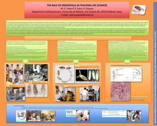
The Core Lab
We provided fruit flies (Fig. 2B): one vial of each type of flies for each pair of students..
It takes about one week for the flies to hatch upon fecundation, and two weeks for each
subculture to increase the fly population to reach the size necessary for classroom use.
We proposed to students some activities:
Observation of D.melanogaster morphology
-Drosophilae are moved from the test-tube where they live to "anesthetics" tube“ where
they are left few minutes with CO2 (alka seltzer tablets or dry ice).
-Flies are left on a thin cardboard or a Petri plate and observed under a stereomicroscope.
Students observed Drosophila sexual dimorphism: the female is bigger than male and
abdomen is light .
The male has got a dark spot on posterior extremity of the body and a structure like a
comb on the anterior leg to block the female during the copulation (Fig. 2B).
Other activities:
-Observation of the Drosophila life cycle.
At school students performed crosses. Crossed wild type (wt) male and female
Drosophilae in a test-tube, made a series of passages in other test-tubes on the 2th,
4th, 6th, 8th and 10th day, and, after 10 days, they observed embrions, larvas, pupas
and adults in different test-tubes. . The Drosophila egg is about half a millimiter long.
It takes about one day after fertilisation for the embryo to develop and hatch into a
worm-like larva. (Fig. 3A):
The larva eats and grows continuously, moulting one day, two days, and four days
after hatching (first, second and third instars). (Fig. 3B).
-Observation of D.melanogaster mutants: regarding the colour of the eyes, regarding
the colour of the body, regarding the structure of the wings.
We provided fruit flies mutants and they try and describe the mutations.
Fig. 3A,3B -The Life cycle of Drosophila melanogaster
and the various larval instars
Fig.2A Stereomicroscope
Fig.2B Drosophila sexual dimorphism
Fig.1A Students in the LAB
Fig.1B Vials of flies
embryo
2° and 3° instar larva
Extraction of salivary glands and staining giant chromosomes:
Polytene chromosomes are over-sized chromosomes which have developed from standard
chromosomes and are commonly found in the salivary glands of Drosophila melanogaster.
Specialized cells undergo repeated rounds of DNA replication without cell division
(endomitosis), to increase cell volume, forming a giant polytene chromosome. Polytene
chromosomes form when multiple rounds of replication produce many sister chromatides that
remain synapsed together. In addition to increasing the volume of the cells' nuclei and causing
cell expansion, polytene cells may also have a metabolic advantage as multiple copies of genes
permits a high level of gene expression. In Drosophila melanogaster, for example, the
chromosomes of the larval salivary glands undergo many rounds of endoreduplication, to
produce large amounts of glue before pupation. Polytene chromosomes have characteristic light
and dark banding patterns that can be used to identify chromosomal rearrangements and
deletions. Dark banding frequently corresponds to inactive chromatin, whereas light banding is
usually found at areas with higher transcriptional activity.
We provided the Petri plates with larvae at 3rd instar in acetic acid solution and the students
have extracted and colored cells of salivary glands .
Extraction of salivary glands and staining giant chromosomes:
prepupa
Fig. 4A,4B Observation of D.melanogaster mutants
Polytene chromosomes stained
with acetic orcein and visualized
by light microscopy (100X).
Results and conclusions
To understand a complex problem, it is sometimes useful to exploit an easier "model". Model organisms play an important role in conveying biological concepts.
The proposed activities in the context of various events of scientific communication organized by the University of Milan, result effective for learning about topics in biology, zoology and genetics.
Workshop SCIENCE EDUCATION AND GUIDANCE IN SCHOOLS: THE WAY FORWARD Firenze, October 21-22, 2013
THE ROLE OF DROSOPHILA IN TEACHING LIFE SCIENCES
M. E. Pasini*,Y. Intra, P. Fasano
Department of Biosciences, University di Milano, Via Celoria 26, 20133 Milano, Italy.
E-mail: maria.pasini@unimi.it