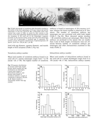
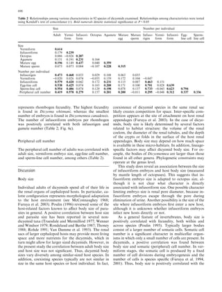
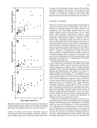
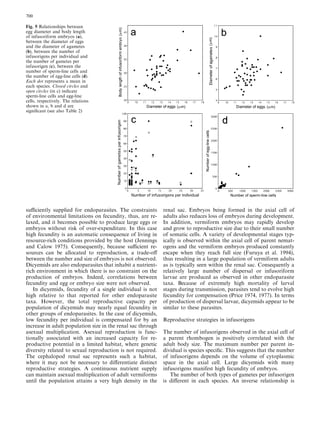
Reproductive traits in dicyemids
Received: 8 July 2002 / Accepted: 15 November 2002 / Published online: 12 February 2003
Ó Springer-Verlag 2003
Abstract Several characters involved in the life cycle in
dicyemids were examined to understand reproductive
strategy and adaptations to cephalopod hosts. In most
dicyemids distinctly small numbers of sperms are pro-
duced in a hermaphroditic gonad (infusorigen). The
number of eggs and sperms are roughly equal (means of
the number of sperm:egg=1:1.58). An inverse propor-
tional relationship was found between the number of
infusorigens and the gametes, suggesting a trade-off
between them. Fecundity was positively correlated with
the body size of adult stages (nematogens and rhomb-
ogens). Fecundity of a single dicyemid is not very high
compared with that of the other endoparasite taxa, but a
total reproductive capacity per community is high, be-
cause a great number of individuals multiply asexually in
the renal sac. The size of mature infusoriform embryos
(dispersal embryos) that develop from fertilized eggs was
not correlated with their adult sizes, but the size of
embryos was correlated with the maximum mantle
length of the host octopus species. Although at present
the process of infecting new hosts is still unknown, the
size of the infusoriform embryo is likely determined by
host-specific factors in this process. The size of vermi-
form embryos that are asexually formed from agametes
was positively correlated with size of the adults; how-
ever, the number of vermiform embryos present in the
axial cell of adults was not correlated with size of the
adults. A correlation was not found between maximum
mantle length of the host cephalopod species and length
of the adult dicyemids. In dicyemid species the size of
adults appears to be constrained by the renal habitat,
including renal-pancreatic complex and branchial hearts
of each host cephalopod species. Size thus may be de-
termined by the volume of the renal sac, the diameter of
the renal tubules, or the depth of folding in the surface
of glandular renal appendages of cephalopods.
Introduction
Dicyemid mesozoans are endosymbionts that typically
are found in the renal sac of benthic cephalopod mol-
lusks (Nouvel 1947; McConnaughey 1951; Hochberg
1990). Although recent studies have revealed that they
might not be truly primitive animals deserving the name
of ‘‘mesozoans’’ (Katayama et al. 1995; Kobayashi et al.
1999), they are still one of the most interesting and
puzzling groups of lower invertebrates. Their body,
consisting of a very small number of cells (usually 10–
40), is organized in a very simple fashion. Their life cycle
is also peculiar. They produce two distinct embryo types:
a vermiform embryo from an asexual agamete and an
infusoriform embryo from a fertilized egg (Furuya et al.
1992a, 1994, 1996, 2001). A unique hermaphroditic go-
nad, the infusorigen, is formed in the cytoplasm of the
axial cell of rhombogens (Lameere 1918; Nouvel 1947;
McConnaughey 1951). The process of gametogenesis
shows several special features (Austin 1964; Short and
Damian 1966; Furuya et al. 1993).
Parasitism is a common way of life in the animal
kingdom. A wide diversity of life histories and habitats
have been documented. The excretory organs of ceph-
alopods are a unique environment providing living
space for a diversity of parasites. The fluid-filled renal
coelom provides an ideal habitat for the establishment
and maintenance of dicyemids (Hochberg 1982, 1983).
Dicyemids are subjected to a number of selecting
pressures due to their unique habitats. In this study, we
Marine Biology (2003) 142: 693–706
DOI 10.1007/s00227-002-0991-6
H. Furuya Æ F.G. Hochberg Æ K. Tsuneki
Communicated by O. Kinne, Oldendorf/Luhe
H. Furuya (&) Æ K. Tsuneki
Department of Biology, Graduate School of Science,
Osaka University, Toyonaka, Osaka 560-0043, Japan
E-mail: hfuruya@bio.sci.osaka-u.ac.jp
F.G. Hochberg
Department of Invertebrate Zoology,
Santa Barbara Museum of Natural History,
2559 Puesta del Sol Road, Santa Barbara, CA 93105, USA
examine the life-history traits of dicyemids to reveal
adaptations to cephalopod renal organs and repro-
ductive strategies.
Materials and methods
In this study several life-history characters of dicyemids were
examined using fixed specimens. A total of 92 species of dicye-
mids were used for our analyses (see Table 1). Specimens in the
authors’ collections, mainly from the northwestern Pacific Ocean,
including the seas off Japan, and the collections of the Depart-
ment of Invertebrate Zoology, Santa Barbara Museum of Nat-
ural History, Santa Barbara, California, USA (SBMNH) were
examined during the course of this study. Slide preparations at
the SBMNH were obtained principally from five sources as
follows: (1) Henri Nouvel (Universite´ Paul-Sabatier, Toulouse,
France), who studied dicyemids in cephalopod hosts collected
throughout the Mediterranean and northeastern Atlantic Ocean
(including the English Channel); (2) Bayard H. McConnaughey
(University of Oregon, Eugene, Oregon, USA), who worked on
dicyemids in cephalopods in the northeastern Pacific Ocean; (3)
Robert B. Short (Florida State University, Tallahassee, Florida,
USA), who examined dicyemids in cephalopods collected in the
northwestern Atlantic Ocean, the Gulf of Mexico, and in the
Southern Ocean off Antarctica; (4) John L. Mohr (University of
Southern California, Los Angeles, California, USA), who pre-
pared smears of cephalopod kidney parasites in Europe at the
Marine Biological Laboratory, Plymouth, England, and the
Stazione Zoologica, Naples, Italy; and (5) F.G. Hochberg (Santa
Barbara Museum of Natural History, Santa Barbara, California,
USA), who prepared dicyemids from cephalopods captured in
the northeastern Pacific Ocean off California, Oregon, Wash-
ington, Canada, and Mexico, the Mediterranean off France and
Italy, and the English Channel.
Additional cephalopod hosts were examined from the seas
around Japan. When dicyemids were detected, small pieces of renal
appendages with attached dicyemids were removed and smeared on
glass microslides. The smears were fixed immediately in Bouin’s
fluid for 24 h and then stored in 70% ethyl alcohol. The majority of
fixed smears were stained in Ehrlich’s hematoxylin and counter-
stained in eosin. Stained smears were mounted using Canada bal-
sam, Damar, Permount or Entellan (Merck).
Dicyemids were observed with a light microscope at magnifi-
cations up to ·2000, using a selector for variable magnifications
(Olympus U-ECA). Measurements and drawings were made with
the aid of an ocular micrometer and a drawing tube (Zeiss or
Olympus U-DA), respectively. A total of 20–100 individuals were
examined in each species. The data analyzed in this study are
shown in the Appendix. The adult body lengths represent means
that were obtained by measurement of large (mature) individuals.
The embryo lengths are means of fully formed embryos within
parent individuals. The numbers of embryos, infusorigens, egg- and
sperm-line cells represent modes that were gained by measurement
of the embryos, infusorigens, and cells produced in the larger
parent individuals, because in these cases the modes were appar-
ently appropriate to represent real characteristics.
Additional data from references also were used. They are shown
in Table 1.
To reveal correlations between reproductive characteristics,
Kendall’s rank correlation coefficients were calculated, and the
significance was determined at a level P<0.05. These numerical
values did not show normal distribution, and therefore drawing of
a regression curve might be inappropriate.
In the case of dicyemids, it is difficult to precisely estimate fe-
cundity. We assumed that the number of embryos found in the
axial cell of an adult individual (nematogen and rhombogen) rep-
resents the potential reproductive capacity. Thus, the number of
gametes and embryos that are detected in a single nematogen or
rhombogen are used throughout to represent fecundity.
Results
General notes
The life cycle of dicyemids consists of two stages of very
different body organization (Fig. 1). Vermiform stages
are observed in renal appendages (Fig. 2a), in which the
dicyemid exists as a vermiform embryo formed asexually
from an agamete, and the adult form, the nematogen or
rhombogen. Gametes are formed in a hermaphroditic
gonad, termed ‘‘an infusorigen’’, in rhombogens. Sper-
matogenesis proceeds within the cytoplasm of the axial
cell of infusorigens, while oogenesis proceeds in the ex-
ternal portion of the axial cell of infusorigens (Fig. 2b).
Mature spermatozoa move to and through the external
surface of the infusorigen and fertilize the oocytes.
Subsequently the infusoriform embryo develops from a
fertilized egg. A high population in the cephalopod
kidney may cause the shift from an asexual mode to a
sexual mode of reproduction (Lapan and Morowitz
1975).
Individuals of the vermiform stage live in the host’s
renal coelom. Nematogens and rhombogens have a
distinct anterior attachment region termed ‘‘a calotte’’.
The vermiforms insert their calottes into renal folds or
crypts of the renal appendages (Ridley 1968; Hochberg
1990; Furuya et al. 1997). The infusoriform embryos
escape from the host into the sea to search for a new host
(Lameere 1922; Gersch 1938; Nouvel 1948; McConn-
aughey 1951). However, it remains to be determined
whether germ cells from infusoriform embryos develop
directly into vermiform stages in the new host or
whether germ cells from an intermediate host are in-
volved.
To date 93 species of dicyemids have been described
from cephalopods in all oceans of the world. The largest
numbers of dicyemids are placed in the genus Dicyema
(47 species), followed by Dicyemennea (36), Dicyemo-
deca (4), and Pseudicyema (2). Four genera, namely
Conocyema, Microcyema, Dodecadicyema, and Kantha-
rella, are monotypic.
Adult body size
Among described dicyemids, the frequency distribution
of mean body length of adult vermiforms ranges from
0.1 to 8.0 mm (mode=1.0 mm; number of species=84).
The majority of all known dicyemid species are smaller
than 3.0 mm (Fig. 3a). The largest dicyemid species are
Dicyemennea eledones, D. gracile, and D. trochoceph-
alum, and these reach 8 mm in body length, whereas the
smallest species, D. curta, never reaches 200 lm. Body
length of vermiform adults is positively correlated with
that of vermiform embryos within the axial cell of
the parent (Table 2; Fig. 4a). Similarly, body length of
the vermiform adult was positively correlated with the
numbers of infusorigens, infusoriform embryos, and
gametes (Table 2; Fig. 4b, c). There was a positive
694
correlation between adult body length and oocyte di-
ameter (Table 2). The correlation between adult body
length and mantle length of host octopus was not sig-
nificant (Table 2). Larger adult individuals demonstrate
higher fecundity in each dicyemid species.
Vermiform embryo size
Mean body length of vermiform embryos ranges from
25 to 350 lm (mode=70 lm; n=62) (Fig. 3d). The
largest known vermiform embryos, which typically ex-
ceed 300 lm, are found in D. minabense, D. antarctic-
ensis, D. gracile, and D. nouveli; the smallest embryos,
which never reach 35 lm, are found in Dicyema apal-
achiensis. The body length of vermiform embryos is
correlated with the numbers of infusorigens, gametes,
infusoriform embryos, and with the length of infusori-
form embryos (Table 2).
Agamete size
The mean diameter of agametes ranges from 4.5 to
10.0 lm (mode=5.4 lm; n=53). The largest agametes
are found in D. trochocephalum, whereas the smallest are
observed in Dicyemennea dolichocephalum. The diameter
of agametes is correlated with oocyte diameter, infu-
soriform embryo length, and the number of both
gametes and infusoriform embryos per rhombogen
Table 1 Dicyemid taxa examined in this study including the loca-
tion of specimens of each species or the source of reference infor-
mation [SBMNH Santa Barbara Museum of Natural History
collection; NP new preparation; HF collection of senior author
(Osaka University, Japan)
DICYEMIDAE
Dicyema acciacatum McConnaughey (1949)
D. acheroni McConnaughey (1949)
D. acuticephalum NP
D. aegira Short and Damian (1966); SBMNH
D. apalachiensis Short (1962); SBMNH
D. apollyoni SBMNH
D. australis Penchaszadeh (1968)
D. banyulensis Furuya and Hochberg (1999)
D. benedeni Furuya and Hochberg (1999)
D. benthoctopi Hochberg and Short (1970)
D. bilobum Short (1964); SBMNH
D. briarei Short (1961); SBMNH
D. caudatum Bogolepova-Dobrokhotova(1960)
D. clavatum Furuya et al. (1992b); HF
D. ganapatii Kalavati et al. (1984)
D. colurum Furuya (1999);HF
D. dolichocephalum Furuya (1999); HF
D. erythrum Furuya (1999); HF
D. hadrum Furuya (1999); HF
D. hypercephalum Short (1962); SBMNH
D. japonicum Furuya et al. (1992b); NP
D. knoxi Short (1971); SBMNH
D. lycidoeceum Furuya (1999); HF
D. macrocephalum SBMNH
D. madrasensis Kalavati et al. (1984)
D. maorum Short (1971); SBMNH
D. megalocephalum Nouvel (1947)
D. microcephalum SBMNH
D. misakiense NP
D. monodi SBMNH
D. moshatum SBMNH
D. nouveli Kalavati et al. (1984)
D. octopusi Kalavati et al. (1984)
D. oligomerum Bogolepova-Dobrokhotova(1960)
D. orientale Furuya et al. (1993); NP
D. paradoxum SBMNH
D. platycephalum Penchaszadeh (1969)
D. rhadinum Furuya (1999); HF
D. robsonellae Short (1971)
D. rondeletiolae SBMNH
D. schulzianum SBMNH
D. shorti Furuya et al. (2002a)
D. sphyrocephalum Furuya (1999); HF
D. sullivani SBMNH
D. typoides SBMNH
D. typus SBMNH
D. whitmani Furuya and Hochberg (1999)
Dicyemenneaabasi SBMNH
D. abbreviata SBMNH
D. abelis SBMNH
D. abreida SBMNH
D. adminicula SBMNH
D. adscita SBMNH
D. antarcticensis Short and Hochberg (1970)
D. bathybenthum Furuya and Hochberg (2002)
D. brevicephala SBMNH
D. brevicephaloides SBMNH
D. californica SBMNH
D. canadensis Furuya et al. (2002b)
D. coromandelensis Kalavati et al. (1978)
D. curta Bogolepova-Dobrokhotova (1962)
D. discocephala Hochberg and Short (1983)
D. dogieli Bogolepova-Dobrokhotova (1962)
D. dorycephalum Furuya and Hochberg (2002)
Table 1 (Contd.)
DICYEMIDAE
D. eledones SBMNH
D. eltanini Short and Powell (1969)
D. filiformis Bogolepova-Dobrokhotova (1962)
D. gracile SBMNH
D. granularis SBMNH
D. gyrinum Furuya (1999); HF
D. kaikouriensis Short and Hochberg (1969)
D. lameerei SBMNH
D. littlei Hochberg and Short (1970)
D. longinucleata Bogolepova-Dobrokhotova (1962)
D. marplatensis Penchaszadeh and Christiansen (1970)
D. mastigoides Furuya (1999); HF
D. minabense Furuya (1999); HF
D. nouveli SBMNH
D. ophioides Furuya (1999); HF
D. parva SBMNH
D. rossiae Bogolepova-Dobrokhotova (1962)
D. rostrata Short and Hochberg (1969)
D. trochocephalum Furuya (1999); HF
Dicyemodecadeca SBMNH
D. dogieli Bogolepova (1957)
D. delamarei Nouvel (1932)
D. anthinocephalum Furuya (1999); HF
Dodecadicyema loligoi Kalavati and Narasimhamurti (1980)
Pseudicyematruncatum SBMNH
P. nakaoi Furuya (1999);HF
CONOCYEMIDAE
Conocyema polymorpha SBMNH
Microcyema vespa SBMNH
695
(Table 2). Although agametes develop into vermiform
embryos, no correlation was found between the agamete
diameter and characteristics of vermiform embryos
(Table 2).
Gamete sizes
Mean oocyte diameter ranges from 9.8 to 17.6 lm
(mode=12.5 lm; n=53). The largest eggs are found in
Dicyemennea gyrinum, whereas the smallest are ob-
served in Dicyema monodi. Oocyte diameter is posi-
tively correlated with the length of infusoriform
embryos, and both sperm and agamete diameter (Ta-
ble 2; Fig. 5a, b).
Sperms of dicyemids are amoeboid (not flagellated).
Mean sperm diameter ranges from 1.8 to 3.0 lm
(mode=2.2 lm; n=52). The largest sperms are present
in D. gyrinum, whereas the smallest are found in Cono-
cyema polymorpha. Sperm diameter was positively cor-
related with both egg and agamete diameter (Table 2).
Infusorigen number
Mean total number of infusorigens found in a rhomb-
ogen parent ranges from 1 to 30 (mode=2; n=58). Most
dicyemid species produce one or two infusorigens.
The two very large dicyemid species, Dicyemennea
gracile and D. trochocephalum, produce very large
numbers of infusorigens. A positive correlation was
found between infusorigen number and the number of
both gamete types (Table 2). There was a negative cur-
vilinear relationship between the number of infusorigens
per rhombogen and the number of gametes per infu-
sorigen (Fig. 5c). Two distinct groups of dicyemid
species were apparent; one type forms a small number of
infusorigens and consists of a relatively large number
(4–70) of gametes per infusorigen as seen in Dicyemen-
nea gyrinum, D. abreida, and Dicyema whitmani, and the
other type tends to produce a large number of infuso-
rigens, each of which has only at most 20 gametes per
infusorigen as seen in Dicyemennea gracile and Dicyema
orientale (Fig. 5c). There are a few exceptional species in
which the rhombogens produce large numbers of infu-
sorigens, and each infusorigen has a large number of
gametes.
Gamete number
Mean total number of egg-line cells (oogonia, primary
oocytes, secondary oocytes, mature eggs) found in a
rhombogen parent ranges from 5 to 2520 (n=54). Most
species produce about ten eggs per parent at a fixed
time. The highest number is observed in Dicyemennea
trochocephalum. The number of sperm-line cells (sper-
matogonia, primary spermatocytes, secondary sperma-
tocytes, mature sperms) found in a rhombogen parent
ranges from 2 to 2640 (mode=44; n=52). A positive
correlation was found between the number of egg-line
cells and the number of sperm-line cells (Table 2;
Fig. 5d). Mean sperm number ranges from 4 to 75 per
infusorigen (mode=6; n=52). In a single infusorigen,
the ratio of the total number of sperm-line cells to total
number of egg-line cells ranges from 0.45 to 3.43
(mode=1.5; n=52) (Fig. 3b).
Infusoriform embryo size
Mean body length of infusoriform embryos present
within the axial cell of parent rhombogens ranges from
22.4 to 45.3 lm (Fig. 3c) (mode=29 lm; n=54). The
smallest embryos are found in Dicyema paradoxum,
whereas the largest are in Dicyemennea abreida.
Infusoriform embryo body length was positively corre-
Fig. 1 Dicyemid life cycle. The life cycle consists of two stages of
different body organization: (1) the vermiform stage, in which the
dicyemid exists as an adult or vermiform embryo that is formed
asexually from an agamete; the adult forms are referred to as
nematogens or rhombogens; (2) the infusoriform embryo, which
develops from a fertilized egg produced by the infusorigen.
Vermiform stages are restricted to the renal sac of cephalopods,
whereas infusoriform embryos escape from the host into the sea to
search for a new host. It remains to be understood how
infusoriform embryos develop into vermiform stages in the new
host
696
lated with egg diameter, agamete diameter, and mantle
length of host octopuses (Table 2; Fig. 6a).
Vermiform embryo number
Mean total number of vermiform embryos found in an
axial cell of a parent nematogen ranges from 7 to 60
(mode=20; n=49). The highest number of vermiform
embryos is found in nematogens of Dicyemennea gyri-
num, whereas the lowest number is observed in D. min-
abense. The number of vermiform embryos per
nematogen was not correlated with adult body length
(Table 2; Fig. 6b). Most dicyemid species produce
<40 vermiform embryos per nematogen. More than 40
embryos are produced in five species that are smaller
than 3 mm in body length. Correlations were not ob-
served between number of vermiform embryos per
nematogen and other characteristics examined in this
study (Table 2).
Infusoriform embryo number
Mean total number of infusoriform embryos found in
the axial cell of a parent rhombogen ranges from 9 to
350 (mode=40; n=60). Infusoriform embryo number
Fig. 3 Frequency distributions
of: a mean adult body length,
b mean sperm number per egg
produced in an infusorigen,
c mean body length of
infusoriform embryos, and
d mean body length of
vermiform embryos. Length of
vermiforms is skewed to the left
(a, d). Sperm number is
occasionally smaller than egg
number
Fig. 2 Light micrograph of vermiform adult dicyemids attached to
the surface of the renal appendages of Octopus vulgaris (a) and the
infusorigen of Dicyema japonicum (b). a Dicyemids insert their
calottes into crypts or folds, or attach by their calottes to the
surface of the renal appendage. b Spermatogenesis proceeds within
the cytoplasm of the axial cell of the infusorigen; oogenesis
proceeds on the external surface of the axial cell of the infusorigen
(A axial cell of infusorigen; F fertilized egg; O oogonium; PO
primary oocyte; PS primary spermatocyte; S spermatogonium; SP
sperm). Scale bars: 100 lm (a); 5 lm (b)
697
represents rhombogen fecundity. The highest fecundity
is found in Dicyema whitmani, whereas the smallest
number of embryos is found in Dicyemennea canadensis.
The number of infusoriform embryos per rhombogen
was positively correlated with both infusorigen and
gamete number (Table 2; Fig. 6c).
Peripheral cell number
The peripheral cell number of adults was correlated with
adult size, vermiform embryo size, egg-line cell number,
and sperm-line cell number, among others (Table 2).
Discussion
Body size
Individual adults of dicyemids spend all of their life in
the renal organs of cephalopod hosts. In particular, ca-
lotte configuration represents morphological adaptation
to the host environment (see McConnaughey 1968;
Furuya et al. 2003). Poulin (1996) reviewed some of the
host-related factors known to affect body size of para-
sites in general. A positive correlation between host size
and parasite size has been reported in several non-
dicyemid taxa (Truesdale and Mermilliod 1977; Wenner
and Windsor 1979; Rondelaud and Barthe 1987; Thoney
1988; Rohde 1991; Van Damme et al. 1993). The renal
sacs of larger cephalopod hosts may provide more living
space and more nutrients for the dicyemids, which in
turn might allow for larger sized dicyemids. However, in
the present study the correlation between adult body size
and host size was not significant. Thus, dicyemid body
sizes vary diversely among similar-sized host species. In
addition, coexisting species typically are not similar in
size in the same host species or host individual. In fact,
coexistence of dicyemid species in the same renal sac
likely creates competition for space. Inter-specific com-
petition appears at the site of attachment on host renal
appendages (Furuya et al. 2003). In the case of dicye-
mids, body size is likely determined by several factors
related to habitat structure: the volume of the renal
coelom, the diameter of the renal tubules, and the depth
of the crypts or folds in the surface of the host renal
appendages. Body size may depend on how much space
is available in these micro-habitats. In addition, lineage-
specific factors may affect dicyemid body size. For ex-
ample, the bodies of Dicyemennea are larger than those
found in all other genera. Phylogenetic constraints may
operate at the genus level.
This study does reveal an association between the size
of infusoriform embryos and host body size (measured
by mantle length of octopuses). This suggests that in-
fusoriform embryo size is adapted to octopus size, al-
though it is not clear what character is directly
associated with infusoriform size. One possible character
limiting embryo size is renal pore diameter, because in-
fusoriform embryos escape through the pore during
elimination of urine. Another possibility is the size of the
site where infusoriform embryos first enter a new host,
although it is unknown whether infusoriform embryos
infect new hosts directly or not.
As a general feature of invertebrates, body size is
positively correlated with fecundity, both within and
across species (Poulin 1995). Typically larger bodies
consist of a larger number of somatic cells. Somatic cell
number is a significant character in multicellar organ-
isms in which only a small number of cells are present. In
dicyemids, a positive correlation was found between
body size and somatic (peripheral) cell number. In ver-
miform stages, the somatic cell is produced by a fixed
number of cell divisions during embryogenesis and the
number of cells is species specific (Furuya et al. 1994,
2001). Thus, body size is positively correlated with the
Table 2 Relationships among various characteristics in 92 species of dicyemids examined. Relationships among characteristics were tested
using Kendall’s test of concordance (s). Bold numerals denote statistical significance at P<0.05
Size Number per individual
Adult Vermi-
form
Infusori-
form
Octopus Agamete Mature
egg
Mature
sperm
Infuso-
rigen
Vermi-
form
Infusori-
form
Egg-
line cell
Sperm-
line cell
Size
Vermiform 0.614
Infusoriform 0.179 0.239
Octopus 0.086 )0.026 0.287
Agamete 0.151 0.191 0.231 0.164
Mature egg 0.196 0.149 0.437 0.040 0.359
Mature sperm 0.073 0.073 0.084 )0.107 0.228 0.315
Number per individual
Infusorigen 0.473 0.468 0.033 0.029 0.108 0.063 0.035
Vermiform )0.020 0.026 0.074 )0.055 0.159 0.172 0.104 )0.047
Infusoriform 0.579 0.420 0.042 0.172 0.231 0.115 0.087 0.463 0.151
Egg-line cell 0.538 0.425 0.074 0.165 0.208 0.171 0.100 0.701 0.028 0.639
Sperm-line cell 0.554 0.486 0.074 0.129 0.198 0.078 0.137 0.733 )0.043 0.623 0.794
Peripheral cell number 0.419 0.578 0.279 0.157 0.201 0.200 )0.011 0.295 )0.048 0.312 0.337 0.336
698
number of somatic cell divisions. Therefore, large di-
cyemid species with a large number of somatic cells may
have a higher capacity for cell divisions than small ones.
In terms of cell production, there seems to be a positive
correlation between the number of cell divisions and
fecundity. Peripheral cell number of vermiform stages
actually was positively correlated with several life-
history characters involved in reproduction (see Table 2).
Fecundity of dicyemids
Alteration of sexual and asexual modes of reproduction
occurs in the life cycle of all dicyemids. Two types of
embryos are formed during two distinct modes of re-
production. The relationship between adult size and
embryo number varies with each mode. In the sexual
mode, which produces infusoriform embryos, adult
body size is positively correlated with embryo number
(fecundity). Infusoriform embryos represent the dis-
persal stage, and high fecundity may have evolved to
increase the number of new hosts infected. In contrast to
sexual reproduction, the asexual mode of reproduction,
which produces vermiform embryos, does not show a
positive correlation between adult body size and embryo
number. The size of fully grown vermiform embryos just
prior to eclosion is proportional to adult size and is
species specific. A trade-off between number and size of
vermiform embryos does not appear to be present. This
may be due to differences in the role of each embryo type
(dispersal to another host vs. multiplication of individ-
uals within the renal sac).
Size-dependent fecundity has been reported in a wide
range of free-living invertebrate taxa (Sibly and Calow
1986; Godfray 1987), and also in various parasite taxa,
namely monogeneans (Kearn 1985), cestodes (Shostak
and Dick 1987), nematodes (Mo¨ ssinger and Wenk 1986;
Sinniah and Subramaniam 1991; Sorci et al. 1997), co-
pepods (Tedla and Fernando 1970; Van Damme et al.
1993), bopyrid isopods (Wenner and Windsor 1979),
and ticks (Honzakova et al. 1975; Iwuala and Okapala
1977). Some endoparasitic digeneans and cestodes for
instance may produce more than ten million eggs (Jen-
nings and Calow 1975; Rohde 1993). Many parasites,
including dicyemids, are host specific. When a narrow
taxonomic range of suitable hosts is present, the possi-
bility of finding appropriate host individuals may not be
high. Host specificity typically implies massive losses of
infective stages during transmission to new host indi-
viduals. Production of large numbers of dispersal stages,
thus, may have evolved to increase the possibility of
infection of the next generation of hosts.
Generally all life-history characters involved in re-
productive success cannot be simultaneously maximized
(Stearns 1992). Resources for reproduction are divided
either into many small embryos or a few large embryos,
if an individual has a fixed amount of resources. In the
case of endoparasites or endosymbionts, such a trade-off
between the size and the number of eggs or embryos is
not seen. For instance, some flatworm endoparasites not
only have high fecundity, but also produce large eggs
(Jennings and Calow 1975). In this case, nutrients are
Fig. 4 Relationships between adult body length and the length of
vermiform embryos (a), the number of infusoriform embryos (b),
and the number of egg-line cells (c). Each dot represents a mean in
each species. These relations are positive as shown in Table 2
699
sufficiently supplied for endoparasites. The constraints
of environmental limitations on fecundity, thus, are re-
laxed, and it becomes possible to produce large eggs or
embryos without risk of over-expenditure. In this case
high fecundity is an automatic consequence of living in
resource-rich conditions provided by the host (Jennings
and Calow 1975). Consequently, because sufficient re-
sources can be allocated to reproduction, a trade-off
between the number and size of embryos is not observed.
Dicyemids are also endoparasites that inhabit a nutrient-
rich environment in which there is no constraint on the
production of embryos. Indeed, correlations between
fecundity and egg or embryo size were not observed.
In dicyemids, fecundity of a single individual is not
high relative to that reported for other endoparasite
taxa. However, the total reproductive capacity per
population of dicyemids may nearly equal fecundity in
other groups of endoparasites. In the case of dicyemids,
low fecundity per individual is compensated for by an
increase in adult population size in the renal sac through
asexual multiplication. Asexual reproduction is func-
tionally associated with an increased capacity for re-
productive potential in a limited habitat, where genetic
diversity related to sexual reproduction is not required.
The cephalopod renal sac represents such a habitat,
where it may not be necessary to differentiate distinct
reproductive strategies. A continuous nutrient supply
can maintain asexual multiplication of adult vermiforms
until the population attains a very high density in the
renal sac. Embryos being formed in the axial cell of
adults also reduces loss of embryos during development.
In addition, vermiform embryos may rapidly develop
and grow to reproductive size due to their small number
of somatic cells. A variety of developmental stages typ-
ically is observed within the axial cell of parent nemat-
ogens and the vermiform embryos produced constantly
escape when they reach full size (Furuya et al. 1994),
thus resulting in a large population of vermiform adults
as is typically seen within the renal sac. Consequently a
relatively large number of dispersal or infusoriform
larvae are produced as observed in other endoparasite
taxa. Because of extremely high mortality of larval
stages during transmission, parasites tend to evolve high
fecundity for compensation (Price 1974, 1977). In terms
of production of dispersal larvae, dicyemids appear to be
similar to these parasites.
Reproductive strategies in infusorigens
The number of infusorigens observed in the axial cell of
a parent rhombogen is positively correlated with the
adult body size. The maximum number per parent in-
dividual is species specific. This suggests that the number
of infusorigens depends on the volume of cytoplasmic
space in the axial cell. Large dicyemids with many
infusorigens manifest high fecundity of embryos.
The number of both types of gametes per infusorigen
is different in each species. An inverse relationship is
Fig. 5 Relationships between
egg diameter and body length
of infusoriform embryos (a),
between the diameter of eggs
and the diameter of agametes
(b), between the number of
infusorigens per individual and
the number of gametes per
infusorigen (c), between the
number of sperm-line cells and
the number of egg-line cells (d).
Each dot represents a mean in
each species. Closed circles and
open circles (in c) indicate
sperm-line cells and egg-line
cells, respectively. The relations
shown in a, b and d are
significant (see also Table 2)
700
found between the number of infusorigens per adult and
the number of gametes per infusorigen. There seems to
be a trade-off between infusorigen number and gamete
number. Two distinct types are recognized: (1) large
numbers of infusorigens, with a small number of ga-
metes and (2) small numbers of infusorigens, with a large
number of gametes. In the dicyemids of similar adult
sizes and even though there are two opposing types of
gamete production, in the end there is little difference in
total number of gametes produced by these two types.
The costs of producing gametes also seem to be nearly
equal.
In addition to the two types mentioned above, we
found a few species in which there was a positive cor-
relation between the number and size of infusorigens,
namely, a large number of infusorigens that produced a
large number of gametes. This third type is found in only
two middle- to large-sized species and may not represent
as a strategy.
Dicyemids, thus, are marine invertebrates that pro-
duce small numbers of gametes. In particular, only a
very few sperm are produced. In Dicyema sullivani, the
number of sperm may even be smaller than the number
of oocytes (McConnaughey 1983). In our study we dis-
covered that nearly 10% of all species examined pro-
duced fewer sperm than eggs. The rates of development
of both sperm and oocytes appear to be similar (Furuya
et al. 1993). As a consequence, a few oocytes probably
remain unfertilized due to the disproportional ratio of
both gametes. In such situations, polyspermy does not
occur. Indeed, no fertilization membranes are found in
dicyemids (Furuya, personal observation). Because of
the unique organization of hermaphroditic gonads,
spermatogenesis occurs within the cytoplasm of an in-
fusorigen’s axial cell. In many dicyemid species, the
number of sperm is possibly restricted by cytoplasmic
space, although the number is positively correlated with
egg number.
Adaptation of life cycle
Dicyemids most likely evolved from free-living ancestors
(Hyman 1940; Nouvel 1947; Stunkard 1954). The com-
plicated diphasic life cycle of dicyemids probably
evolved as an adaptation to parasitism. It must have
been developed concomitantly with their unique habitat
in the renal organs of cephalopod hosts. One of the re-
markable characters that make the life cycle complicated
is asexual reproduction, as has been observed in many
endoparasitic groups, namely, protozoans (Grell 1956;
Hochberg 1990; Smyth 1994), cestodes (Hyman 1940,
1949; Stunkard 1975; Rohde 1993), trematodes (Hyman
1940, 1949; Stunkard 1975; Rohde 1993), and ortho-
nectids (Kozloff 1990). Because of the similarity in
alteration of sexual and asexual generations, some
workers previously postulated a close phylogenetic
relationship between trematodes and dicyemids (Stun-
kard 1954; Bogolepova-Dobrokhotova 1963; Ginet-
Fig. 6 Relationships between mantle length of octopus hosts and
body length of infusoriform embryos (a), between adult body
length and number of vermiform embryos (b), between number of
infusoriform embryos and number of egg-line cells (c). Each dot
represents a mean in each species. Relations shown in a and c are
significant as shown in Table 2
701
sinskaya 1988). However, molecular study based on
analysis of 18S rDNA nucleotide sequences does not
support a close phylogenetic relationship between
trematodes and dicyemids (Katayama et al. 1995); these
authors suggest that dicyemids are a sister group to
nematodes, myxozoans and acoel turbellarians. In ad-
dition, differences are present in the pattern of asexual
reproduction between trematodes and dicyemids. Asex-
ual reproduction or parthenogenesis is well known in
trematodes. It occurs in the body cavity of various lar-
vae in different developmental stages. In contrast asex-
ual reproduction in dicyemids occurs within the
cytoplasm of the parent’s axial cell. In orthonectids
asexual reproduction occurs within the cytoplasm of the
host cells, where germinal cells multiply to form male
and female adult individuals (Kozloff 1997). Compari-
sons of nucleotide sequences of 18S rRNA in the di-
cyemids and orthonectids have shown the two groups to
have separate origins (Powlowski et al. 1996). Thus,
asexual reproduction in all three groups of parasites
seems to develop independently in each lineage. In these
endoparasites, asexual reproduction appears to be an
adaptation for similar niches in different hosts.
In aquatic animals, taxa with small adults are
commonly brooders with embryos held on or in the
adult body. However, in species with larger adults,
offspring typically are either not cared for or are re-
leased at an earlier stage (Strathmann 1990). Adult
dicyemids are small in size, and embryos are formed
within the adult body. Full grown embryos are re-
leased. This essentially equates to brooding. Brooding
is common among colonial animals that are composed
of many small modules (Strathmann and Strathmann
1982; Jackson 1986), although brooding style is diverse
among bryozoans, pterobranch hemichordates, com-
pound ascidians, and several kinds of hard and soft
corals. A population or community of dicyemids
formed in the renal sac is similar to a colony, although
individuals are monozoic.
In dicyemids, the community may develop from a
small number of individuals (one or few) at the initiation
of the infection of the renal sac, because success of in-
fecting new non-gregarious hosts is apparently low at
the level of individual infusoriforms. Dicyemids are oc-
casionally found in only one of the two renal sacs in a
host octopus. Two different dicyemid species are occa-
sionally detected, one each in the right and left renal sacs
of respective hosts (Furuya et al. 1992b). These cases
suggest that only a small number of propagules may
infect an individual host. Subsequent asexual multipli-
cation forms a large population in the renal sac. Under
such conditions, cross)fertilization is of little advantage.
Thus, self-fertilization via a hermaphroditic gonad
might be settled for in dicyemids.
A very short larval stage in the plankton also is
typical in colonial benthic animals (Strathmann 1990).
Infusoriform larvae actively swim close to the bottom
for only a few days in vitro (McConnaughey 1951;
Furuya, unpublished data). In the anterior region of
an embryo, there is a pair of unique cells called the
apical cells, each containing a refringent body com-
posed of a hydrated magnesium salt of inositol hexa-
phosphate (Lapan and Morowitz 1975). Its high
specific gravity imparts a negative buoyancy to the
dispersal larvae. McConnaughey (1951) and Lapan
(1975) suggested the role of refringent bodies is to help
the larvae remain near the sea bottom, where they can
encounter another host. Dicyemids eventually enter the
excretory organs and apparently do not move when
once attached. The analogy between colonial animals
and dicyemids can be attributed to their sedentary life
styles.
Relationship among reproductive traits in dicyemids
In this paper we have summarized relationships among
several reproductive traits of the dicyemids (Fig. 7). An
agamete is a germ cell, and it generates two different
reproductive types: adult vermiform stages and infuso-
rigens. The change of phase in the dicyemid life cycle
probably is triggered by population density within the
renal environment. Thus, agamete size is regarded as a
representative of cell size of dicyemids and is significant
for reproductive traits. Its size is correlated with both
egg size and egg number. Evolutionary changes in aga-
mete size likely exert considerable influence on several
reproductive characters.
Acknowledgements We wish to express our gratitude to the late Dr.
Y. Koshida, Professor Emeritus of Osaka University for his con-
tinual advice and suggestions on the biology of dicyemids. We also
would like to thank Drs. B.H. McConnaughey, R.B. Short, and
J.L. Mohr who donated their collections of dicyemids, which we
examined during the course of this study, to the SBMNH. The
dicyemid collection of Henri Nouvel (Universite´ Paul-Sabatier)
is currently housed in Geneve, Switzerland, at the Muse´ um
d’Histoire Naturelle. Portions of his collection were made available
to us through the courtesy of S. v. Boletzky (Laboratoire Arago
Banyuls, France) and C. Combs (Universite´ de Perpignan, France).
This study was supported by grants from the Nakayama
Foundation for Human Science, the Research Institute of Marine
Invertebrates Foundation, the Japan Society for the Promotion of
Science (research grant nos. 12740468 and 14540645), and visiting
researcher funds from the Santa Barbara Museum of Natural
History.
Fig. 7 Summary of relationships among dicyemid life-history
characters. Arrows indicate positive correlations at the level
P<0.05
702
Table 3 The data used in analysis in the present study
Taxa Mean length Mean diameter Mode number Mantle length
of hostb
(cm)
Adult
body
(mm)
Vermi-
form
embryo
(lm)
Infusori-
form
embryo
(lm)
Agamete
(lm)
Mature
egg (lm)
Mature
sperm
(lm)
Vermi-
form
embryo
Infusori-
form
embryo
Infuso-
rigen
Egg-
line-
cell
Sperm-
line-
cell
Peri-
pheral
cell
CONOCYEMIDAE
Conocyema
polymorpha
0.4 31 25.3 6.6 11.0 1.8 30 10 2 5 9 12 12
Microcyema vespa 0.8 25 25.2 5.8 11.3 2.3 20 80 10 9 8 10 13
DICYEMIDAE
Dicyemaacciacatum 1.0a
) ) ) ) ) ) ) 2a
) ) 22 18
D. acheroni 1.5a
) ) ) ) ) ) ) ) ) ) 28a
18
D. acuticephalum 0.8 50 29.8 6.4 12.5 2.8 15 15 1 9 16 18a
12
D. aegira 1.5 50 32.5 5.4 12.5 2.2 20 15 1a
6 12.5 22a
12
D. apalachiensis 0.7a
25a
) 5.4 ) ) ) ) ) ) ) 14a
5
D. apollyoni 3.0a
110 29.3 6.3 13.3 2.6 30 90 7 11 16 22a
10
D. australis 3.0a
) ) ) ) ) ) ) ) ) ) 39a
)
D. banyulensis 1.0a
70a
30.0a
7.1a
13.6a
2.2a
30a
40a
2a
20a
14a
22a
12
D. benedeni 1.0a
48a
26.7a
5.7a
12.8a
2.2a
33a
45a
1a
50a
51a
18a
12
D. benthoctopi 1.6a
) ) ) ) ) ) ) ) ) 22a
)
D. bilobum 0.8a
60 ) 6.0 12.0 2.2 30 ) 1 8 16 18a
20
D. briarei 1.0a
70a
38.6 6.3 15.0 2.2 40 20 2a
6 13 22a
13
D. caudatum 1.6a
) ) ) ) ) ) ) ) ) ) 16a
7.5
D. clavatum 1.0a
100a
24.1a
5.8 12.1 ) 30 15 2a
6 ) 22a
8
D. colurum 1.0a
80a
29.3a
8.6 12.0a
2.7a
40a
50a
1a
10a
15a
22a
6
D. dolichocephalum 0.8a
50a
28.0a
4.5 12.0a
2.0a
30a
20a
1a
6.5a
13a
20a
8
D. erythrum 2.5a
130a
31.5a
6.9 14.1a
2.6a
30a
50a
4a
9a
23a
34a
6
D. ganapatii 1.2a
) ) ) ) ) ) ) ) ) ) 32a
)
D. hadrum 1.0a
100a
28.5a
7.4 12.3a
2.5a
50a
50a
4a
8a
16a
22a
18
D. hypercephalum 0.7a
50a
) ) ) ) ) ) 1a
) ) 14a
5
D. japonicum 1.5a
70a
23.7a
5.4 12.3 2.6 40a
30a
2a
15a
22a
22a
12
D. knoxi 1.3a
55a
29.0 ) ) ) ) ) 1a
6 ) 16a
)
D. lycidoeceum 3.0a
150a
29.1a
5.4 11.4a
2.3a
15a
40a
15a
6a
16a
32a
38
D. macrocephalum 7.0a
142 30.0 6.9 12.9 2.5 30 90 4 27 40.5 31a
9
D. madrasensis 2.4a
128a
) ) ) ) ) ) ) ) ) 31a
)
D. maorum 1.6a
65a
28.0a
) ) ) ) ) 1a
6a
) 16a
)
D. megalocephalum 0.4a
) ) ) ) ) ) ) ) ) ) 16a
20
D. microcephalum 3.5a
167a
) 5.4 ) ) ) ) ) ) ) 26a
5
D. misakiense 1.5a
70a
24.6a
5.8 11.5 2.6a
30a
40a
2a
15a
21a
22a
12
D. monodi 0.6a
40 31.0 5.1 9.8 1.9 20 20 1 5 7 16a
20
D. moshatum 6.0a
120 24.6 6.3 11.5 2.0 ) 90 2 36.5 55.5 24a
10
D. nouveli 1.8a
65a
) ) ) ) ) ) ) ) ) 28a
)
D. octopusi 1.8a
200a
) ) ) ) ) ) ) ) ) 20a
)
D. oligomerum 2.0a
) ) ) ) ) ) ) ) ) ) 16a
)
D. orientale 4.0a
150 25.7 6.1 11.2 2.5 20 250 25 5 9 22a
45
D. paradoxum 3.0a
95 22.4 5.3 10.4 2.0 20 100 2 11 18 28a
18
D. platycephalum 1.5a
) ) ) ) ) ) ) ) ) ) 18a
)
D. rhadinum 4.0a
200a
34.5a
6.6 13.3a
2.7a
20a
20a
6a
6a
9a
26a
18
D. robsonellae 1.5a
90a
) ) ) ) ) ) 2a
9a
) 20a
4
D. rondeletiolae 2.0a
75 28.9 5.6 12.3 2.4 20 70 14 10 13 22a
1.5
D. schulzianum 1.0a
70 32.0 6.7 13.7 2.6 20 40 2 7 14.5 22a
9
D. shorti 0.5a
35a
) ) ) ) 10a
) ) ) ) 18a
)
D. sphyrocephalum 1.0a
80a
24.1a
4.8 12.1a
2.0a
40a
25a
1a
7a
9a
22a
8
D. sullivani 1.5a
130 36.5 5.9 13.4 2.0 25 30 11 9 12 32a
18
D. typoides 0.7a
35 25.0 5.2 10.0 1.9 10 10 1 5 11 18a
20
D. typus 0.9a
50 36.3 7.2 11.9 2.5 50 20 1 11 13 18a
20
D. whitmani 7.0a
) 24.4a
5.0 11.8a
2.5a
) 350a
4a
64a
28.5a
28a
12
Dicyemennea abasi 1.0a
) ) ) ) ) ) ) ) ) ) 26a
)
D. abbreviata 1.0a
) ) ) ) ) ) ) ) ) ) 25a
18
D. abelis 2.5a
100 35.9 6.3 12.9 2.2 20 40 1 7 24 27a
18
D. abreida 1.0a
120 45.3 7.9 15.7 2.3 50 90 2 52.5 62.5 24a
50
D. adminicula 2.0a
55 29.2 7.3 13.1 2.3 20 50 7 10.5 8 17a
10
D. adscita 3.0a
130 38.4 6.6 16.6 2.8 50 40 1 10.5 19.5 23a
10
D. antarcticensis 5.5a
300a
) ) ) ) ) ) ) ) ) 36a
)
D. bathybentum 0.8a
70a
) ) ) ) ) ) ) ) ) 23a
)
D. brevicephala 1.0a
70 27.5 7.3 11.9 2.3 15 30 2 14.5 23 27a
10
D. brevicephaloides 3.0a
85 29.3 7.1 13.3 2.9 20 300 3 63 42 23a
7.5
703
Appendix
Table 3 shows the data used in analysis in the present
study.
References
Austin CR (1964) Gametogensis and fertilization in the mesozoan
Dicyema aegira. Parasitology 54:597–600
Bogolepova II (1957) Concerning the existence of Dicyemodeca
Wheeler, 1897. Trans Leningrad Soc Nat 73:52–57
Bogolepova-Dobrokhotova II (1960) Dicyemidae of the far-eastern
seas. I. New species of the genus Dicyema. Zool Zh 39:1293–
1302
Bogolepova-Dobrokhotova II (1962) Dicyemidae of the far-eastern
seas. II. New species of the genus Dicyemennea. Zool Zh
41:503–518
Bogolepova-Dobrokhotova II (1963) The current classification of
dicyemids. Parazit Sb 21:259–271
Furuya H (1999) Fourteen new species of dicyemid mesozoans
from six Japanese cephalopods, with comments on host speci-
ficity. Spec Divers 4:257–319
Furuya H, Hochberg FG (1999) Three new species of Dicyema
(phylum: Dicyemida) from cephalopods in the western Medi-
terranean. Vie Milieu 49:117–128
Furuya H, Hochberg FG (2002) New species of Dicyemennea
(phylum: Dicyemida) in deep-water Graneledone (Mollusca:
Cephalopoda: Octopoda) from the Antarctic. J Parasitol
88:330–336
Furuya H, Tsuneki K, Koshida Y (1992a) Development of the
infusoriform embryo of Dicyema japonicum (Mesozoa: Dicy-
emidae). Biol Bull (Woods Hole) 183:248–257
Furuya H, Tsuneki K, Koshida Y (1992b) Two new species of the
genus Dicyema (Mesozoa) from octopuses of Japan with notes
on D. misakiense and D. acuticephalum. Zool Sci (Tokyo)
9:423–437
Table 3 (Cond)
Taxa Mean length Mean diameter Mode number Mantle length
of hostb
(cm)
Adult
body
(mm)
Vermi-
form
embryo
(lm)
Infusori-
form
embryo
(lm)
Agamete
(lm)
Mature
egg (lm)
Mature
sperm
(lm)
Vermi-
form
embryo
Infusori-
form
embryo
Infuso-
rigen
Egg-
line-
cell
Sperm-
line-
cell
Peri-
pheral
cell
DICYEMIDAE
D. californica 4.0a
250 35.1 7.9 15.6 2.0 25 100 2 37.5 35.5 36a
18
D. canadensis 0.5a
50a
24.8a
5.9 13.0a
2.6a
10a
9a
1a
8a
10a
21a
)
D. coromandelensis 1.3a
) ) ) ) ) ) ) ) ) ) 23a
)
D. curta 0.2a
) ) ) ) ) ) ) ) ) ) 21a
7.5a
D. discocephala 1.5a
190a
) ) ) ) ) ) ) ) ) 23a
40
D. dogieli 1.4a
) ) ) ) ) ) ) ) ) ) 23a
8
D. dorycephalum 3.0a
160 32.8a
) 15.0a
2.0a
15a
15a
2a
8a
16a
27a
)
D. eledones 8.0a
149 30.5 7.0 12.5 2.0 40 100 12 45 68 23a
10
D. eltanini 1.5a
80 ) ) ) ) ) ) ) ) ) 23a
)
D. filiformis 0.6a
) ) ) ) ) ) ) ) ) ) 23a
7.5
D. gracile 8.0a
290 36.4 5.4 13.0 2.3 30 250 30 20 24 23a
13
D. granularis 4.0a
100 34.7 7.3 15.0 2.7 30 100 2 49 34 35a
18
D. gyrinum 2.5a
80a
29.9a
8.8 17.6a
3.0a
60a
300a
10a
90a
125a
21a
30
D. kaikouriensis 2.0a
120a
) ) ) ) 2a
) ) 23a
)
D. lameerei 1.0a
80 31.8 5.9 12.5 2.8 20 30 2 9 25.5 23a
10
D. littlei 5.0a
) ) ) ) ) ) ) ) ) ) 23a
)
D. longinucleata 0.7a
) ) ) ) ) ) ) ) ) ) 22a
)
D. marplatensis 2.0a
) ) ) ) ) ) ) ) ) ) 27a
)
D. mastigoides 5.0a
) 31.0a
) 13.9a
2.6a
) 40a
4a
43a
67a
21a
18
D. minabense 5.0a
350a
31.4a
7.6 11.9a
2.2a
7a
50a
4a
20a
51a
23a
18
D. nouveli 5.0a
300 31.1 6.9 11.3 2.1 30 100 4 53 27 35a
50
D. ophioides 5.0a
170a
34.9a
6.2 11.7a
2.2a
15a
150a
5a
20a
49a
23a
30
D. parva 1.5a
75 29.3 7.9 12.5 2.4 20 40 2 28 27 25a
7.5
D. rossiae 0.5a
) ) ) ) ) ) ) ) ) ) 23a
7.5
D. rostrata 2.5a
230a
) ) ) ) ) ) 4a
) ) 23a
4
D. trochocephalum 8.0a
) 30.6a
10.0 14.1a
2.1a
) 300a
30a
84a
88a
29a
30
Dicyemodeca
anthinocephalum
3.0a
70a
32.5a
7.1 13.1a
2.8a
35a
70a
1a
18a
12a
24a
50
D. deca 2.5a
35 32.5 5.9 12.9 2.1 20 40 2 16 15 24a
50
D. delamarei 1.3a
) ) ) ) ) ) ) ) ) ) 24a
10
D. dogieli 2.5a
) ) ) ) ) ) ) ) ) ) 24a
50
Dodecadicyema loligoi 1.7 ) ) ) ) ) ) ) ) ) ) 30a
)
Pseudicyema truncatum 3.0a
107 32.9 7.8 13.8 2.8 20 40 2 7 12.5 22a
13
P. nakaoi 1.0a
90a
29.6a
6.5 12.2a
2.2a
25a
40a
2a
5a
12a
22a
18
a
These values were taken from references shown in Table 1; the others represent values of measurements from the present study
(20–100 individuals were studied in each species)
b
According to Nesis (1982)
704
Furuya H, Tsuneki K, Koshida Y (1993) The development of the
hermaphroditic gonad in four species of dicyemid mesozoans.
Zool Sci (Tokyo) 10:455–466
Furuya H, Tsuneki K, Koshida Y (1994) The development of the
vermiform embryos of two mesozoans, Dicyema acuticephalum
and Dicyema japonicum. Zool Sci (Tokyo) 11:235–246
Furuya H, Tsuneki K, Koshida Y (1996) The cell lineages of two
types of embryo and a hermaphroditic gonad in dicyemid
mesozoans. Dev Growth Differ 38:453–463
Furuya H, Tsuneki K, Koshida Y (1997) Fine structure of a
dicyemid mesozoan, Dicyema acuticephalum, with special
reference to cell junctions. J Morphol 231:297–305
Furuya H, Hochberg FG, Tsuneki K (2001) Developmental
patterns and cell lineages of vermiform embryos in dicyemid
mesozoans. Biol Bull (Woods Hole) 201:405–416
Furuya H, Damian RT, Hochberg FG (2002a) A new species of
Dicyema (phylum Dicyemida) from Octopus burryi (Mollusca:
Cephalopoda) in the Gulf of Mexico. J Parasitol 88:325–329
Furuya H, Hochberg FG, Short RB (2002b) Dicyemennea canad-
ensis n. sp. (phylum Dicyemida) from Bathypolypus arcticus
(Mollusca: Cephalopoda: Octopoda). J Parasitol 88:119–123
Furuya H, Hochberg FG, Tsuneki K (2003) Calotte morphology
in the phylum Dicyemida: niche separation and convergence.
J Zool (Lond) 259:(in press)
Gersch J (1938) Der Entwicklungszyklus der Dicyemiden. Z Wiss
Zool 151:515–605
Ginetsinskaya TA (1988) Trematodes: their life cycles, biology
and evolution (translation of original Russian edition, 1968).
Amerind, New Delhi
Godfray HCJ (1987) The evolution of clutch size in vertebrates.
Oxf Surv Evol Biol 4:117–154
Grell KG (1956) Protozoologie. Springer, Berlin Heidelberg New
York
Hochberg FG (1982) The "kidneys" of cephalopods: a unique
habitat for parasites. Malacologia 23:121–134
Hochberg FG (1983) The parasite of cephalopods: a review. Mem
Natl Mus Vict 44:109–145
Hochberg FG (1990) Diseases caused by protistans and mesozoans.
In: Kinne O (ed) Diseases of marine animals, vol III. Biologi-
sche Anstalt Helgoland, Hamburg, pp 47–202
Hochberg FG, Short RB (1970) Dicyemennea littlei sp. n. and
Dicyema benthoctopi sp. n.: dicyemid Mesozoa from Benthoc-
topus megellanicus. Trans Am Microsc Soc 89:216–224
Hochberg FG, Short RB (1983) Dicyemennea discocephala sp. n.
(Mesozoa: Dicyemidae) in a finned octopod from the Antarctic.
J Parasitol 69:963–966
Honzakova E, Olejincek J, Cerny V, Daniel M, Dusbabek F (1975)
Relationship between number of eggs deposited and body
weight of engorged Ixodes ricinus female. Folia Parasitol (Ceske
Budejovice) 22:37)43
Hyman LH (1940) The invertebrates, vol I. Protozoa through
Ctenophora. McGraw Hill, New York, pp 233–247
Hyman LH (1949) The invertebrates, vol II. Platyhelminthes and
Rhynchocoela. McGraw Hill, New York, pp 219–458
Iwuala MOE, Okapala I (1977) Egg output in the weights and
states of engorgement of Amblyomma variegatum (Fabr.) and
Boophilus annulatus (Say): (Ixodoidea: Ixodidae). Folia Parasi-
tol (Ceske Budejovice) 24:162–172
Jackson JBC (1986) Modes of dispersal of clonal benthic inverte-
brates: consequences for species distributions and genetic
structure of local populations. Bull Mar Sci 39:588–606
Jennings JB, Calow P (1975) The relationship between high
fecundity and the evolution of entoparasitism. Oecologia
21:109–115
Kalavati C, Narasimhamurti CC (1980) A new dicyemid mesozoan,
Dodecadicyema loligoi n. gen., n. sp. from the renal appendages
of Loligo sp. Proc Indian Acad Sci Anim Sci 89:287–292
Kalavati C, Narasimhamurti CC, Suseela T (1978) A new species of
Dicyemennea, D. coromandelensis n. sp. from Sepia elliptica
Hoyle. Proc Indian Acad Sci Anim Sci 87:161–167
Kalavati C, Narasimhamurti CC, Suseela T (1984) Four new spe-
cies of mesozoan parasites (Mesozoa: Dicyemidae) from ceph-
alopods of Bay of Bengal. Proc Indian Acad Sci Anim Sci
93:639–654
Katayama T, Wada H, Furuya H, Satoh N, Yamamoto M (1995)
Phylogenetic position of the dicyemid Mesozoa inferred from
18S rDNA sequences. Biol Bull (Woods Hole) 189:81–90
Kearn GC (1985) Observations on egg production in the
monogenean Entobdella soleae. Int J Parasitol 15:187–194
Kobayashi M, Furuya H, Holland WH (1999) Dicyemids are
higher animals. Nature 401:762
Kozloff EN (1990) Invertebrates. Saunders, Philadelphia, pp 216–
220
Kozloff EN (1997) Studies on the so-called plasmodium of Cilio-
cincta sabellariae (phylum Orthonectida), with notes on an
associated microsporan parasite. Cah Biol Mar 38:151–159
Lameere A (1918) Contributions a` la connaissance des Dicye´ mides.
Bull Biol Fr Belg 51:347–390
Lameere A (1922) L’histoire naturelle des Dicye´ mides. Bull Acad
Belg Cl Sci 8:779–792
Lapan EA (1975) Inositol polyphosphate deposits in the dense
bodies of mesozoan dispersal larvae. Exp Cell Res 83:143–151
Lapan EA, Morowitz HJ (1975) The dicyemid Mesozoa as an in-
tegrated system for morphogenetic studies. 1. Description,
isolation and maintenance. J Exp Zool 193:147–160
McConnaughey BH (1949) Mesozoa of the family Dicyemidae
from California. Univ Calif Publ Zool 55:1–34
McConnaughey BH (1951) The life cycle of the dicyemid Mesozoa.
Univ Calif Publ Zool 55:295–336
McConnaughey BH (1968) The Mesozoa. In: Florkin M, Scheer
BT (eds) Polifera, Coelenterata, and Platyhelminthes. Chemical
zoology, vol II. Academic Press, New York, pp 557–570
McConnaughey BH (1983) Mesozoa. In: Adiyodi KG, Adiyodi
RG (eds) Spermatogenesis and sperm function. Repro-
ductive biology of invertebrates, vol II. Wiley, New York,
pp 151–157
Mo¨ ssinger J, Wenk P (1986) Fecundity of Litomosoides carinii
(Nematoda, Filarioidea): ecological and phylogenetic influences.
Evolution 41:882–891
Nesis KN (1982) Cephalopods of the world. T.F.H. Publica-
tions,Neptune City, N.J.
Nouvel H (1932) Un Dicye´ mide nouveau de poulpe Dicyemennea
lameerei n. sp. Bull Soc Zool Fr 57:217–223
Nouvel H (1947) Les Dicye´ mides. 1re partie: syste´ matique, ge´ ne´ -
rations, vermiformes, infusorige` ne et sexualite´ . Arch Biol
58:59–220
Nouvel H (1948) Les Dicye´ mides. 2e´ partie: infusoriforme, te´ ra-
tologie, spe´ cificite´ du parasitisme, affinite´ s. Arch Biol 59:147–
223
Penchaszadeh PE (1968) Dicie´ moidos (Mesozoa) en cefalopodos
de Argentina. Dicyema australis sp. nov. parasito del pulpo
Octopus tehuelchus D’Orb. Neotropica (la Plata) 14:127–131
Penchaszadeh PE (1969) Una nueva especie de Dicyemidae (Mes-
ozoa) parasito del pulpo Octopus tehuelchus D’Orb., Dicyema
platycephalum sp. nov. Neotropica (la Plata) 15:1–6
Penchaszadeh PE, Christiansen HE (1970) Conocyema marplatensis
sp. nov. (Mesozoa, Dicyemidae) parasito del pulpo Octopus
tehuelchus D’Orbigny. Neotropica (la Plata) 16:119–123
Poulin R (1995) Evolution of parasite life history traits: myths and
reality. Parasitol Today 11:342–345
Poulin R (1996) The evolution of life history strategies in parasitic
animals. Adv Parasitol 37:107–134
Powlowski J, Montoya-Burgos JI, Fahrni JF, West J, Zaninetti L
(1996) Origin of the Mesozoa inferred from 18S rRNA gene
sequences. Mol Biol Evol 13:1128–1132
Price PW (1974) Strategies for egg production. Evolution 28:76–84
Price PW (1977) General concepts on the evolutionary biology of
parasites. Evolution 31:405–420
Ridley RK (1968) Electron microscopic studies on dicyemid Mes-
ozoa. I. Vermiform stages. J Parasitol 54:975–998
705
Rohde K (1991) Size differences in hamuli of Kuhnia scombri
(Monogenea: Polyophisthocotylea) from different geographical
areas not due to differences in host size. Int J Parasitol 21:113–
114
Rohde K (1993) Ecology of marine parasites. CAB International,
Wallingford, pp 16–67
Rondelaud D, Barthe D (1987) Fasciola hepatica L.: e´ tude de le
productivite´ d’un sporocyste en fonction de la taille de Lymnaea
truncatula Mu¨ ller. Parasitol Res 74:155–160
Short RB (1961) A new mesozoan from the Florida Keys.
J Parasitol 47:273–278
Short RB (1962) Two new dicyemid mesozoans from the Gulf of
Mexico. Tulane Stud Zool 9:101–11
Short RB (1964) Dicyema typoides sp. n. (Mesozoa: Dicyemidae)
from the northern Gulf of Mexico. J Parasitol 50:646–651
Short RB (1971) Three new species of Dicyema (Mesozoa: Dicy-
emidae) from New Zealand. Antarct Res Ser 17:231–249
Short RB, Damian RT (1966) Morphology of the infusoriform
larva of Dicyema aegira (Mesozoa: Dicyemidae). J Parasitol
52:746–751
Short RB, Hochberg FG (1969) Two new species of Dicyema
(Mesozoa: Dicyemidae) from Kaikoura, New Zealand.
J Parasitol 55:583–596
Short RB, Hochberg FG (1970) A new species of Dicyemennea
(Mesozoa: Dicyemidae) from near the Antarctic Peninsula.
J Parasitol 56:517–522
Short RB, Powell EC (1969) Dicyemennea eltanini sp. n. (Mesozoa:
Dicyemeidae) from Antarctic waters. J Parasitol 55:794–799
Shostak AW, Dick TA (1987) Effect of food intake by Cyclops
bicuspidatus thomasi (Copepoda) on growth of procercoids of
Triaenophorus crassus (Pseudophyllidea) and on host fecundity.
Am Midl Nat 115:225–233
Sibly RM, Calow P (1986) Physiological ecology of animals: an
evolutionary approach. Blackwell, London
Sinniah B, Subramaniam K (1991) Factors influencing the egg
production of Ascaris lumbricoides: relationship to weight,
length and diameter of worms. J Helminthol 65:141–147
Smyth JD (1994) Introduction to animal parasitology. Cambridge
University Press, Cambridge, pp 22–154
Sorci G, Morand S, Hugot J-P (1997) Host)parasite coevolution:
comparative evidence for covariation of life history traits in
primates and oxyurid parasites. Proc R Soc Lond B Biol Sci
264:285–289
Stearns SC (1992) The evolution of life histories. Oxford University
Press, Oxford
Strathmann RR (1990) Why life histories evolve differently in the
sea. Am Zool 30:197–207
Strathmann RR, Strathmann MF (1982) The relationship between
adult size and brooding in marine invertebrates. Am Nat
119:91–101
Stunkard HW (1954) The life history and systematic relations of
the Mesozoa. Q Rev Biol 29:230–244
Stunkard HW (1975) Life-histories and systematics of parasitic
flatworms. Syst Zool 24:378–385
Tedla S, Fernando CH (1970) On the biology of Ergasilus confusus
Bere, 1931 (Copepoda), infesting yellow perch, Perca flaverscens
L. in the Bay of Quinte, Lake Ontario, Canada. Crustaceana
19:1–14
Thoney DA (1988) Developmental variation of Heteraxinoides
xanthophilis (Monogenea) on hosts of different sizes. J Parasitol
74:999–1003
Truesdale FM, Mermilliod WJ (1977) Some observations on the
host)parasite relationship of Macrobrachium ohione (Smith)
(Decapoda, Palaemonidae) and Probopyrus bithynis Richard-
son (Isopoda, Bopyridae). Crustaceana 32:216–220
Van Damme PA, Maertens D, Arrumm A, Hamerlynck O, Ollevier
F (1993) The role of Callionymus lyra and C. reticulatus in the
life cycle of Lernaeocera lusci in Belgian coastal waters
(Southern Bight of the North Sea). J Fish Biol 42:395–401
Wenner EL, Windsor NT (1979) Parasitism of galatheid crusta-
ceans from the Norfolk Canyon and Middle Atlantic Bight by
bopyrid isopods. Crustaceana 37:293–303
706
Reproductive traits in dicyemids

Reproductive traits in dicyemids

  • 1.
    Reproductive traits indicyemids Received: 8 July 2002 / Accepted: 15 November 2002 / Published online: 12 February 2003 Ó Springer-Verlag 2003 Abstract Several characters involved in the life cycle in dicyemids were examined to understand reproductive strategy and adaptations to cephalopod hosts. In most dicyemids distinctly small numbers of sperms are pro- duced in a hermaphroditic gonad (infusorigen). The number of eggs and sperms are roughly equal (means of the number of sperm:egg=1:1.58). An inverse propor- tional relationship was found between the number of infusorigens and the gametes, suggesting a trade-off between them. Fecundity was positively correlated with the body size of adult stages (nematogens and rhomb- ogens). Fecundity of a single dicyemid is not very high compared with that of the other endoparasite taxa, but a total reproductive capacity per community is high, be- cause a great number of individuals multiply asexually in the renal sac. The size of mature infusoriform embryos (dispersal embryos) that develop from fertilized eggs was not correlated with their adult sizes, but the size of embryos was correlated with the maximum mantle length of the host octopus species. Although at present the process of infecting new hosts is still unknown, the size of the infusoriform embryo is likely determined by host-specific factors in this process. The size of vermi- form embryos that are asexually formed from agametes was positively correlated with size of the adults; how- ever, the number of vermiform embryos present in the axial cell of adults was not correlated with size of the adults. A correlation was not found between maximum mantle length of the host cephalopod species and length of the adult dicyemids. In dicyemid species the size of adults appears to be constrained by the renal habitat, including renal-pancreatic complex and branchial hearts of each host cephalopod species. Size thus may be de- termined by the volume of the renal sac, the diameter of the renal tubules, or the depth of folding in the surface of glandular renal appendages of cephalopods. Introduction Dicyemid mesozoans are endosymbionts that typically are found in the renal sac of benthic cephalopod mol- lusks (Nouvel 1947; McConnaughey 1951; Hochberg 1990). Although recent studies have revealed that they might not be truly primitive animals deserving the name of ‘‘mesozoans’’ (Katayama et al. 1995; Kobayashi et al. 1999), they are still one of the most interesting and puzzling groups of lower invertebrates. Their body, consisting of a very small number of cells (usually 10– 40), is organized in a very simple fashion. Their life cycle is also peculiar. They produce two distinct embryo types: a vermiform embryo from an asexual agamete and an infusoriform embryo from a fertilized egg (Furuya et al. 1992a, 1994, 1996, 2001). A unique hermaphroditic go- nad, the infusorigen, is formed in the cytoplasm of the axial cell of rhombogens (Lameere 1918; Nouvel 1947; McConnaughey 1951). The process of gametogenesis shows several special features (Austin 1964; Short and Damian 1966; Furuya et al. 1993). Parasitism is a common way of life in the animal kingdom. A wide diversity of life histories and habitats have been documented. The excretory organs of ceph- alopods are a unique environment providing living space for a diversity of parasites. The fluid-filled renal coelom provides an ideal habitat for the establishment and maintenance of dicyemids (Hochberg 1982, 1983). Dicyemids are subjected to a number of selecting pressures due to their unique habitats. In this study, we Marine Biology (2003) 142: 693–706 DOI 10.1007/s00227-002-0991-6 H. Furuya Æ F.G. Hochberg Æ K. Tsuneki Communicated by O. Kinne, Oldendorf/Luhe H. Furuya (&) Æ K. Tsuneki Department of Biology, Graduate School of Science, Osaka University, Toyonaka, Osaka 560-0043, Japan E-mail: hfuruya@bio.sci.osaka-u.ac.jp F.G. Hochberg Department of Invertebrate Zoology, Santa Barbara Museum of Natural History, 2559 Puesta del Sol Road, Santa Barbara, CA 93105, USA
  • 2.
    examine the life-historytraits of dicyemids to reveal adaptations to cephalopod renal organs and repro- ductive strategies. Materials and methods In this study several life-history characters of dicyemids were examined using fixed specimens. A total of 92 species of dicye- mids were used for our analyses (see Table 1). Specimens in the authors’ collections, mainly from the northwestern Pacific Ocean, including the seas off Japan, and the collections of the Depart- ment of Invertebrate Zoology, Santa Barbara Museum of Nat- ural History, Santa Barbara, California, USA (SBMNH) were examined during the course of this study. Slide preparations at the SBMNH were obtained principally from five sources as follows: (1) Henri Nouvel (Universite´ Paul-Sabatier, Toulouse, France), who studied dicyemids in cephalopod hosts collected throughout the Mediterranean and northeastern Atlantic Ocean (including the English Channel); (2) Bayard H. McConnaughey (University of Oregon, Eugene, Oregon, USA), who worked on dicyemids in cephalopods in the northeastern Pacific Ocean; (3) Robert B. Short (Florida State University, Tallahassee, Florida, USA), who examined dicyemids in cephalopods collected in the northwestern Atlantic Ocean, the Gulf of Mexico, and in the Southern Ocean off Antarctica; (4) John L. Mohr (University of Southern California, Los Angeles, California, USA), who pre- pared smears of cephalopod kidney parasites in Europe at the Marine Biological Laboratory, Plymouth, England, and the Stazione Zoologica, Naples, Italy; and (5) F.G. Hochberg (Santa Barbara Museum of Natural History, Santa Barbara, California, USA), who prepared dicyemids from cephalopods captured in the northeastern Pacific Ocean off California, Oregon, Wash- ington, Canada, and Mexico, the Mediterranean off France and Italy, and the English Channel. Additional cephalopod hosts were examined from the seas around Japan. When dicyemids were detected, small pieces of renal appendages with attached dicyemids were removed and smeared on glass microslides. The smears were fixed immediately in Bouin’s fluid for 24 h and then stored in 70% ethyl alcohol. The majority of fixed smears were stained in Ehrlich’s hematoxylin and counter- stained in eosin. Stained smears were mounted using Canada bal- sam, Damar, Permount or Entellan (Merck). Dicyemids were observed with a light microscope at magnifi- cations up to ·2000, using a selector for variable magnifications (Olympus U-ECA). Measurements and drawings were made with the aid of an ocular micrometer and a drawing tube (Zeiss or Olympus U-DA), respectively. A total of 20–100 individuals were examined in each species. The data analyzed in this study are shown in the Appendix. The adult body lengths represent means that were obtained by measurement of large (mature) individuals. The embryo lengths are means of fully formed embryos within parent individuals. The numbers of embryos, infusorigens, egg- and sperm-line cells represent modes that were gained by measurement of the embryos, infusorigens, and cells produced in the larger parent individuals, because in these cases the modes were appar- ently appropriate to represent real characteristics. Additional data from references also were used. They are shown in Table 1. To reveal correlations between reproductive characteristics, Kendall’s rank correlation coefficients were calculated, and the significance was determined at a level P<0.05. These numerical values did not show normal distribution, and therefore drawing of a regression curve might be inappropriate. In the case of dicyemids, it is difficult to precisely estimate fe- cundity. We assumed that the number of embryos found in the axial cell of an adult individual (nematogen and rhombogen) rep- resents the potential reproductive capacity. Thus, the number of gametes and embryos that are detected in a single nematogen or rhombogen are used throughout to represent fecundity. Results General notes The life cycle of dicyemids consists of two stages of very different body organization (Fig. 1). Vermiform stages are observed in renal appendages (Fig. 2a), in which the dicyemid exists as a vermiform embryo formed asexually from an agamete, and the adult form, the nematogen or rhombogen. Gametes are formed in a hermaphroditic gonad, termed ‘‘an infusorigen’’, in rhombogens. Sper- matogenesis proceeds within the cytoplasm of the axial cell of infusorigens, while oogenesis proceeds in the ex- ternal portion of the axial cell of infusorigens (Fig. 2b). Mature spermatozoa move to and through the external surface of the infusorigen and fertilize the oocytes. Subsequently the infusoriform embryo develops from a fertilized egg. A high population in the cephalopod kidney may cause the shift from an asexual mode to a sexual mode of reproduction (Lapan and Morowitz 1975). Individuals of the vermiform stage live in the host’s renal coelom. Nematogens and rhombogens have a distinct anterior attachment region termed ‘‘a calotte’’. The vermiforms insert their calottes into renal folds or crypts of the renal appendages (Ridley 1968; Hochberg 1990; Furuya et al. 1997). The infusoriform embryos escape from the host into the sea to search for a new host (Lameere 1922; Gersch 1938; Nouvel 1948; McConn- aughey 1951). However, it remains to be determined whether germ cells from infusoriform embryos develop directly into vermiform stages in the new host or whether germ cells from an intermediate host are in- volved. To date 93 species of dicyemids have been described from cephalopods in all oceans of the world. The largest numbers of dicyemids are placed in the genus Dicyema (47 species), followed by Dicyemennea (36), Dicyemo- deca (4), and Pseudicyema (2). Four genera, namely Conocyema, Microcyema, Dodecadicyema, and Kantha- rella, are monotypic. Adult body size Among described dicyemids, the frequency distribution of mean body length of adult vermiforms ranges from 0.1 to 8.0 mm (mode=1.0 mm; number of species=84). The majority of all known dicyemid species are smaller than 3.0 mm (Fig. 3a). The largest dicyemid species are Dicyemennea eledones, D. gracile, and D. trochoceph- alum, and these reach 8 mm in body length, whereas the smallest species, D. curta, never reaches 200 lm. Body length of vermiform adults is positively correlated with that of vermiform embryos within the axial cell of the parent (Table 2; Fig. 4a). Similarly, body length of the vermiform adult was positively correlated with the numbers of infusorigens, infusoriform embryos, and gametes (Table 2; Fig. 4b, c). There was a positive 694
  • 3.
    correlation between adultbody length and oocyte di- ameter (Table 2). The correlation between adult body length and mantle length of host octopus was not sig- nificant (Table 2). Larger adult individuals demonstrate higher fecundity in each dicyemid species. Vermiform embryo size Mean body length of vermiform embryos ranges from 25 to 350 lm (mode=70 lm; n=62) (Fig. 3d). The largest known vermiform embryos, which typically ex- ceed 300 lm, are found in D. minabense, D. antarctic- ensis, D. gracile, and D. nouveli; the smallest embryos, which never reach 35 lm, are found in Dicyema apal- achiensis. The body length of vermiform embryos is correlated with the numbers of infusorigens, gametes, infusoriform embryos, and with the length of infusori- form embryos (Table 2). Agamete size The mean diameter of agametes ranges from 4.5 to 10.0 lm (mode=5.4 lm; n=53). The largest agametes are found in D. trochocephalum, whereas the smallest are observed in Dicyemennea dolichocephalum. The diameter of agametes is correlated with oocyte diameter, infu- soriform embryo length, and the number of both gametes and infusoriform embryos per rhombogen Table 1 Dicyemid taxa examined in this study including the loca- tion of specimens of each species or the source of reference infor- mation [SBMNH Santa Barbara Museum of Natural History collection; NP new preparation; HF collection of senior author (Osaka University, Japan) DICYEMIDAE Dicyema acciacatum McConnaughey (1949) D. acheroni McConnaughey (1949) D. acuticephalum NP D. aegira Short and Damian (1966); SBMNH D. apalachiensis Short (1962); SBMNH D. apollyoni SBMNH D. australis Penchaszadeh (1968) D. banyulensis Furuya and Hochberg (1999) D. benedeni Furuya and Hochberg (1999) D. benthoctopi Hochberg and Short (1970) D. bilobum Short (1964); SBMNH D. briarei Short (1961); SBMNH D. caudatum Bogolepova-Dobrokhotova(1960) D. clavatum Furuya et al. (1992b); HF D. ganapatii Kalavati et al. (1984) D. colurum Furuya (1999);HF D. dolichocephalum Furuya (1999); HF D. erythrum Furuya (1999); HF D. hadrum Furuya (1999); HF D. hypercephalum Short (1962); SBMNH D. japonicum Furuya et al. (1992b); NP D. knoxi Short (1971); SBMNH D. lycidoeceum Furuya (1999); HF D. macrocephalum SBMNH D. madrasensis Kalavati et al. (1984) D. maorum Short (1971); SBMNH D. megalocephalum Nouvel (1947) D. microcephalum SBMNH D. misakiense NP D. monodi SBMNH D. moshatum SBMNH D. nouveli Kalavati et al. (1984) D. octopusi Kalavati et al. (1984) D. oligomerum Bogolepova-Dobrokhotova(1960) D. orientale Furuya et al. (1993); NP D. paradoxum SBMNH D. platycephalum Penchaszadeh (1969) D. rhadinum Furuya (1999); HF D. robsonellae Short (1971) D. rondeletiolae SBMNH D. schulzianum SBMNH D. shorti Furuya et al. (2002a) D. sphyrocephalum Furuya (1999); HF D. sullivani SBMNH D. typoides SBMNH D. typus SBMNH D. whitmani Furuya and Hochberg (1999) Dicyemenneaabasi SBMNH D. abbreviata SBMNH D. abelis SBMNH D. abreida SBMNH D. adminicula SBMNH D. adscita SBMNH D. antarcticensis Short and Hochberg (1970) D. bathybenthum Furuya and Hochberg (2002) D. brevicephala SBMNH D. brevicephaloides SBMNH D. californica SBMNH D. canadensis Furuya et al. (2002b) D. coromandelensis Kalavati et al. (1978) D. curta Bogolepova-Dobrokhotova (1962) D. discocephala Hochberg and Short (1983) D. dogieli Bogolepova-Dobrokhotova (1962) D. dorycephalum Furuya and Hochberg (2002) Table 1 (Contd.) DICYEMIDAE D. eledones SBMNH D. eltanini Short and Powell (1969) D. filiformis Bogolepova-Dobrokhotova (1962) D. gracile SBMNH D. granularis SBMNH D. gyrinum Furuya (1999); HF D. kaikouriensis Short and Hochberg (1969) D. lameerei SBMNH D. littlei Hochberg and Short (1970) D. longinucleata Bogolepova-Dobrokhotova (1962) D. marplatensis Penchaszadeh and Christiansen (1970) D. mastigoides Furuya (1999); HF D. minabense Furuya (1999); HF D. nouveli SBMNH D. ophioides Furuya (1999); HF D. parva SBMNH D. rossiae Bogolepova-Dobrokhotova (1962) D. rostrata Short and Hochberg (1969) D. trochocephalum Furuya (1999); HF Dicyemodecadeca SBMNH D. dogieli Bogolepova (1957) D. delamarei Nouvel (1932) D. anthinocephalum Furuya (1999); HF Dodecadicyema loligoi Kalavati and Narasimhamurti (1980) Pseudicyematruncatum SBMNH P. nakaoi Furuya (1999);HF CONOCYEMIDAE Conocyema polymorpha SBMNH Microcyema vespa SBMNH 695
  • 4.
    (Table 2). Althoughagametes develop into vermiform embryos, no correlation was found between the agamete diameter and characteristics of vermiform embryos (Table 2). Gamete sizes Mean oocyte diameter ranges from 9.8 to 17.6 lm (mode=12.5 lm; n=53). The largest eggs are found in Dicyemennea gyrinum, whereas the smallest are ob- served in Dicyema monodi. Oocyte diameter is posi- tively correlated with the length of infusoriform embryos, and both sperm and agamete diameter (Ta- ble 2; Fig. 5a, b). Sperms of dicyemids are amoeboid (not flagellated). Mean sperm diameter ranges from 1.8 to 3.0 lm (mode=2.2 lm; n=52). The largest sperms are present in D. gyrinum, whereas the smallest are found in Cono- cyema polymorpha. Sperm diameter was positively cor- related with both egg and agamete diameter (Table 2). Infusorigen number Mean total number of infusorigens found in a rhomb- ogen parent ranges from 1 to 30 (mode=2; n=58). Most dicyemid species produce one or two infusorigens. The two very large dicyemid species, Dicyemennea gracile and D. trochocephalum, produce very large numbers of infusorigens. A positive correlation was found between infusorigen number and the number of both gamete types (Table 2). There was a negative cur- vilinear relationship between the number of infusorigens per rhombogen and the number of gametes per infu- sorigen (Fig. 5c). Two distinct groups of dicyemid species were apparent; one type forms a small number of infusorigens and consists of a relatively large number (4–70) of gametes per infusorigen as seen in Dicyemen- nea gyrinum, D. abreida, and Dicyema whitmani, and the other type tends to produce a large number of infuso- rigens, each of which has only at most 20 gametes per infusorigen as seen in Dicyemennea gracile and Dicyema orientale (Fig. 5c). There are a few exceptional species in which the rhombogens produce large numbers of infu- sorigens, and each infusorigen has a large number of gametes. Gamete number Mean total number of egg-line cells (oogonia, primary oocytes, secondary oocytes, mature eggs) found in a rhombogen parent ranges from 5 to 2520 (n=54). Most species produce about ten eggs per parent at a fixed time. The highest number is observed in Dicyemennea trochocephalum. The number of sperm-line cells (sper- matogonia, primary spermatocytes, secondary sperma- tocytes, mature sperms) found in a rhombogen parent ranges from 2 to 2640 (mode=44; n=52). A positive correlation was found between the number of egg-line cells and the number of sperm-line cells (Table 2; Fig. 5d). Mean sperm number ranges from 4 to 75 per infusorigen (mode=6; n=52). In a single infusorigen, the ratio of the total number of sperm-line cells to total number of egg-line cells ranges from 0.45 to 3.43 (mode=1.5; n=52) (Fig. 3b). Infusoriform embryo size Mean body length of infusoriform embryos present within the axial cell of parent rhombogens ranges from 22.4 to 45.3 lm (Fig. 3c) (mode=29 lm; n=54). The smallest embryos are found in Dicyema paradoxum, whereas the largest are in Dicyemennea abreida. Infusoriform embryo body length was positively corre- Fig. 1 Dicyemid life cycle. The life cycle consists of two stages of different body organization: (1) the vermiform stage, in which the dicyemid exists as an adult or vermiform embryo that is formed asexually from an agamete; the adult forms are referred to as nematogens or rhombogens; (2) the infusoriform embryo, which develops from a fertilized egg produced by the infusorigen. Vermiform stages are restricted to the renal sac of cephalopods, whereas infusoriform embryos escape from the host into the sea to search for a new host. It remains to be understood how infusoriform embryos develop into vermiform stages in the new host 696
  • 5.
    lated with eggdiameter, agamete diameter, and mantle length of host octopuses (Table 2; Fig. 6a). Vermiform embryo number Mean total number of vermiform embryos found in an axial cell of a parent nematogen ranges from 7 to 60 (mode=20; n=49). The highest number of vermiform embryos is found in nematogens of Dicyemennea gyri- num, whereas the lowest number is observed in D. min- abense. The number of vermiform embryos per nematogen was not correlated with adult body length (Table 2; Fig. 6b). Most dicyemid species produce <40 vermiform embryos per nematogen. More than 40 embryos are produced in five species that are smaller than 3 mm in body length. Correlations were not ob- served between number of vermiform embryos per nematogen and other characteristics examined in this study (Table 2). Infusoriform embryo number Mean total number of infusoriform embryos found in the axial cell of a parent rhombogen ranges from 9 to 350 (mode=40; n=60). Infusoriform embryo number Fig. 3 Frequency distributions of: a mean adult body length, b mean sperm number per egg produced in an infusorigen, c mean body length of infusoriform embryos, and d mean body length of vermiform embryos. Length of vermiforms is skewed to the left (a, d). Sperm number is occasionally smaller than egg number Fig. 2 Light micrograph of vermiform adult dicyemids attached to the surface of the renal appendages of Octopus vulgaris (a) and the infusorigen of Dicyema japonicum (b). a Dicyemids insert their calottes into crypts or folds, or attach by their calottes to the surface of the renal appendage. b Spermatogenesis proceeds within the cytoplasm of the axial cell of the infusorigen; oogenesis proceeds on the external surface of the axial cell of the infusorigen (A axial cell of infusorigen; F fertilized egg; O oogonium; PO primary oocyte; PS primary spermatocyte; S spermatogonium; SP sperm). Scale bars: 100 lm (a); 5 lm (b) 697
  • 6.
    represents rhombogen fecundity.The highest fecundity is found in Dicyema whitmani, whereas the smallest number of embryos is found in Dicyemennea canadensis. The number of infusoriform embryos per rhombogen was positively correlated with both infusorigen and gamete number (Table 2; Fig. 6c). Peripheral cell number The peripheral cell number of adults was correlated with adult size, vermiform embryo size, egg-line cell number, and sperm-line cell number, among others (Table 2). Discussion Body size Individual adults of dicyemids spend all of their life in the renal organs of cephalopod hosts. In particular, ca- lotte configuration represents morphological adaptation to the host environment (see McConnaughey 1968; Furuya et al. 2003). Poulin (1996) reviewed some of the host-related factors known to affect body size of para- sites in general. A positive correlation between host size and parasite size has been reported in several non- dicyemid taxa (Truesdale and Mermilliod 1977; Wenner and Windsor 1979; Rondelaud and Barthe 1987; Thoney 1988; Rohde 1991; Van Damme et al. 1993). The renal sacs of larger cephalopod hosts may provide more living space and more nutrients for the dicyemids, which in turn might allow for larger sized dicyemids. However, in the present study the correlation between adult body size and host size was not significant. Thus, dicyemid body sizes vary diversely among similar-sized host species. In addition, coexisting species typically are not similar in size in the same host species or host individual. In fact, coexistence of dicyemid species in the same renal sac likely creates competition for space. Inter-specific com- petition appears at the site of attachment on host renal appendages (Furuya et al. 2003). In the case of dicye- mids, body size is likely determined by several factors related to habitat structure: the volume of the renal coelom, the diameter of the renal tubules, and the depth of the crypts or folds in the surface of the host renal appendages. Body size may depend on how much space is available in these micro-habitats. In addition, lineage- specific factors may affect dicyemid body size. For ex- ample, the bodies of Dicyemennea are larger than those found in all other genera. Phylogenetic constraints may operate at the genus level. This study does reveal an association between the size of infusoriform embryos and host body size (measured by mantle length of octopuses). This suggests that in- fusoriform embryo size is adapted to octopus size, al- though it is not clear what character is directly associated with infusoriform size. One possible character limiting embryo size is renal pore diameter, because in- fusoriform embryos escape through the pore during elimination of urine. Another possibility is the size of the site where infusoriform embryos first enter a new host, although it is unknown whether infusoriform embryos infect new hosts directly or not. As a general feature of invertebrates, body size is positively correlated with fecundity, both within and across species (Poulin 1995). Typically larger bodies consist of a larger number of somatic cells. Somatic cell number is a significant character in multicellar organ- isms in which only a small number of cells are present. In dicyemids, a positive correlation was found between body size and somatic (peripheral) cell number. In ver- miform stages, the somatic cell is produced by a fixed number of cell divisions during embryogenesis and the number of cells is species specific (Furuya et al. 1994, 2001). Thus, body size is positively correlated with the Table 2 Relationships among various characteristics in 92 species of dicyemids examined. Relationships among characteristics were tested using Kendall’s test of concordance (s). Bold numerals denote statistical significance at P<0.05 Size Number per individual Adult Vermi- form Infusori- form Octopus Agamete Mature egg Mature sperm Infuso- rigen Vermi- form Infusori- form Egg- line cell Sperm- line cell Size Vermiform 0.614 Infusoriform 0.179 0.239 Octopus 0.086 )0.026 0.287 Agamete 0.151 0.191 0.231 0.164 Mature egg 0.196 0.149 0.437 0.040 0.359 Mature sperm 0.073 0.073 0.084 )0.107 0.228 0.315 Number per individual Infusorigen 0.473 0.468 0.033 0.029 0.108 0.063 0.035 Vermiform )0.020 0.026 0.074 )0.055 0.159 0.172 0.104 )0.047 Infusoriform 0.579 0.420 0.042 0.172 0.231 0.115 0.087 0.463 0.151 Egg-line cell 0.538 0.425 0.074 0.165 0.208 0.171 0.100 0.701 0.028 0.639 Sperm-line cell 0.554 0.486 0.074 0.129 0.198 0.078 0.137 0.733 )0.043 0.623 0.794 Peripheral cell number 0.419 0.578 0.279 0.157 0.201 0.200 )0.011 0.295 )0.048 0.312 0.337 0.336 698
  • 7.
    number of somaticcell divisions. Therefore, large di- cyemid species with a large number of somatic cells may have a higher capacity for cell divisions than small ones. In terms of cell production, there seems to be a positive correlation between the number of cell divisions and fecundity. Peripheral cell number of vermiform stages actually was positively correlated with several life- history characters involved in reproduction (see Table 2). Fecundity of dicyemids Alteration of sexual and asexual modes of reproduction occurs in the life cycle of all dicyemids. Two types of embryos are formed during two distinct modes of re- production. The relationship between adult size and embryo number varies with each mode. In the sexual mode, which produces infusoriform embryos, adult body size is positively correlated with embryo number (fecundity). Infusoriform embryos represent the dis- persal stage, and high fecundity may have evolved to increase the number of new hosts infected. In contrast to sexual reproduction, the asexual mode of reproduction, which produces vermiform embryos, does not show a positive correlation between adult body size and embryo number. The size of fully grown vermiform embryos just prior to eclosion is proportional to adult size and is species specific. A trade-off between number and size of vermiform embryos does not appear to be present. This may be due to differences in the role of each embryo type (dispersal to another host vs. multiplication of individ- uals within the renal sac). Size-dependent fecundity has been reported in a wide range of free-living invertebrate taxa (Sibly and Calow 1986; Godfray 1987), and also in various parasite taxa, namely monogeneans (Kearn 1985), cestodes (Shostak and Dick 1987), nematodes (Mo¨ ssinger and Wenk 1986; Sinniah and Subramaniam 1991; Sorci et al. 1997), co- pepods (Tedla and Fernando 1970; Van Damme et al. 1993), bopyrid isopods (Wenner and Windsor 1979), and ticks (Honzakova et al. 1975; Iwuala and Okapala 1977). Some endoparasitic digeneans and cestodes for instance may produce more than ten million eggs (Jen- nings and Calow 1975; Rohde 1993). Many parasites, including dicyemids, are host specific. When a narrow taxonomic range of suitable hosts is present, the possi- bility of finding appropriate host individuals may not be high. Host specificity typically implies massive losses of infective stages during transmission to new host indi- viduals. Production of large numbers of dispersal stages, thus, may have evolved to increase the possibility of infection of the next generation of hosts. Generally all life-history characters involved in re- productive success cannot be simultaneously maximized (Stearns 1992). Resources for reproduction are divided either into many small embryos or a few large embryos, if an individual has a fixed amount of resources. In the case of endoparasites or endosymbionts, such a trade-off between the size and the number of eggs or embryos is not seen. For instance, some flatworm endoparasites not only have high fecundity, but also produce large eggs (Jennings and Calow 1975). In this case, nutrients are Fig. 4 Relationships between adult body length and the length of vermiform embryos (a), the number of infusoriform embryos (b), and the number of egg-line cells (c). Each dot represents a mean in each species. These relations are positive as shown in Table 2 699
  • 8.
    sufficiently supplied forendoparasites. The constraints of environmental limitations on fecundity, thus, are re- laxed, and it becomes possible to produce large eggs or embryos without risk of over-expenditure. In this case high fecundity is an automatic consequence of living in resource-rich conditions provided by the host (Jennings and Calow 1975). Consequently, because sufficient re- sources can be allocated to reproduction, a trade-off between the number and size of embryos is not observed. Dicyemids are also endoparasites that inhabit a nutrient- rich environment in which there is no constraint on the production of embryos. Indeed, correlations between fecundity and egg or embryo size were not observed. In dicyemids, fecundity of a single individual is not high relative to that reported for other endoparasite taxa. However, the total reproductive capacity per population of dicyemids may nearly equal fecundity in other groups of endoparasites. In the case of dicyemids, low fecundity per individual is compensated for by an increase in adult population size in the renal sac through asexual multiplication. Asexual reproduction is func- tionally associated with an increased capacity for re- productive potential in a limited habitat, where genetic diversity related to sexual reproduction is not required. The cephalopod renal sac represents such a habitat, where it may not be necessary to differentiate distinct reproductive strategies. A continuous nutrient supply can maintain asexual multiplication of adult vermiforms until the population attains a very high density in the renal sac. Embryos being formed in the axial cell of adults also reduces loss of embryos during development. In addition, vermiform embryos may rapidly develop and grow to reproductive size due to their small number of somatic cells. A variety of developmental stages typ- ically is observed within the axial cell of parent nemat- ogens and the vermiform embryos produced constantly escape when they reach full size (Furuya et al. 1994), thus resulting in a large population of vermiform adults as is typically seen within the renal sac. Consequently a relatively large number of dispersal or infusoriform larvae are produced as observed in other endoparasite taxa. Because of extremely high mortality of larval stages during transmission, parasites tend to evolve high fecundity for compensation (Price 1974, 1977). In terms of production of dispersal larvae, dicyemids appear to be similar to these parasites. Reproductive strategies in infusorigens The number of infusorigens observed in the axial cell of a parent rhombogen is positively correlated with the adult body size. The maximum number per parent in- dividual is species specific. This suggests that the number of infusorigens depends on the volume of cytoplasmic space in the axial cell. Large dicyemids with many infusorigens manifest high fecundity of embryos. The number of both types of gametes per infusorigen is different in each species. An inverse relationship is Fig. 5 Relationships between egg diameter and body length of infusoriform embryos (a), between the diameter of eggs and the diameter of agametes (b), between the number of infusorigens per individual and the number of gametes per infusorigen (c), between the number of sperm-line cells and the number of egg-line cells (d). Each dot represents a mean in each species. Closed circles and open circles (in c) indicate sperm-line cells and egg-line cells, respectively. The relations shown in a, b and d are significant (see also Table 2) 700
  • 9.
    found between thenumber of infusorigens per adult and the number of gametes per infusorigen. There seems to be a trade-off between infusorigen number and gamete number. Two distinct types are recognized: (1) large numbers of infusorigens, with a small number of ga- metes and (2) small numbers of infusorigens, with a large number of gametes. In the dicyemids of similar adult sizes and even though there are two opposing types of gamete production, in the end there is little difference in total number of gametes produced by these two types. The costs of producing gametes also seem to be nearly equal. In addition to the two types mentioned above, we found a few species in which there was a positive cor- relation between the number and size of infusorigens, namely, a large number of infusorigens that produced a large number of gametes. This third type is found in only two middle- to large-sized species and may not represent as a strategy. Dicyemids, thus, are marine invertebrates that pro- duce small numbers of gametes. In particular, only a very few sperm are produced. In Dicyema sullivani, the number of sperm may even be smaller than the number of oocytes (McConnaughey 1983). In our study we dis- covered that nearly 10% of all species examined pro- duced fewer sperm than eggs. The rates of development of both sperm and oocytes appear to be similar (Furuya et al. 1993). As a consequence, a few oocytes probably remain unfertilized due to the disproportional ratio of both gametes. In such situations, polyspermy does not occur. Indeed, no fertilization membranes are found in dicyemids (Furuya, personal observation). Because of the unique organization of hermaphroditic gonads, spermatogenesis occurs within the cytoplasm of an in- fusorigen’s axial cell. In many dicyemid species, the number of sperm is possibly restricted by cytoplasmic space, although the number is positively correlated with egg number. Adaptation of life cycle Dicyemids most likely evolved from free-living ancestors (Hyman 1940; Nouvel 1947; Stunkard 1954). The com- plicated diphasic life cycle of dicyemids probably evolved as an adaptation to parasitism. It must have been developed concomitantly with their unique habitat in the renal organs of cephalopod hosts. One of the re- markable characters that make the life cycle complicated is asexual reproduction, as has been observed in many endoparasitic groups, namely, protozoans (Grell 1956; Hochberg 1990; Smyth 1994), cestodes (Hyman 1940, 1949; Stunkard 1975; Rohde 1993), trematodes (Hyman 1940, 1949; Stunkard 1975; Rohde 1993), and ortho- nectids (Kozloff 1990). Because of the similarity in alteration of sexual and asexual generations, some workers previously postulated a close phylogenetic relationship between trematodes and dicyemids (Stun- kard 1954; Bogolepova-Dobrokhotova 1963; Ginet- Fig. 6 Relationships between mantle length of octopus hosts and body length of infusoriform embryos (a), between adult body length and number of vermiform embryos (b), between number of infusoriform embryos and number of egg-line cells (c). Each dot represents a mean in each species. Relations shown in a and c are significant as shown in Table 2 701
  • 10.
    sinskaya 1988). However,molecular study based on analysis of 18S rDNA nucleotide sequences does not support a close phylogenetic relationship between trematodes and dicyemids (Katayama et al. 1995); these authors suggest that dicyemids are a sister group to nematodes, myxozoans and acoel turbellarians. In ad- dition, differences are present in the pattern of asexual reproduction between trematodes and dicyemids. Asex- ual reproduction or parthenogenesis is well known in trematodes. It occurs in the body cavity of various lar- vae in different developmental stages. In contrast asex- ual reproduction in dicyemids occurs within the cytoplasm of the parent’s axial cell. In orthonectids asexual reproduction occurs within the cytoplasm of the host cells, where germinal cells multiply to form male and female adult individuals (Kozloff 1997). Compari- sons of nucleotide sequences of 18S rRNA in the di- cyemids and orthonectids have shown the two groups to have separate origins (Powlowski et al. 1996). Thus, asexual reproduction in all three groups of parasites seems to develop independently in each lineage. In these endoparasites, asexual reproduction appears to be an adaptation for similar niches in different hosts. In aquatic animals, taxa with small adults are commonly brooders with embryos held on or in the adult body. However, in species with larger adults, offspring typically are either not cared for or are re- leased at an earlier stage (Strathmann 1990). Adult dicyemids are small in size, and embryos are formed within the adult body. Full grown embryos are re- leased. This essentially equates to brooding. Brooding is common among colonial animals that are composed of many small modules (Strathmann and Strathmann 1982; Jackson 1986), although brooding style is diverse among bryozoans, pterobranch hemichordates, com- pound ascidians, and several kinds of hard and soft corals. A population or community of dicyemids formed in the renal sac is similar to a colony, although individuals are monozoic. In dicyemids, the community may develop from a small number of individuals (one or few) at the initiation of the infection of the renal sac, because success of in- fecting new non-gregarious hosts is apparently low at the level of individual infusoriforms. Dicyemids are oc- casionally found in only one of the two renal sacs in a host octopus. Two different dicyemid species are occa- sionally detected, one each in the right and left renal sacs of respective hosts (Furuya et al. 1992b). These cases suggest that only a small number of propagules may infect an individual host. Subsequent asexual multipli- cation forms a large population in the renal sac. Under such conditions, cross)fertilization is of little advantage. Thus, self-fertilization via a hermaphroditic gonad might be settled for in dicyemids. A very short larval stage in the plankton also is typical in colonial benthic animals (Strathmann 1990). Infusoriform larvae actively swim close to the bottom for only a few days in vitro (McConnaughey 1951; Furuya, unpublished data). In the anterior region of an embryo, there is a pair of unique cells called the apical cells, each containing a refringent body com- posed of a hydrated magnesium salt of inositol hexa- phosphate (Lapan and Morowitz 1975). Its high specific gravity imparts a negative buoyancy to the dispersal larvae. McConnaughey (1951) and Lapan (1975) suggested the role of refringent bodies is to help the larvae remain near the sea bottom, where they can encounter another host. Dicyemids eventually enter the excretory organs and apparently do not move when once attached. The analogy between colonial animals and dicyemids can be attributed to their sedentary life styles. Relationship among reproductive traits in dicyemids In this paper we have summarized relationships among several reproductive traits of the dicyemids (Fig. 7). An agamete is a germ cell, and it generates two different reproductive types: adult vermiform stages and infuso- rigens. The change of phase in the dicyemid life cycle probably is triggered by population density within the renal environment. Thus, agamete size is regarded as a representative of cell size of dicyemids and is significant for reproductive traits. Its size is correlated with both egg size and egg number. Evolutionary changes in aga- mete size likely exert considerable influence on several reproductive characters. Acknowledgements We wish to express our gratitude to the late Dr. Y. Koshida, Professor Emeritus of Osaka University for his con- tinual advice and suggestions on the biology of dicyemids. We also would like to thank Drs. B.H. McConnaughey, R.B. Short, and J.L. Mohr who donated their collections of dicyemids, which we examined during the course of this study, to the SBMNH. The dicyemid collection of Henri Nouvel (Universite´ Paul-Sabatier) is currently housed in Geneve, Switzerland, at the Muse´ um d’Histoire Naturelle. Portions of his collection were made available to us through the courtesy of S. v. Boletzky (Laboratoire Arago Banyuls, France) and C. Combs (Universite´ de Perpignan, France). This study was supported by grants from the Nakayama Foundation for Human Science, the Research Institute of Marine Invertebrates Foundation, the Japan Society for the Promotion of Science (research grant nos. 12740468 and 14540645), and visiting researcher funds from the Santa Barbara Museum of Natural History. Fig. 7 Summary of relationships among dicyemid life-history characters. Arrows indicate positive correlations at the level P<0.05 702
  • 11.
    Table 3 Thedata used in analysis in the present study Taxa Mean length Mean diameter Mode number Mantle length of hostb (cm) Adult body (mm) Vermi- form embryo (lm) Infusori- form embryo (lm) Agamete (lm) Mature egg (lm) Mature sperm (lm) Vermi- form embryo Infusori- form embryo Infuso- rigen Egg- line- cell Sperm- line- cell Peri- pheral cell CONOCYEMIDAE Conocyema polymorpha 0.4 31 25.3 6.6 11.0 1.8 30 10 2 5 9 12 12 Microcyema vespa 0.8 25 25.2 5.8 11.3 2.3 20 80 10 9 8 10 13 DICYEMIDAE Dicyemaacciacatum 1.0a ) ) ) ) ) ) ) 2a ) ) 22 18 D. acheroni 1.5a ) ) ) ) ) ) ) ) ) ) 28a 18 D. acuticephalum 0.8 50 29.8 6.4 12.5 2.8 15 15 1 9 16 18a 12 D. aegira 1.5 50 32.5 5.4 12.5 2.2 20 15 1a 6 12.5 22a 12 D. apalachiensis 0.7a 25a ) 5.4 ) ) ) ) ) ) ) 14a 5 D. apollyoni 3.0a 110 29.3 6.3 13.3 2.6 30 90 7 11 16 22a 10 D. australis 3.0a ) ) ) ) ) ) ) ) ) ) 39a ) D. banyulensis 1.0a 70a 30.0a 7.1a 13.6a 2.2a 30a 40a 2a 20a 14a 22a 12 D. benedeni 1.0a 48a 26.7a 5.7a 12.8a 2.2a 33a 45a 1a 50a 51a 18a 12 D. benthoctopi 1.6a ) ) ) ) ) ) ) ) ) 22a ) D. bilobum 0.8a 60 ) 6.0 12.0 2.2 30 ) 1 8 16 18a 20 D. briarei 1.0a 70a 38.6 6.3 15.0 2.2 40 20 2a 6 13 22a 13 D. caudatum 1.6a ) ) ) ) ) ) ) ) ) ) 16a 7.5 D. clavatum 1.0a 100a 24.1a 5.8 12.1 ) 30 15 2a 6 ) 22a 8 D. colurum 1.0a 80a 29.3a 8.6 12.0a 2.7a 40a 50a 1a 10a 15a 22a 6 D. dolichocephalum 0.8a 50a 28.0a 4.5 12.0a 2.0a 30a 20a 1a 6.5a 13a 20a 8 D. erythrum 2.5a 130a 31.5a 6.9 14.1a 2.6a 30a 50a 4a 9a 23a 34a 6 D. ganapatii 1.2a ) ) ) ) ) ) ) ) ) ) 32a ) D. hadrum 1.0a 100a 28.5a 7.4 12.3a 2.5a 50a 50a 4a 8a 16a 22a 18 D. hypercephalum 0.7a 50a ) ) ) ) ) ) 1a ) ) 14a 5 D. japonicum 1.5a 70a 23.7a 5.4 12.3 2.6 40a 30a 2a 15a 22a 22a 12 D. knoxi 1.3a 55a 29.0 ) ) ) ) ) 1a 6 ) 16a ) D. lycidoeceum 3.0a 150a 29.1a 5.4 11.4a 2.3a 15a 40a 15a 6a 16a 32a 38 D. macrocephalum 7.0a 142 30.0 6.9 12.9 2.5 30 90 4 27 40.5 31a 9 D. madrasensis 2.4a 128a ) ) ) ) ) ) ) ) ) 31a ) D. maorum 1.6a 65a 28.0a ) ) ) ) ) 1a 6a ) 16a ) D. megalocephalum 0.4a ) ) ) ) ) ) ) ) ) ) 16a 20 D. microcephalum 3.5a 167a ) 5.4 ) ) ) ) ) ) ) 26a 5 D. misakiense 1.5a 70a 24.6a 5.8 11.5 2.6a 30a 40a 2a 15a 21a 22a 12 D. monodi 0.6a 40 31.0 5.1 9.8 1.9 20 20 1 5 7 16a 20 D. moshatum 6.0a 120 24.6 6.3 11.5 2.0 ) 90 2 36.5 55.5 24a 10 D. nouveli 1.8a 65a ) ) ) ) ) ) ) ) ) 28a ) D. octopusi 1.8a 200a ) ) ) ) ) ) ) ) ) 20a ) D. oligomerum 2.0a ) ) ) ) ) ) ) ) ) ) 16a ) D. orientale 4.0a 150 25.7 6.1 11.2 2.5 20 250 25 5 9 22a 45 D. paradoxum 3.0a 95 22.4 5.3 10.4 2.0 20 100 2 11 18 28a 18 D. platycephalum 1.5a ) ) ) ) ) ) ) ) ) ) 18a ) D. rhadinum 4.0a 200a 34.5a 6.6 13.3a 2.7a 20a 20a 6a 6a 9a 26a 18 D. robsonellae 1.5a 90a ) ) ) ) ) ) 2a 9a ) 20a 4 D. rondeletiolae 2.0a 75 28.9 5.6 12.3 2.4 20 70 14 10 13 22a 1.5 D. schulzianum 1.0a 70 32.0 6.7 13.7 2.6 20 40 2 7 14.5 22a 9 D. shorti 0.5a 35a ) ) ) ) 10a ) ) ) ) 18a ) D. sphyrocephalum 1.0a 80a 24.1a 4.8 12.1a 2.0a 40a 25a 1a 7a 9a 22a 8 D. sullivani 1.5a 130 36.5 5.9 13.4 2.0 25 30 11 9 12 32a 18 D. typoides 0.7a 35 25.0 5.2 10.0 1.9 10 10 1 5 11 18a 20 D. typus 0.9a 50 36.3 7.2 11.9 2.5 50 20 1 11 13 18a 20 D. whitmani 7.0a ) 24.4a 5.0 11.8a 2.5a ) 350a 4a 64a 28.5a 28a 12 Dicyemennea abasi 1.0a ) ) ) ) ) ) ) ) ) ) 26a ) D. abbreviata 1.0a ) ) ) ) ) ) ) ) ) ) 25a 18 D. abelis 2.5a 100 35.9 6.3 12.9 2.2 20 40 1 7 24 27a 18 D. abreida 1.0a 120 45.3 7.9 15.7 2.3 50 90 2 52.5 62.5 24a 50 D. adminicula 2.0a 55 29.2 7.3 13.1 2.3 20 50 7 10.5 8 17a 10 D. adscita 3.0a 130 38.4 6.6 16.6 2.8 50 40 1 10.5 19.5 23a 10 D. antarcticensis 5.5a 300a ) ) ) ) ) ) ) ) ) 36a ) D. bathybentum 0.8a 70a ) ) ) ) ) ) ) ) ) 23a ) D. brevicephala 1.0a 70 27.5 7.3 11.9 2.3 15 30 2 14.5 23 27a 10 D. brevicephaloides 3.0a 85 29.3 7.1 13.3 2.9 20 300 3 63 42 23a 7.5 703
  • 12.
    Appendix Table 3 showsthe data used in analysis in the present study. References Austin CR (1964) Gametogensis and fertilization in the mesozoan Dicyema aegira. Parasitology 54:597–600 Bogolepova II (1957) Concerning the existence of Dicyemodeca Wheeler, 1897. Trans Leningrad Soc Nat 73:52–57 Bogolepova-Dobrokhotova II (1960) Dicyemidae of the far-eastern seas. I. New species of the genus Dicyema. Zool Zh 39:1293– 1302 Bogolepova-Dobrokhotova II (1962) Dicyemidae of the far-eastern seas. II. New species of the genus Dicyemennea. Zool Zh 41:503–518 Bogolepova-Dobrokhotova II (1963) The current classification of dicyemids. Parazit Sb 21:259–271 Furuya H (1999) Fourteen new species of dicyemid mesozoans from six Japanese cephalopods, with comments on host speci- ficity. Spec Divers 4:257–319 Furuya H, Hochberg FG (1999) Three new species of Dicyema (phylum: Dicyemida) from cephalopods in the western Medi- terranean. Vie Milieu 49:117–128 Furuya H, Hochberg FG (2002) New species of Dicyemennea (phylum: Dicyemida) in deep-water Graneledone (Mollusca: Cephalopoda: Octopoda) from the Antarctic. J Parasitol 88:330–336 Furuya H, Tsuneki K, Koshida Y (1992a) Development of the infusoriform embryo of Dicyema japonicum (Mesozoa: Dicy- emidae). Biol Bull (Woods Hole) 183:248–257 Furuya H, Tsuneki K, Koshida Y (1992b) Two new species of the genus Dicyema (Mesozoa) from octopuses of Japan with notes on D. misakiense and D. acuticephalum. Zool Sci (Tokyo) 9:423–437 Table 3 (Cond) Taxa Mean length Mean diameter Mode number Mantle length of hostb (cm) Adult body (mm) Vermi- form embryo (lm) Infusori- form embryo (lm) Agamete (lm) Mature egg (lm) Mature sperm (lm) Vermi- form embryo Infusori- form embryo Infuso- rigen Egg- line- cell Sperm- line- cell Peri- pheral cell DICYEMIDAE D. californica 4.0a 250 35.1 7.9 15.6 2.0 25 100 2 37.5 35.5 36a 18 D. canadensis 0.5a 50a 24.8a 5.9 13.0a 2.6a 10a 9a 1a 8a 10a 21a ) D. coromandelensis 1.3a ) ) ) ) ) ) ) ) ) ) 23a ) D. curta 0.2a ) ) ) ) ) ) ) ) ) ) 21a 7.5a D. discocephala 1.5a 190a ) ) ) ) ) ) ) ) ) 23a 40 D. dogieli 1.4a ) ) ) ) ) ) ) ) ) ) 23a 8 D. dorycephalum 3.0a 160 32.8a ) 15.0a 2.0a 15a 15a 2a 8a 16a 27a ) D. eledones 8.0a 149 30.5 7.0 12.5 2.0 40 100 12 45 68 23a 10 D. eltanini 1.5a 80 ) ) ) ) ) ) ) ) ) 23a ) D. filiformis 0.6a ) ) ) ) ) ) ) ) ) ) 23a 7.5 D. gracile 8.0a 290 36.4 5.4 13.0 2.3 30 250 30 20 24 23a 13 D. granularis 4.0a 100 34.7 7.3 15.0 2.7 30 100 2 49 34 35a 18 D. gyrinum 2.5a 80a 29.9a 8.8 17.6a 3.0a 60a 300a 10a 90a 125a 21a 30 D. kaikouriensis 2.0a 120a ) ) ) ) 2a ) ) 23a ) D. lameerei 1.0a 80 31.8 5.9 12.5 2.8 20 30 2 9 25.5 23a 10 D. littlei 5.0a ) ) ) ) ) ) ) ) ) ) 23a ) D. longinucleata 0.7a ) ) ) ) ) ) ) ) ) ) 22a ) D. marplatensis 2.0a ) ) ) ) ) ) ) ) ) ) 27a ) D. mastigoides 5.0a ) 31.0a ) 13.9a 2.6a ) 40a 4a 43a 67a 21a 18 D. minabense 5.0a 350a 31.4a 7.6 11.9a 2.2a 7a 50a 4a 20a 51a 23a 18 D. nouveli 5.0a 300 31.1 6.9 11.3 2.1 30 100 4 53 27 35a 50 D. ophioides 5.0a 170a 34.9a 6.2 11.7a 2.2a 15a 150a 5a 20a 49a 23a 30 D. parva 1.5a 75 29.3 7.9 12.5 2.4 20 40 2 28 27 25a 7.5 D. rossiae 0.5a ) ) ) ) ) ) ) ) ) ) 23a 7.5 D. rostrata 2.5a 230a ) ) ) ) ) ) 4a ) ) 23a 4 D. trochocephalum 8.0a ) 30.6a 10.0 14.1a 2.1a ) 300a 30a 84a 88a 29a 30 Dicyemodeca anthinocephalum 3.0a 70a 32.5a 7.1 13.1a 2.8a 35a 70a 1a 18a 12a 24a 50 D. deca 2.5a 35 32.5 5.9 12.9 2.1 20 40 2 16 15 24a 50 D. delamarei 1.3a ) ) ) ) ) ) ) ) ) ) 24a 10 D. dogieli 2.5a ) ) ) ) ) ) ) ) ) ) 24a 50 Dodecadicyema loligoi 1.7 ) ) ) ) ) ) ) ) ) ) 30a ) Pseudicyema truncatum 3.0a 107 32.9 7.8 13.8 2.8 20 40 2 7 12.5 22a 13 P. nakaoi 1.0a 90a 29.6a 6.5 12.2a 2.2a 25a 40a 2a 5a 12a 22a 18 a These values were taken from references shown in Table 1; the others represent values of measurements from the present study (20–100 individuals were studied in each species) b According to Nesis (1982) 704
  • 13.
    Furuya H, TsunekiK, Koshida Y (1993) The development of the hermaphroditic gonad in four species of dicyemid mesozoans. Zool Sci (Tokyo) 10:455–466 Furuya H, Tsuneki K, Koshida Y (1994) The development of the vermiform embryos of two mesozoans, Dicyema acuticephalum and Dicyema japonicum. Zool Sci (Tokyo) 11:235–246 Furuya H, Tsuneki K, Koshida Y (1996) The cell lineages of two types of embryo and a hermaphroditic gonad in dicyemid mesozoans. Dev Growth Differ 38:453–463 Furuya H, Tsuneki K, Koshida Y (1997) Fine structure of a dicyemid mesozoan, Dicyema acuticephalum, with special reference to cell junctions. J Morphol 231:297–305 Furuya H, Hochberg FG, Tsuneki K (2001) Developmental patterns and cell lineages of vermiform embryos in dicyemid mesozoans. Biol Bull (Woods Hole) 201:405–416 Furuya H, Damian RT, Hochberg FG (2002a) A new species of Dicyema (phylum Dicyemida) from Octopus burryi (Mollusca: Cephalopoda) in the Gulf of Mexico. J Parasitol 88:325–329 Furuya H, Hochberg FG, Short RB (2002b) Dicyemennea canad- ensis n. sp. (phylum Dicyemida) from Bathypolypus arcticus (Mollusca: Cephalopoda: Octopoda). J Parasitol 88:119–123 Furuya H, Hochberg FG, Tsuneki K (2003) Calotte morphology in the phylum Dicyemida: niche separation and convergence. J Zool (Lond) 259:(in press) Gersch J (1938) Der Entwicklungszyklus der Dicyemiden. Z Wiss Zool 151:515–605 Ginetsinskaya TA (1988) Trematodes: their life cycles, biology and evolution (translation of original Russian edition, 1968). Amerind, New Delhi Godfray HCJ (1987) The evolution of clutch size in vertebrates. Oxf Surv Evol Biol 4:117–154 Grell KG (1956) Protozoologie. Springer, Berlin Heidelberg New York Hochberg FG (1982) The "kidneys" of cephalopods: a unique habitat for parasites. Malacologia 23:121–134 Hochberg FG (1983) The parasite of cephalopods: a review. Mem Natl Mus Vict 44:109–145 Hochberg FG (1990) Diseases caused by protistans and mesozoans. In: Kinne O (ed) Diseases of marine animals, vol III. Biologi- sche Anstalt Helgoland, Hamburg, pp 47–202 Hochberg FG, Short RB (1970) Dicyemennea littlei sp. n. and Dicyema benthoctopi sp. n.: dicyemid Mesozoa from Benthoc- topus megellanicus. Trans Am Microsc Soc 89:216–224 Hochberg FG, Short RB (1983) Dicyemennea discocephala sp. n. (Mesozoa: Dicyemidae) in a finned octopod from the Antarctic. J Parasitol 69:963–966 Honzakova E, Olejincek J, Cerny V, Daniel M, Dusbabek F (1975) Relationship between number of eggs deposited and body weight of engorged Ixodes ricinus female. Folia Parasitol (Ceske Budejovice) 22:37)43 Hyman LH (1940) The invertebrates, vol I. Protozoa through Ctenophora. McGraw Hill, New York, pp 233–247 Hyman LH (1949) The invertebrates, vol II. Platyhelminthes and Rhynchocoela. McGraw Hill, New York, pp 219–458 Iwuala MOE, Okapala I (1977) Egg output in the weights and states of engorgement of Amblyomma variegatum (Fabr.) and Boophilus annulatus (Say): (Ixodoidea: Ixodidae). Folia Parasi- tol (Ceske Budejovice) 24:162–172 Jackson JBC (1986) Modes of dispersal of clonal benthic inverte- brates: consequences for species distributions and genetic structure of local populations. Bull Mar Sci 39:588–606 Jennings JB, Calow P (1975) The relationship between high fecundity and the evolution of entoparasitism. Oecologia 21:109–115 Kalavati C, Narasimhamurti CC (1980) A new dicyemid mesozoan, Dodecadicyema loligoi n. gen., n. sp. from the renal appendages of Loligo sp. Proc Indian Acad Sci Anim Sci 89:287–292 Kalavati C, Narasimhamurti CC, Suseela T (1978) A new species of Dicyemennea, D. coromandelensis n. sp. from Sepia elliptica Hoyle. Proc Indian Acad Sci Anim Sci 87:161–167 Kalavati C, Narasimhamurti CC, Suseela T (1984) Four new spe- cies of mesozoan parasites (Mesozoa: Dicyemidae) from ceph- alopods of Bay of Bengal. Proc Indian Acad Sci Anim Sci 93:639–654 Katayama T, Wada H, Furuya H, Satoh N, Yamamoto M (1995) Phylogenetic position of the dicyemid Mesozoa inferred from 18S rDNA sequences. Biol Bull (Woods Hole) 189:81–90 Kearn GC (1985) Observations on egg production in the monogenean Entobdella soleae. Int J Parasitol 15:187–194 Kobayashi M, Furuya H, Holland WH (1999) Dicyemids are higher animals. Nature 401:762 Kozloff EN (1990) Invertebrates. Saunders, Philadelphia, pp 216– 220 Kozloff EN (1997) Studies on the so-called plasmodium of Cilio- cincta sabellariae (phylum Orthonectida), with notes on an associated microsporan parasite. Cah Biol Mar 38:151–159 Lameere A (1918) Contributions a` la connaissance des Dicye´ mides. Bull Biol Fr Belg 51:347–390 Lameere A (1922) L’histoire naturelle des Dicye´ mides. Bull Acad Belg Cl Sci 8:779–792 Lapan EA (1975) Inositol polyphosphate deposits in the dense bodies of mesozoan dispersal larvae. Exp Cell Res 83:143–151 Lapan EA, Morowitz HJ (1975) The dicyemid Mesozoa as an in- tegrated system for morphogenetic studies. 1. Description, isolation and maintenance. J Exp Zool 193:147–160 McConnaughey BH (1949) Mesozoa of the family Dicyemidae from California. Univ Calif Publ Zool 55:1–34 McConnaughey BH (1951) The life cycle of the dicyemid Mesozoa. Univ Calif Publ Zool 55:295–336 McConnaughey BH (1968) The Mesozoa. In: Florkin M, Scheer BT (eds) Polifera, Coelenterata, and Platyhelminthes. Chemical zoology, vol II. Academic Press, New York, pp 557–570 McConnaughey BH (1983) Mesozoa. In: Adiyodi KG, Adiyodi RG (eds) Spermatogenesis and sperm function. Repro- ductive biology of invertebrates, vol II. Wiley, New York, pp 151–157 Mo¨ ssinger J, Wenk P (1986) Fecundity of Litomosoides carinii (Nematoda, Filarioidea): ecological and phylogenetic influences. Evolution 41:882–891 Nesis KN (1982) Cephalopods of the world. T.F.H. Publica- tions,Neptune City, N.J. Nouvel H (1932) Un Dicye´ mide nouveau de poulpe Dicyemennea lameerei n. sp. Bull Soc Zool Fr 57:217–223 Nouvel H (1947) Les Dicye´ mides. 1re partie: syste´ matique, ge´ ne´ - rations, vermiformes, infusorige` ne et sexualite´ . Arch Biol 58:59–220 Nouvel H (1948) Les Dicye´ mides. 2e´ partie: infusoriforme, te´ ra- tologie, spe´ cificite´ du parasitisme, affinite´ s. Arch Biol 59:147– 223 Penchaszadeh PE (1968) Dicie´ moidos (Mesozoa) en cefalopodos de Argentina. Dicyema australis sp. nov. parasito del pulpo Octopus tehuelchus D’Orb. Neotropica (la Plata) 14:127–131 Penchaszadeh PE (1969) Una nueva especie de Dicyemidae (Mes- ozoa) parasito del pulpo Octopus tehuelchus D’Orb., Dicyema platycephalum sp. nov. Neotropica (la Plata) 15:1–6 Penchaszadeh PE, Christiansen HE (1970) Conocyema marplatensis sp. nov. (Mesozoa, Dicyemidae) parasito del pulpo Octopus tehuelchus D’Orbigny. Neotropica (la Plata) 16:119–123 Poulin R (1995) Evolution of parasite life history traits: myths and reality. Parasitol Today 11:342–345 Poulin R (1996) The evolution of life history strategies in parasitic animals. Adv Parasitol 37:107–134 Powlowski J, Montoya-Burgos JI, Fahrni JF, West J, Zaninetti L (1996) Origin of the Mesozoa inferred from 18S rRNA gene sequences. Mol Biol Evol 13:1128–1132 Price PW (1974) Strategies for egg production. Evolution 28:76–84 Price PW (1977) General concepts on the evolutionary biology of parasites. Evolution 31:405–420 Ridley RK (1968) Electron microscopic studies on dicyemid Mes- ozoa. I. Vermiform stages. J Parasitol 54:975–998 705
  • 14.
    Rohde K (1991)Size differences in hamuli of Kuhnia scombri (Monogenea: Polyophisthocotylea) from different geographical areas not due to differences in host size. Int J Parasitol 21:113– 114 Rohde K (1993) Ecology of marine parasites. CAB International, Wallingford, pp 16–67 Rondelaud D, Barthe D (1987) Fasciola hepatica L.: e´ tude de le productivite´ d’un sporocyste en fonction de la taille de Lymnaea truncatula Mu¨ ller. Parasitol Res 74:155–160 Short RB (1961) A new mesozoan from the Florida Keys. J Parasitol 47:273–278 Short RB (1962) Two new dicyemid mesozoans from the Gulf of Mexico. Tulane Stud Zool 9:101–11 Short RB (1964) Dicyema typoides sp. n. (Mesozoa: Dicyemidae) from the northern Gulf of Mexico. J Parasitol 50:646–651 Short RB (1971) Three new species of Dicyema (Mesozoa: Dicy- emidae) from New Zealand. Antarct Res Ser 17:231–249 Short RB, Damian RT (1966) Morphology of the infusoriform larva of Dicyema aegira (Mesozoa: Dicyemidae). J Parasitol 52:746–751 Short RB, Hochberg FG (1969) Two new species of Dicyema (Mesozoa: Dicyemidae) from Kaikoura, New Zealand. J Parasitol 55:583–596 Short RB, Hochberg FG (1970) A new species of Dicyemennea (Mesozoa: Dicyemidae) from near the Antarctic Peninsula. J Parasitol 56:517–522 Short RB, Powell EC (1969) Dicyemennea eltanini sp. n. (Mesozoa: Dicyemeidae) from Antarctic waters. J Parasitol 55:794–799 Shostak AW, Dick TA (1987) Effect of food intake by Cyclops bicuspidatus thomasi (Copepoda) on growth of procercoids of Triaenophorus crassus (Pseudophyllidea) and on host fecundity. Am Midl Nat 115:225–233 Sibly RM, Calow P (1986) Physiological ecology of animals: an evolutionary approach. Blackwell, London Sinniah B, Subramaniam K (1991) Factors influencing the egg production of Ascaris lumbricoides: relationship to weight, length and diameter of worms. J Helminthol 65:141–147 Smyth JD (1994) Introduction to animal parasitology. Cambridge University Press, Cambridge, pp 22–154 Sorci G, Morand S, Hugot J-P (1997) Host)parasite coevolution: comparative evidence for covariation of life history traits in primates and oxyurid parasites. Proc R Soc Lond B Biol Sci 264:285–289 Stearns SC (1992) The evolution of life histories. Oxford University Press, Oxford Strathmann RR (1990) Why life histories evolve differently in the sea. Am Zool 30:197–207 Strathmann RR, Strathmann MF (1982) The relationship between adult size and brooding in marine invertebrates. Am Nat 119:91–101 Stunkard HW (1954) The life history and systematic relations of the Mesozoa. Q Rev Biol 29:230–244 Stunkard HW (1975) Life-histories and systematics of parasitic flatworms. Syst Zool 24:378–385 Tedla S, Fernando CH (1970) On the biology of Ergasilus confusus Bere, 1931 (Copepoda), infesting yellow perch, Perca flaverscens L. in the Bay of Quinte, Lake Ontario, Canada. Crustaceana 19:1–14 Thoney DA (1988) Developmental variation of Heteraxinoides xanthophilis (Monogenea) on hosts of different sizes. J Parasitol 74:999–1003 Truesdale FM, Mermilliod WJ (1977) Some observations on the host)parasite relationship of Macrobrachium ohione (Smith) (Decapoda, Palaemonidae) and Probopyrus bithynis Richard- son (Isopoda, Bopyridae). Crustaceana 32:216–220 Van Damme PA, Maertens D, Arrumm A, Hamerlynck O, Ollevier F (1993) The role of Callionymus lyra and C. reticulatus in the life cycle of Lernaeocera lusci in Belgian coastal waters (Southern Bight of the North Sea). J Fish Biol 42:395–401 Wenner EL, Windsor NT (1979) Parasitism of galatheid crusta- ceans from the Norfolk Canyon and Middle Atlantic Bight by bopyrid isopods. Crustaceana 37:293–303 706