Downloaded 527 times
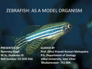


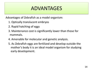


The document provides an overview of zebrafish (Danio rerio) as a model organism, detailing its taxonomy, genetic similarities to humans, and various applications in biological research, such as cancer studies and regenerative medicine. Key advantages for using zebrafish include their translucent embryos, rapid growth, and genetic similarity to humans, which make them ideal for studying development and disease. The document also highlights the historical context of zebrafish research and its future prospects in various scientific disciplines.