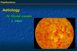
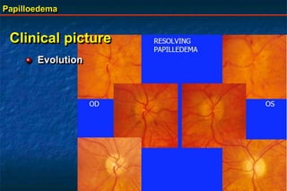
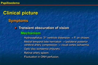
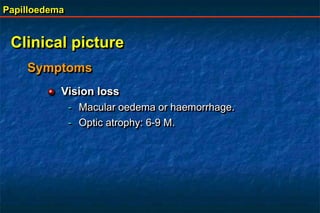
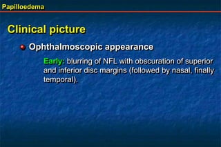
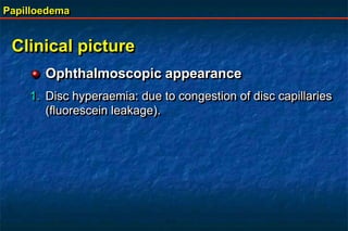
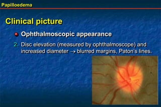
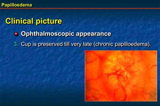
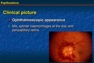
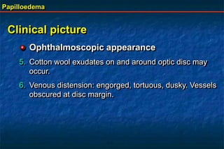
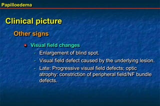
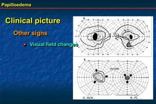
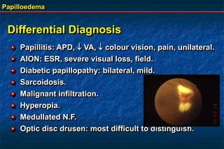
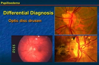
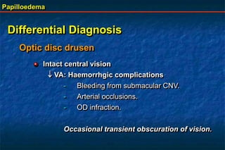
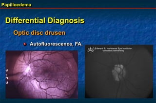
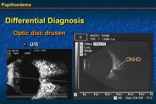
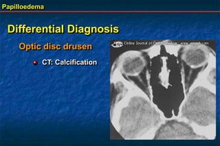
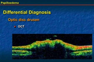
Papilloedema is swelling of the optic disc due to increased intracranial pressure. It is usually bilateral with normal vision. Common causes include brain tumors, subarachnoid hemorrhage, and idiopathic intracranial hypertension. On examination, papilloedema appears as blurred disc margins and venous engorgement. Grading scales are used to describe the degree of swelling. Differential diagnoses include papillitis, anterior ischemic optic neuropathy, and optic disc drusen.
This document discusses papilloedema, which refers to optic disc swelling caused by increased intracranial pressure. It defines various types of optic disc swelling including unilateral, bilateral, and papilloedema. It also discusses pseudo-papilloedema. The causes, signs, symptoms, diagnostic workup, and grading scale of papilloedema are explained in detail. Treatment options for increased intracranial pressure including idiopathic intracranial hypertension involve medical management with carbonic anhydrase inhibitors and weight loss as well as potential surgical interventions.
Papilloedema is swelling of the optic nerve head caused by increased intracranial pressure (ICP). It is nearly always bilateral and patients should be evaluated for an intracranial mass. Papilloedema can be caused by idiopathic intracranial hypertension, obstructive lesions, space occupying lesions, impaired CSF absorption, cerebral venous sinus thrombosis, cerebral edema from head trauma, or severe hypertension. Treatment involves reducing ICP through measures like mannitol infusion, CSF withdrawal, sedation, or surgery to remove masses. Complications can include visual loss or chronic headaches.
This document discusses optic disc swelling (papilledema) caused by increased intracranial pressure. It presents a case of a 35-year-old woman with severe headaches and vision issues. Examination found bilateral disc edema. CT scan revealed a brain tumor causing pressure. The patient was diagnosed with papilledema from the tumor and underwent surgery. The document then discusses the causes, presentation, stages, histopathology, and treatment of papilledema, emphasizing the importance of an eye exam for patients with headaches to identify potential intracranial issues.
This document provides an overview of hydrocephalus. It defines hydrocephalus as an abnormal enlargement of the head due to impaired circulation or absorption of cerebrospinal fluid. Hydrocephalus can be communicating or obstructive. It discusses the history, causes, types, symptoms, investigations and management of hydrocephalus. For management, it describes medical options to decrease CSF production or increase absorption. It also discusses various surgical procedures like shunt placement and endoscopic third ventriculostomy to divert CSF and bypass obstructions. Complications of shunt surgery like infections, mechanical issues and overdrainage are also summarized.
This document defines and describes papilledema, providing information on its pathophysiology, causes, symptoms, signs, grading, investigations, and treatment. Papilledema is passive swelling of the optic nerve head due to increased intracranial pressure. It is usually bilateral but can occasionally be unilateral. Increased ICP leads to increased optic nerve tissue pressure, altering the pressure gradient and causing swelling. Common causes include space occupying lesions, idiopathic intracranial hypertension, and cerebral edema. Signs include elevation and blurring of the optic disc margin. Treatment is directed at the underlying cause, with resolution of papilledema typically occurring within weeks of treatment.
Benign intracranial hypertension by Dr.Syed Alam ZebSyed Alam Zeb
This document summarizes a case of benign intracranial hypertension (BIH) in a 14-year-old boy. He presented with headaches and transient visual issues. Imaging showed sagittal sinus thrombosis and elevated cerebrospinal fluid pressure. He was diagnosed with pseudotumor cerebri due to an underlying protein deficiency. Treatment with medications improved his symptoms and papilledema. The document then discusses the pathophysiology, causes, signs/symptoms, diagnosis and treatment of BIH.
Papilledema refers to swelling of the optic discs due to increased intracranial pressure. If left untreated, it can cause vision loss or damage. Papillitis is inflammation of the optic nerve head, which can be caused by infections, autoimmune disorders, or other issues. It often causes reduced vision and color perception. Retrobulbar neuritis involves inflammation behind the eye affecting the optic nerve, and is a type of optic neuritis. It can be idiopathic or associated with conditions like multiple sclerosis.
This document contains descriptions of various physical examination findings and their associated diagnoses or causes. It includes descriptions and relevant information about clubbing fingers, splinter hemorrhages, xanthomata, pitting edema, pectus excavatum, carcinoma of the breast, Peutz-Jegher syndrome, hereditary hemorrhagic telangiectasia, porphyria cutanea tarda, spider nevi, scleral icterus, periorbital purpura, abdominal distention, caput medusa, and spleenomegaly. For each finding, it provides the description, associated diagnoses or causes, and sometimes additional relevant details.
Central retinal vein occlusion occurs when the central retinal vein becomes blocked, disrupting blood flow out of the retina. It can be caused by physical blockage at the lamina cribrosa or hemodynamic factors that obstruct blood flow. Histopathology shows occlusion at or behind the lamina cribrosa. Risk factors include hypertension, diabetes, glaucoma, and low physical activity. Investigations may include blood tests, imaging like fluorescein angiography, and screening for thrombophilias in younger patients. Features include retinal hemorrhages, edema, and delayed venous filling on angiography. Prognosis is generally poor for ischemic cases due to vision loss from macular edema, nonperfusion, and neovascular
1. The document discusses various eye conditions and symptoms, including how light is transmitted through the visual pathway and how vision is tested.
2. It describes papilledema, a sign of increased intracranial pressure that results in swelling of the optic disc. Causes include brain tumors and increased cerebral spinal fluid pressure.
3. Optic neuritis and optic atrophy are discussed as inflammatory and degenerative conditions of the optic nerve that can cause vision loss.
4. Hypertensive and diabetic retinopathies are summarized, noting how they are graded based on clinical appearance and prognosis. Diabetic retinopathy symptoms include blurred vision and can progress to vision loss if untreated.
Hydrocephalus is a condition caused by excess cerebrospinal fluid (CSF) building up in the cerebral ventricles. It can be caused by increased CSF production or decreased absorption. Symptoms include headache, vomiting, and vision changes. Diagnosis involves imaging tests like CT or MRI scans of the head. Treatment depends on the underlying cause but often involves surgically placing a shunt to drain fluid from the brain ventricles to other parts of the body like the abdomen. Prognosis depends on the individual case but hydrocephalus is associated with developmental delays and neurological complications.
Polypoidal choroidal vasculopathy (PCV) is a type of macular degeneration characterized by abnormal choroidal blood vessels that bulge outward forming polyp-like structures. It shares similarities with neovascular age-related macular degeneration but may have distinct pathogenesis involving disruption of choroidal vascular smooth muscle cells. PCV predominantly affects Asians and is a leading cause of vision loss in this population. Diagnosis is made using indocyanine green angiography which clearly visualizes the abnormal polypoidal choroidal vessels. Treatment options include laser photocoagulation, photodynamic therapy, anti-VEGF drugs, and combinations with the goal of resolving fluid, hemorrhage and regressing the poly
The document discusses the posterior fossa, including its boundaries, contents, blood supply, clinical presentation of lesions, and considerations for anesthesia. The posterior fossa is bounded anteriorly by the clivus and petrous bone, posteriorly by the occipital bone, and laterally by the temporal bone. It contains the cerebellar hemispheres, brainstem, and cranial nerves III-XII. Lesions can cause a variety of signs and symptoms depending on location, including ataxia, nystagmus, limb weakness, and cranial nerve deficits. Anesthesia for posterior fossa surgery requires careful monitoring and positioning to maintain stability while allowing surgical access.
This document discusses various types of edema (abnormal accumulation of fluid in tissues), including their causes and characteristics. It covers topics such as Starling's forces that govern fluid movement between blood vessels and tissues, the mechanisms leading to edema formation like increased hydrostatic pressure or reduced colloid osmotic pressure, and different forms of edema like pulmonary edema or cerebral edema. Specific conditions that can cause edema like nephrotic syndrome or congestive heart failure are also described.
This document discusses papilledema, which is swelling of the optic disc due to increased intracranial pressure. It defines papilledema and outlines its causes, signs, symptoms, grading, histopathology, investigations, differential diagnosis, and treatment. The main points are that papilledema is caused by increased intracranial pressure, it can be graded on a scale from 0-5 based on severity, and treatment involves addressing the underlying cause of pressure increase as well as surgical decompression in severe cases to prevent vision loss.
This document discusses optic disc edema (papilledema), which is swelling of the optic disc due to increased intracranial pressure. It defines papilledema and differentiates it from pseudopapilledema. Symptoms, signs, investigations and treatment are described. Papilledema is caused by increased intracranial pressure disrupting axoplasmic flow in the optic nerve. Signs include blurred disc margins, retinal folds, and fullness of the optic cup. Treatment involves addressing the underlying cause to reduce pressure and prevent vision loss.
Hydrocephalus is a condition characterized by the excessive accumulation of cerebral spinal fluid in the ventricles of the brain, causing their enlargement. It occurs due to an imbalance between CSF production and absorption. CSF is produced by the choroid plexus within the ventricles and circulates through the ventricular system before being absorbed into the venous system. Hydrocephalus can be obstructive, caused by a blockage in CSF flow within the ventricles, or communicating, caused by impaired absorption of CSF out of the ventricles. It is diagnosed through imaging like CT scans and MRIs showing ventricle dilation and treated through surgical implantation of shunts to drain CSF or endoscopic procedures to open blockages.
The document discusses the anatomy and clinical assessment of the visual pathway, including the optic nerve, chiasm, and cranial nerves involved (oculomotor, trochlear, abducent). It describes testing such as visual fields and perimetry to localize lesions. Specific conditions are examined like optic neuritis, papilledema, Horner's syndrome, and third, fourth, and sixth nerve palsies. Clinical features and causes are outlined for localized evaluation of disorders affecting the anterior visual pathway.
This document discusses various causes of optic disc edema. It begins by defining disc edema as swelling of the optic disc that can be caused by active or passive factors other than papilledema. Several pseudoedemas are described including drusen, myelinated fibers, tilted discs, and hypoplastic discs. True disc edemas can result from inflammation, vascular issues like CRVO, infiltrative diseases, or papilledema from increased intracranial pressure. Papilledema is usually bilateral non-inflammatory swelling caused by conditions that raise ICP like brain tumors, infections, pseudotumor cerebri, or venous sinus thrombosis. The pathogenesis and features of optic neuritis, multiple sclerosis, and papille
This document provides information about hydrocephalus, including:
1) Hydrocephalus is a condition where excess cerebrospinal fluid accumulates in the brain's ventricles and spinal cord spaces, dilating them.
2) It can be caused by obstruction of CSF flow, inadequate CSF absorption, or overproduction of CSF.
3) Symptoms vary depending on age but include headaches, vomiting, blurred vision, and gait or cognitive difficulties.
Hydrocephalus is a disorder where there is excessive cerebrospinal fluid in the brain ventricles causing increased pressure. It can be congenital due to neural tube defects, infections, or genetic syndromes. Symptoms include headache, vomiting, blurred vision. Diagnosis is made through neuroimaging and lumbar puncture. Treatment involves surgically placing a shunt to drain fluid from the brain to the abdomen. Complications can include infection or shunt malfunction.
Neurological Conditions and Diseases (At birth)Liew Boon Seng
This document discusses various neurological conditions and diseases that can cause macrocephaly in infants and children. It describes conditions present at birth such as caput succedaneum, subgaleal hemorrhage, cephalohematoma, osteopetrosis, subdural hematomas, benign enlargement of the subarachnoid space, megalencephaly, vein of Galen aneurysm, and hydrocephalus. Hydrocephalus and its causes, clinical presentation, assessment, treatments including shunts, and complications are discussed in detail. Posthemorrhagic hydrocephalus as a consequence of intraventricular hemorrhage is also outlined.
This document provides information on anterior ischemic optic neuropathy (AION), which is the most common cause of acute optic neuropathy in older age groups. It can be divided into two types: arteritic AION, which is due to giant cell arteritis; and non-arteritic AION, which makes up most cases. Both types present with sudden painless vision loss and optic disc swelling. Arteritic AION carries a worse prognosis and requires high-dose steroid treatment to prevent loss of vision in the fellow eye. Non-arteritic AION has a variable course but generally a poor rate of recovery without any proven effective treatments.
Angioid streaks are irregular, spoke-like lesions that radiate from the optic disc. They represent cracks in the calcified Bruch's membrane and were first described in 1889. While often asymptomatic, they can be associated with vision loss due to complications like choroidal neovascularization or macular degeneration. The most common systemic associations are pseudoxanthoma elasticum, Paget's disease, and hemoglobinopathies. Treatment may include observation, laser photocoagulation, photodynamic therapy, or surgery depending on complications.
This document discusses thyroid eye disease (TED), also known as Graves' ophthalmopathy. It covers the pathology, signs and symptoms, investigations, and treatment approaches for TED. Key points include that TED is an autoimmune disorder characterized by infiltrative orbitopathy, common signs include lid retraction, proptosis, restrictive myopathy, and optic neuropathy. Investigations include serological tests, CT or MRI imaging, and visual field testing. Treatment focuses on managing acute congestive orbitopathy, compressive optic neuropathy, motility disorders, and eyelid abnormalities, and may include corticosteroids, radiation therapy, or decompression surgery.
This document discusses hydrocephalus, including its classification, causes, investigation, and management. It begins with definitions and classifications of hydrocephalus. Key points include that it results from imbalance in CSF production and absorption, and can be communicating or non-communicating. Management options are then summarized, including ventricular shunting, which has high failure rates, and endoscopic third ventriculostomy as an alternative with fewer complications. Risk factors for shunt failure like infection are also outlined.
- Papilloedema is edema of the optic disc caused by increased intracranial pressure. It is a non-inflammatory condition where the optic nerve swells within the confined space of the optic nerve sheath.
- Increased intracranial pressure from brain tumors, abscesses, venous thrombosis, aneurysms or pseudotumor cerebri can cause papilloedema.
- On examination, the optic disc margins blur, the disc swells and veins become congested. Late stages show macular star/fan exudates and optic atrophy if untreated.
- Treatment is aimed at relieving intracranial pressure through tumor removal, shunting procedures or medication for pseud
This document provides an overview of the anatomy, physiology, clinical evaluation and treatment of various conditions affecting the eyelids, orbit, and lacrimal system. It discusses the basic anatomy of the eyelids including the 7 layers, muscles and glands. Common benign and malignant eyelid lesions are described along with disorders like entropion, ectropion and ptosis. The anatomy and clinical evaluation of orbital and lacrimal diseases are also reviewed. Surgical techniques for various eyelid, orbital and lacrimal conditions are mentioned.
TEST BANK For Community Health Nursing A Canadian Perspective, 5th Edition by...Donc Test
TEST BANK For Community Health Nursing A Canadian Perspective, 5th Edition by Stamler, Verified Chapters 1 - 33, Complete Newest Version Community Health Nursing A Canadian Perspective, 5th Edition by Stamler, Verified Chapters 1 - 33, Complete Newest Version Community Health Nursing A Canadian Perspective, 5th Edition by Stamler Community Health Nursing A Canadian Perspective, 5th Edition TEST BANK by Stamler Test Bank For Community Health Nursing A Canadian Perspective, 5th Edition Pdf Chapters Download Test Bank For Community Health Nursing A Canadian Perspective, 5th Edition Pdf Download Stuvia Test Bank For Community Health Nursing A Canadian Perspective, 5th Edition Study Guide Test Bank For Community Health Nursing A Canadian Perspective, 5th Edition Ebook Download Stuvia Test Bank For Community Health Nursing A Canadian Perspective, 5th Edition Questions and Answers Quizlet Test Bank For Community Health Nursing A Canadian Perspective, 5th Edition Studocu Test Bank For Community Health Nursing A Canadian Perspective, 5th Edition Quizlet Test Bank For Community Health Nursing A Canadian Perspective, 5th Edition Stuvia Community Health Nursing A Canadian Perspective, 5th Edition Pdf Chapters Download Community Health Nursing A Canadian Perspective, 5th Edition Pdf Download Course Hero Community Health Nursing A Canadian Perspective, 5th Edition Answers Quizlet Community Health Nursing A Canadian Perspective, 5th Edition Ebook Download Course hero Community Health Nursing A Canadian Perspective, 5th Edition Questions and Answers Community Health Nursing A Canadian Perspective, 5th Edition Studocu Community Health Nursing A Canadian Perspective, 5th Edition Quizlet Community Health Nursing A Canadian Perspective, 5th Edition Stuvia Community Health Nursing A Canadian Perspective, 5th Edition Test Bank Pdf Chapters Download Community Health Nursing A Canadian Perspective, 5th Edition Test Bank Pdf Download Stuvia Community Health Nursing A Canadian Perspective, 5th Edition Test Bank Study Guide Questions and Answers Community Health Nursing A Canadian Perspective, 5th Edition Test Bank Ebook Download Stuvia Community Health Nursing A Canadian Perspective, 5th Edition Test Bank Questions Quizlet Community Health Nursing A Canadian Perspective, 5th Edition Test Bank Studocu Community Health Nursing A Canadian Perspective, 5th Edition Test Bank Quizlet Community Health Nursing A Canadian Perspective, 5th Edition Test Bank Stuvia
Basavarajeeyam is a Sreshta Sangraha grantha (Compiled book ), written by Neelkanta kotturu Basavaraja Virachita. It contains 25 Prakaranas, First 24 Chapters related to Rogas& 25th to Rasadravyas.
Rasamanikya is a excellent preparation in the field of Rasashastra, it is used in various Kushtha Roga, Shwasa, Vicharchika, Bhagandara, Vatarakta, and Phiranga Roga. In this article Preparation& Comparative analytical profile for both Formulationon i.e Rasamanikya prepared by Kushmanda swarasa & Churnodhaka Shodita Haratala. The study aims to provide insights into the comparative efficacy and analytical aspects of these formulations for enhanced therapeutic outcomes.
Does Over-Masturbation Contribute to Chronic Prostatitis.pptxwalterHu5
In some case, your chronic prostatitis may be related to over-masturbation. Generally, natural medicine Diuretic and Anti-inflammatory Pill can help mee get a cure.
Local Advanced Lung Cancer: Artificial Intelligence, Synergetics, Complex Sys...Oleg Kshivets
Overall life span (LS) was 1671.7±1721.6 days and cumulative 5YS reached 62.4%, 10 years – 50.4%, 20 years – 44.6%. 94 LCP lived more than 5 years without cancer (LS=2958.6±1723.6 days), 22 – more than 10 years (LS=5571±1841.8 days). 67 LCP died because of LC (LS=471.9±344 days). AT significantly improved 5YS (68% vs. 53.7%) (P=0.028 by log-rank test). Cox modeling displayed that 5YS of LCP significantly depended on: N0-N12, T3-4, blood cell circuit, cell ratio factors (ratio between cancer cells-CC and blood cells subpopulations), LC cell dynamics, recalcification time, heparin tolerance, prothrombin index, protein, AT, procedure type (P=0.000-0.031). Neural networks, genetic algorithm selection and bootstrap simulation revealed relationships between 5YS and N0-12 (rank=1), thrombocytes/CC (rank=2), segmented neutrophils/CC (3), eosinophils/CC (4), erythrocytes/CC (5), healthy cells/CC (6), lymphocytes/CC (7), stick neutrophils/CC (8), leucocytes/CC (9), monocytes/CC (10). Correct prediction of 5YS was 100% by neural networks computing (error=0.000; area under ROC curve=1.0).
Osteoporosis - Definition , Evaluation and Management .pdfJim Jacob Roy
Osteoporosis is an increasing cause of morbidity among the elderly.
In this document , a brief outline of osteoporosis is given , including the risk factors of osteoporosis fractures , the indications for testing bone mineral density and the management of osteoporosis
ABDOMINAL TRAUMA in pediatrics part one.drhasanrajab
Abdominal trauma in pediatrics refers to injuries or damage to the abdominal organs in children. It can occur due to various causes such as falls, motor vehicle accidents, sports-related injuries, and physical abuse. Children are more vulnerable to abdominal trauma due to their unique anatomical and physiological characteristics. Signs and symptoms include abdominal pain, tenderness, distension, vomiting, and signs of shock. Diagnosis involves physical examination, imaging studies, and laboratory tests. Management depends on the severity and may involve conservative treatment or surgical intervention. Prevention is crucial in reducing the incidence of abdominal trauma in children.
share - Lions, tigers, AI and health misinformation, oh my!.pptxTina Purnat
• Pitfalls and pivots needed to use AI effectively in public health
• Evidence-based strategies to address health misinformation effectively
• Building trust with communities online and offline
• Equipping health professionals to address questions, concerns and health misinformation
• Assessing risk and mitigating harm from adverse health narratives in communities, health workforce and health system
Cell Therapy Expansion and Challenges in Autoimmune DiseaseHealth Advances
There is increasing confidence that cell therapies will soon play a role in the treatment of autoimmune disorders, but the extent of this impact remains to be seen. Early readouts on autologous CAR-Ts in lupus are encouraging, but manufacturing and cost limitations are likely to restrict access to highly refractory patients. Allogeneic CAR-Ts have the potential to broaden access to earlier lines of treatment due to their inherent cost benefits, however they will need to demonstrate comparable or improved efficacy to established modalities.
In addition to infrastructure and capacity constraints, CAR-Ts face a very different risk-benefit dynamic in autoimmune compared to oncology, highlighting the need for tolerable therapies with low adverse event risk. CAR-NK and Treg-based therapies are also being developed in certain autoimmune disorders and may demonstrate favorable safety profiles. Several novel non-cell therapies such as bispecific antibodies, nanobodies, and RNAi drugs, may also offer future alternative competitive solutions with variable value propositions.
Widespread adoption of cell therapies will not only require strong efficacy and safety data, but also adapted pricing and access strategies. At oncology-based price points, CAR-Ts are unlikely to achieve broad market access in autoimmune disorders, with eligible patient populations that are potentially orders of magnitude greater than the number of currently addressable cancer patients. Developers have made strides towards reducing cell therapy COGS while improving manufacturing efficiency, but payors will inevitably restrict access until more sustainable pricing is achieved.
Despite these headwinds, industry leaders and investors remain confident that cell therapies are poised to address significant unmet need in patients suffering from autoimmune disorders. However, the extent of this impact on the treatment landscape remains to be seen, as the industry rapidly approaches an inflection point.
3. Papilloedema
Definition
It is a passive oedema of the optic disc due to
increased ICP. It is almost always bilateral without
visual deficits.
Disc oedema is used for other forms of optic disc
swelling caused by inflammation, stasis or toxic
substances.
Pathologically papilloedema closely resembles disc
oedema not related to increased ICP.
5. Papilloedema
Aetiology
I. Increased intracranial pressure
1. Increased intracranial total mass volume:
Space occupying lesion: Brain tumours
Infratentorial: cerebellum & 4th ventricle
(obstruction through aqueduct).
Supratentorial: 3rd ventricle tumours.
6. Papilloedema
Aetiology
I. Increased intracranial pressure
1. Increased intracranial total mass volume
Less likely with brainstem and base of skull
tumours, parietal, occipital and parasagittal
tumours.
Rare with pituitary tumours.
Only 60% of patients with cerebral tumours
have papilloedema.
7. Papilloedema
Aetiology
I. Increased intracranial pressure
2. Increased protein content of CSF
Increased CSF osomotic pressure and
blocking absorptive pathways.
e.g. spinal tumours, Guillain-Barre syndrome,
meningitis, encephalitis.
19. Papilloedema
Pathogenesis
Controversial
Mechanical rather than vascular phenomenon.
Old theory
Increased CSF pressure vaginal sheaths of ON
CRV compression in perineural subarachnoid
space stagnation of venous return vascular
congestion and oedema.
20. Papilloedema
Pathogenesis:
Controversial
Hayreh
Increased ICP p. in perineural subarachnooid
space CSF diffuses into ON tissue pressure
axoplasmic flow block at lamina cribrosa ON
fiber swelling in ONH & leakage of H2O, proteins &
other axoplasmic contents into the extracellular
space of prelaminar region of ON.
44. Papilloedema
Clinical picture
Ophthalmoscopic appearance
The Friesen scale for grading papilledema:
Stage 0. Blurring of superior and inferior poles of disc.
Stage 1. Obscuration of nasal borders of disc, graying opacity of radial NFL,
concentric folds.
Stage 2. Obscuration of all borders and nasal elevation.
Stage 3. Moderate obscuration and elevation of all borders and some vessels,
leaving the disc with an early peripapillary halo.
Stage 4. Dome-shaped protrusion (champagne cork appearance) of the disc
with smoothly demarcated peripapillary halo and total obliteration of disc vessels
as well as the cup. There may also be marked hyperemia, vessel tortuosity,
hemorrhages, hard exudates, and cotton wool spots
47. Papilloedema
Clinical picture
Visual field changes
- Enlargement of blind spot.
- Visual field defect caused by the underlying lesion.
- Late: Progressive visual field defects; optic
atrophy: constriction of peripheral field/NF bundle
defects.
Other signs
49. Papilloedema
Clinical picture
E.O. muscle palsies
- 6th nerve; false localizing
Unilateral/bilateral
Long course, P at petrous temporal bone
diplopia, ET.
Sometimes intermittent.
Other signs
50. Papilloedema
Clinical picture
Other signs
E.O. muscle palsies
- 3rd, 4th nerves; rare.
Supratentorial mass cerebral tissue
displacement in a caudal direction nerve
stretching, displacement, compression, ischaemia.
51. Papilloedema
Differential Diagnosis
Papillitis: APD, VA, colour vision, pain, unilateral.
AION: ESR, severe visual loss, field.
Diabetic papillopathy: bilateral, mild.
Sarcoidosis.
Malignant infiltration.
Hyperopia.
Medullated N.F.
Optic disc drusen: most difficult to distinguish.
52. Papilloedema
Differential Diagnosis
Optic disc drusen
AD, irregular.
Onset: 2nd – 3rd decade.
Bilateral: 73%.
Small mulberry-like mass or waxy tumour
crenated edge.
Hidden drusen ≠ papilloedema examine parents.
Central cup: absent.
63. Papilloedema
Lumbar puncture
Increased pressure:
- Normal <200 mm H2O
- Borderline 200-250 mm H2O
- Abnormal >250 mm H2O
No abnormalities in protein level or cell count.
DD: meningitis, encephalitis.
Idiopathic Intracranial Hypertension (IIH)
65. Papilloedema
Diagnosis (Dandy’s)
Symptoms & signs of increased ICP: Headache,
papilloedema ….
No focal neurological signs except for 6th n. palsy.
Normal radiological studies (small ventricles).
Increased CSF pressure, normal contents.
Idiopathic Intracranial Hypertension (IIH)
66. Papilloedema
Causes: U-Devils
U Uraemia
D Drugs: steroids & steroid withdrawal, tetracycline, nalidixic acid,
lithium, amiodarone, heavy metal poisoning.
E Endocrine: obesity, Addison’s, DM, hypoparathyroidism.
V Vitamin A intoxication.
I Iron deficiency anaemia, pernicious anaemia.
L Last menstrual period: female, menarche, pregnancy, eclampsia.
S Sleep apnoea, chronic pulmonary disease.
Cerebral dural sinus obstruction.
In most cases, no clearly identifiable cause.
Idiopathic Intracranial Hypertension (IIH)
67. Papilloedema
Management
Frequent careful perimetry /M /3 M
Weight reduction (obese patients)
FU only:
- No visual symptoms or deficits.
- Minimal headache.
Idiopathic Intracranial Hypertension (IIH)
68. Papilloedema
Management
Severe headache
- Medical treatment
Diamox 500 mg 1x2 up to 2 gm/d.
Or Lasix (furosemide) 80-120 mg/d taper over several M.
Corticosteroids: less useful.
- Several lumbar punctures for several days.
- Visual field defect: initial/develops
Surgery:
o Lumbar peritoneal shunt
o ON sheath decompression
Idiopathic Intracranial Hypertension (IIH)
69. Papilloedema
Therapeutic success
Relief of headaches.
frequency of transient visual obscurations.
Regression of papilloedema.
Stability/improvement of field defects.
Weight reduction.
Idiopathic Intracranial Hypertension (IIH)