Downloaded 645 times
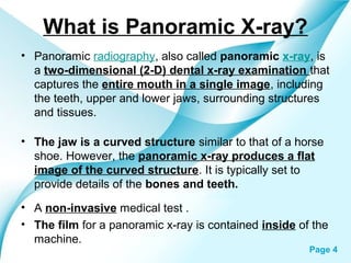
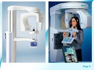
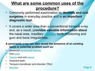


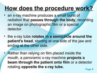


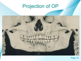

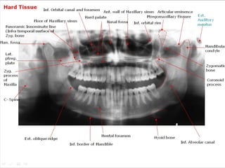


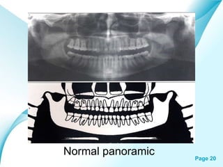

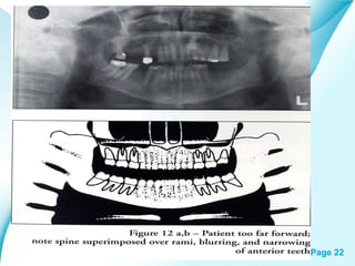


Panoramic radiography, also called a panoramic x-ray, captures a single image of the entire mouth, including teeth, jaws, and surrounding structures. It is commonly used by dentists and oral surgeons to evaluate bones, teeth, and check for issues like tumors, cysts, or impacted teeth. The procedure involves a rotating x-ray tube that projects a beam through the patient's head and onto a rotating film or detector. It is painless, fast, and provides a wider view than intraoral x-rays. While it does not show the same detail as other imaging tests, panoramic x-rays are useful for initial evaluation of dental problems.


