CBCT imaging allows dentists to visualize anatomy in 3 dimensions. It has many applications including implant planning, assessing impacted teeth, orthodontic evaluation, and examining maxillofacial trauma and lesions. CBCT provides important information such as bone quantity and quality, location of vital structures, and relationship of pathologies to surrounding tissues. It also allows for accurate pre-surgical planning through tools like radiographic tracing and implant simulation. CBCT has advantages over medical CT such as smaller size, lower radiation dose, and software tailored for dentistry.
Hey Guys, this presentation is all that a BDS graduate needs to know. A very basic yet important facts about CBCT.
Stay Safe
Regards
Battisi - Dr. Jasmine Singh
This presentation will give you a detailed knowledge about the various techniques that can be performed for imaging various aspects and diseases of TM Joint.
This presentation deals with radiographic imaging of three important phases of implant placement; PHASE 1: PRE-PROSTHETIC IMPLANT IMAGING
PHASE 2: SURGICAL AND INTERVENTIONAL IMPLANT IMAGING
PHASE 3: POST-PROSTHETIC IMPLANT IMAGING
is a diagnostic imaging modality that provide high quality ,CBCT uses systems that are ideal in capturing images of hard tissues especially in the maxillofacial region
brief description about CONTENTS Introduction Principles of panoramic imaging Image layer Panoramic machines Panoramic film Patient positioning Interpreting the panoramic imaging INDICATION Advantages Disadvantages Conclusion References
3. INTRODUCTION • Panoramic imaging also called pantomography is a technique for producing a single tomographic image of facial structures that includes both the maxillary and mandibular dental arches and their supporting structures . • This is a curvilinear variant of conventional tomography.
4. PRINCIPLES OF PANORAMIC IMAGE FORMATION • Patero and Numata - describe the principles of panoramic radiography • based on the principle of reciprocal movement of x-ray source and an image receptor around a central point or plane called the image layer, in which the OBJECT of image is located. • OBJECT in front or behind this image are not clearly captured because of their movement relative to the centre of rotation of the receptor and the x-ray source.
5. The film and x-ray tubehead move around the patient in opposite directions in panoramic radiography
6. ROTATION CENTER The pivotal point or axis around which the cassette carrier and tube head rotate is termed rotation center Three basic rotation center used in panoramic radiography Double centre rotation Triple centre rotation moving centre rotation The location and number of rotational centers INFLUENCE size and shape of focal trough
7. IMAGE LAYER • Also known as focal trough • It is a three dimensional curved zone where the structures lying within this layer are reasonably well defined on final panoramic image. • The structures seen on a panoramic image are primarily those located within image layer. • OBJECTSoutside the image layer are blurred magnified are reduced in size. Even distorted to the extent of not being recognizable. • This shape of image layer varies with the brand of equipment used.
8. FOCAL TROUGH
9. FACTORS AFFECTING SIZE OF IMAGE LAYER: Arc path Velocity of receptor and X-ray tube head Alignment of x-ray beam Collimator width The location of image layer change with extensive machine used so recalibration may be necessary if consistently suboptimal images are produced. As a position of object is moved within the image layer size and shape of image layer change.
10. PANORAMIC UNIT
11. A, Orthophos XG Plus extraoral x-ray machine. B, Orthoralix 8500 extraoral x-ray machine. C, Example of a digital panoramic system
12. PARTS OF PANORAMIC UNITS a. x-ray tube head b. head positioner: chin rest notched bite block forehead rest lateral head support c. exposure controls
13. X-RAY TUBE HEAD: • Similar to intraoral x-ray tube head • Each has a filament to produce electrons and a target to produce x-rays • Collimator is a lead plate with narrow vertical slit • Narrow x-ray beam emerges from collimator minimize patient exposure to radiation
1
This is a presentation describing in brief regarding the physics behind MRI and it's application from dental point of view. It contains few videos as well.
Application of ct & cone beam scan in dental implants /prosthodontic cou...Indian dental academy
The Indian Dental Academy is the Leader in continuing dental education , training dentists in all aspects of dentistry and
offering a wide range of dental certified courses in different formats.
Dental Cone Beam CT scans (CBCT Scans) provide high resolution, 3D volumetric images, used in Dental Implant Planning, Orthodontics and Maxillofacial surgery. allowing for more accurate analysis of bone structure and dental/tooth orientation. The accuracy of a CBCT scan is comparable to medical CT scans but uses a much lower radiation dose but with the added advantage of greater accuracy of bone structures and dental/tooth orientation. Please visit: https://ct-dent.co.uk/cone-beam-cbct-scans/
Cone beam computed tomography.DR. ANUBHUTI Dental Institute RIMS Anubhuti Singh
Cone beam computed tomography
Carm CT
Cone beam volume CT
Flat panel CT
Extra-oral imaging system specifically designed for three dimensional imaging of the oral and maxillofacial structures
ALARA Principle
Principal of cbct- Field of view
voxel
Diagnosis of Vertical Root Fracture Using Digital Radiography, Helical Comput...iosrjce
IOSR Journal of Dental and Medical Sciences is one of the speciality Journal in Dental Science and Medical Science published by International Organization of Scientific Research (IOSR). The Journal publishes papers of the highest scientific merit and widest possible scope work in all areas related to medical and dental science. The Journal welcome review articles, leading medical and clinical research articles, technical notes, case reports and others.
NVBDCP.pptx Nation vector borne disease control programSapna Thakur
NVBDCP was launched in 2003-2004 . Vector-Borne Disease: Disease that results from an infection transmitted to humans and other animals by blood-feeding arthropods, such as mosquitoes, ticks, and fleas. Examples of vector-borne diseases include Dengue fever, West Nile Virus, Lyme disease, and malaria.
TEST BANK for Operations Management, 14th Edition by William J. Stevenson, Ve...kevinkariuki227
TEST BANK for Operations Management, 14th Edition by William J. Stevenson, Verified Chapters 1 - 19, Complete Newest Version.pdf
TEST BANK for Operations Management, 14th Edition by William J. Stevenson, Verified Chapters 1 - 19, Complete Newest Version.pdf
Ethanol (CH3CH2OH), or beverage alcohol, is a two-carbon alcohol
that is rapidly distributed in the body and brain. Ethanol alters many
neurochemical systems and has rewarding and addictive properties. It
is the oldest recreational drug and likely contributes to more morbidity,
mortality, and public health costs than all illicit drugs combined. The
5th edition of the Diagnostic and Statistical Manual of Mental Disorders
(DSM-5) integrates alcohol abuse and alcohol dependence into a single
disorder called alcohol use disorder (AUD), with mild, moderate,
and severe subclassifications (American Psychiatric Association, 2013).
In the DSM-5, all types of substance abuse and dependence have been
combined into a single substance use disorder (SUD) on a continuum
from mild to severe. A diagnosis of AUD requires that at least two of
the 11 DSM-5 behaviors be present within a 12-month period (mild
AUD: 2–3 criteria; moderate AUD: 4–5 criteria; severe AUD: 6–11 criteria).
The four main behavioral effects of AUD are impaired control over
drinking, negative social consequences, risky use, and altered physiological
effects (tolerance, withdrawal). This chapter presents an overview
of the prevalence and harmful consequences of AUD in the U.S.,
the systemic nature of the disease, neurocircuitry and stages of AUD,
comorbidities, fetal alcohol spectrum disorders, genetic risk factors, and
pharmacotherapies for AUD.
- Video recording of this lecture in English language: https://youtu.be/lK81BzxMqdo
- Video recording of this lecture in Arabic language: https://youtu.be/Ve4P0COk9OI
- Link to download the book free: https://nephrotube.blogspot.com/p/nephrotube-nephrology-books.html
- Link to NephroTube website: www.NephroTube.com
- Link to NephroTube social media accounts: https://nephrotube.blogspot.com/p/join-nephrotube-on-social-media.html
micro teaching on communication m.sc nursing.pdfAnurag Sharma
Microteaching is a unique model of practice teaching. It is a viable instrument for the. desired change in the teaching behavior or the behavior potential which, in specified types of real. classroom situations, tends to facilitate the achievement of specified types of objectives.
New Drug Discovery and Development .....NEHA GUPTA
The "New Drug Discovery and Development" process involves the identification, design, testing, and manufacturing of novel pharmaceutical compounds with the aim of introducing new and improved treatments for various medical conditions. This comprehensive endeavor encompasses various stages, including target identification, preclinical studies, clinical trials, regulatory approval, and post-market surveillance. It involves multidisciplinary collaboration among scientists, researchers, clinicians, regulatory experts, and pharmaceutical companies to bring innovative therapies to market and address unmet medical needs.
Tom Selleck Health: A Comprehensive Look at the Iconic Actor’s Wellness Journeygreendigital
Tom Selleck, an enduring figure in Hollywood. has captivated audiences for decades with his rugged charm, iconic moustache. and memorable roles in television and film. From his breakout role as Thomas Magnum in Magnum P.I. to his current portrayal of Frank Reagan in Blue Bloods. Selleck's career has spanned over 50 years. But beyond his professional achievements. fans have often been curious about Tom Selleck Health. especially as he has aged in the public eye.
Follow us on: Pinterest
Introduction
Many have been interested in Tom Selleck health. not only because of his enduring presence on screen but also because of the challenges. and lifestyle choices he has faced and made over the years. This article delves into the various aspects of Tom Selleck health. exploring his fitness regimen, diet, mental health. and the challenges he has encountered as he ages. We'll look at how he maintains his well-being. the health issues he has faced, and his approach to ageing .
Early Life and Career
Childhood and Athletic Beginnings
Tom Selleck was born on January 29, 1945, in Detroit, Michigan, and grew up in Sherman Oaks, California. From an early age, he was involved in sports, particularly basketball. which played a significant role in his physical development. His athletic pursuits continued into college. where he attended the University of Southern California (USC) on a basketball scholarship. This early involvement in sports laid a strong foundation for his physical health and disciplined lifestyle.
Transition to Acting
Selleck's transition from an athlete to an actor came with its physical demands. His first significant role in "Magnum P.I." required him to perform various stunts and maintain a fit appearance. This role, which he played from 1980 to 1988. necessitated a rigorous fitness routine to meet the show's demands. setting the stage for his long-term commitment to health and wellness.
Fitness Regimen
Workout Routine
Tom Selleck health and fitness regimen has evolved. adapting to his changing roles and age. During his "Magnum, P.I." days. Selleck's workouts were intense and focused on building and maintaining muscle mass. His routine included weightlifting, cardiovascular exercises. and specific training for the stunts he performed on the show.
Selleck adjusted his fitness routine as he aged to suit his body's needs. Today, his workouts focus on maintaining flexibility, strength, and cardiovascular health. He incorporates low-impact exercises such as swimming, walking, and light weightlifting. This balanced approach helps him stay fit without putting undue strain on his joints and muscles.
Importance of Flexibility and Mobility
In recent years, Selleck has emphasized the importance of flexibility and mobility in his fitness regimen. Understanding the natural decline in muscle mass and joint flexibility with age. he includes stretching and yoga in his routine. These practices help prevent injuries, improve posture, and maintain mobilit
Flu Vaccine Alert in Bangalore Karnatakaaddon Scans
As flu season approaches, health officials in Bangalore, Karnataka, are urging residents to get their flu vaccinations. The seasonal flu, while common, can lead to severe health complications, particularly for vulnerable populations such as young children, the elderly, and those with underlying health conditions.
Dr. Vidisha Kumari, a leading epidemiologist in Bangalore, emphasizes the importance of getting vaccinated. "The flu vaccine is our best defense against the influenza virus. It not only protects individuals but also helps prevent the spread of the virus in our communities," he says.
This year, the flu season is expected to coincide with a potential increase in other respiratory illnesses. The Karnataka Health Department has launched an awareness campaign highlighting the significance of flu vaccinations. They have set up multiple vaccination centers across Bangalore, making it convenient for residents to receive their shots.
To encourage widespread vaccination, the government is also collaborating with local schools, workplaces, and community centers to facilitate vaccination drives. Special attention is being given to ensuring that the vaccine is accessible to all, including marginalized communities who may have limited access to healthcare.
Residents are reminded that the flu vaccine is safe and effective. Common side effects are mild and may include soreness at the injection site, mild fever, or muscle aches. These side effects are generally short-lived and far less severe than the flu itself.
Healthcare providers are also stressing the importance of continuing COVID-19 precautions. Wearing masks, practicing good hand hygiene, and maintaining social distancing are still crucial, especially in crowded places.
Protect yourself and your loved ones by getting vaccinated. Together, we can help keep Bangalore healthy and safe this flu season. For more information on vaccination centers and schedules, residents can visit the Karnataka Health Department’s official website or follow their social media pages.
Stay informed, stay safe, and get your flu shot today!
These lecture slides, by Dr Sidra Arshad, offer a quick overview of physiological basis of a normal electrocardiogram.
Learning objectives:
1. Define an electrocardiogram (ECG) and electrocardiography
2. Describe how dipoles generated by the heart produce the waveforms of the ECG
3. Describe the components of a normal electrocardiogram of a typical bipolar leads (limb II)
4. Differentiate between intervals and segments
5. Enlist some common indications for obtaining an ECG
Study Resources:
1. Chapter 11, Guyton and Hall Textbook of Medical Physiology, 14th edition
2. Chapter 9, Human Physiology - From Cells to Systems, Lauralee Sherwood, 9th edition
3. Chapter 29, Ganong’s Review of Medical Physiology, 26th edition
4. Electrocardiogram, StatPearls - https://www.ncbi.nlm.nih.gov/books/NBK549803/
5. ECG in Medical Practice by ABM Abdullah, 4th edition
6. ECG Basics, http://www.nataliescasebook.com/tag/e-c-g-basics
New Directions in Targeted Therapeutic Approaches for Older Adults With Mantl...i3 Health
i3 Health is pleased to make the speaker slides from this activity available for use as a non-accredited self-study or teaching resource.
This slide deck presented by Dr. Kami Maddocks, Professor-Clinical in the Division of Hematology and
Associate Division Director for Ambulatory Operations
The Ohio State University Comprehensive Cancer Center, will provide insight into new directions in targeted therapeutic approaches for older adults with mantle cell lymphoma.
STATEMENT OF NEED
Mantle cell lymphoma (MCL) is a rare, aggressive B-cell non-Hodgkin lymphoma (NHL) accounting for 5% to 7% of all lymphomas. Its prognosis ranges from indolent disease that does not require treatment for years to very aggressive disease, which is associated with poor survival (Silkenstedt et al, 2021). Typically, MCL is diagnosed at advanced stage and in older patients who cannot tolerate intensive therapy (NCCN, 2022). Although recent advances have slightly increased remission rates, recurrence and relapse remain very common, leading to a median overall survival between 3 and 6 years (LLS, 2021). Though there are several effective options, progress is still needed towards establishing an accepted frontline approach for MCL (Castellino et al, 2022). Treatment selection and management of MCL are complicated by the heterogeneity of prognosis, advanced age and comorbidities of patients, and lack of an established standard approach for treatment, making it vital that clinicians be familiar with the latest research and advances in this area. In this activity chaired by Michael Wang, MD, Professor in the Department of Lymphoma & Myeloma at MD Anderson Cancer Center, expert faculty will discuss prognostic factors informing treatment, the promising results of recent trials in new therapeutic approaches, and the implications of treatment resistance in therapeutic selection for MCL.
Target Audience
Hematology/oncology fellows, attending faculty, and other health care professionals involved in the treatment of patients with mantle cell lymphoma (MCL).
Learning Objectives
1.) Identify clinical and biological prognostic factors that can guide treatment decision making for older adults with MCL
2.) Evaluate emerging data on targeted therapeutic approaches for treatment-naive and relapsed/refractory MCL and their applicability to older adults
3.) Assess mechanisms of resistance to targeted therapies for MCL and their implications for treatment selection
Pulmonary Thromboembolism - etilogy, types, medical- Surgical and nursing man...VarunMahajani
Disruption of blood supply to lung alveoli due to blockage of one or more pulmonary blood vessels is called as Pulmonary thromboembolism. In this presentation we will discuss its causes, types and its management in depth.
How to Give Better Lectures: Some Tips for Doctors
Basic applications of CBCT
1. Prof. Mohamed Ekram
Cone Beam Computed Tomography (CBCT)
How to improve your practice outcome
and increase your income by using CBCT
2. M.Ekram
Cone Beam Computed Tomography
(CBCT) is the new frontier in dental
radiology.
It is probably the fastest growing area
in our discipline.
The advanced technology in computer
science made it necessary to make use
of it in dental radiography.
3. Objectives of this presentationObjectives of this presentation
Introduce Cone Beam principles
Discuss applications of CBCT
Demonstrate some interesting & unexpected
lesions.
Answer your questions
M.Ekram
4. Why take a 3D image?
2D 3D
When viewingWhen viewing two - dimensionaltwo - dimensional images , theimages , the
three - dimensionalthree - dimensional anatomy responsible foranatomy responsible for
the image must be considered.the image must be considered.
12. Production of a single axial cut.
Each rotation produces another slice
roduction of Conventional CT Image
The default is a slice
M.Ekram
Fan shaped Beam
14. Generation of views in
Conventional Medical
CT
3 - D
Coronal
Saggittal
3. Reconstruction
1. Slice is the
Default
M.Ekram
15. How the CBCT Images (CBCT)
are Generated ?
detector
Cone-shaped x-ray
beam
X-ray source
M.Ekram
16. 1. 3 – D
Default
3. Reconsruction
Axial
Coronal
Sagittal
M.Ekram
Generation of views in
CBCT
SCANORA 3D
17. Complete data can be reformatted to produce
images in other planes
Generation of CBCT Images
Axial
Sagittal
Coronal
M.Ekram
18. 2-D images can, then, be obtained in any plane, including the
M.Ekram
Reformatted panoramic
cross-sectional
cuts of the jaws.
19. X-ray source
object
Line detector
Movement of translation
and axis of rotation
From Multislice CT...
M.Ekram
Fan shape beam
X-ray source
object
axis of rotation
Flat panel
detector
...to real 3D CBCT
Cone
or Rectangle
What is the difference between
conventional CT and CBCT ?
20. M.Ekram
Advantages of CBCT over
Conventional CT
What is the difference between conventional CT and CBVT ?
21. Smaller in size & less space ccupyingSmaller in size & less space ccupying
M.Ekram
27. When the large FOV is the only
available FOV the following
results:
Less safety and more radiation
exposure for the patient.
false bone density results due to
huge amount of scattered
radiation.
When the Small FOV is the only available
FOV the following results:
Limited coverage of the
cranio-facial region with few
amount of information.
28. Dentally more specific than Conventional CTDentally more specific than Conventional CT
M.Ekram
Advantages of CBCT over
Conventional (medical) CT
29. Supplied with more specific softwareSupplied with more specific software
M.Ekram
30. Easier orientation of theEasier orientation of the
occlusal planeocclusal plane
M.Ekram
This is essential for correct slice direction
Face bow + Chair movement
33. Applications of CBCT
Implant site evaluation.
Assessment of impacted teeth.
3D & 2D Orthodontic/ orthognathic evaluation.
Maxillofacial trauma & fractures.
Maxillofacial lesions.
TMJ assessment.
Maxillofacial deformities & alveolar Clefts.
Examination of the Paranasal sinuses
Air way examination.
Other dental applications as endodontics &
Peridontal assessment.
M.Ekram
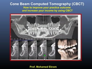
35. Bone quantity With 1 :1 Correspondence
(in all 3 dimensions, vertical, B-L & M-D)
Bone quality (only gives an idea)
Bone anatomy
Location of the vital structures
Presence of any minute bone pathology or small
remaining root fragments which cannot be always
detected by 2D plain radiographs
Useful information obtained from the
CBCT for the dental implantologist:
M.Ekram
36. Mesio-distal dimension: to determine the number
& diameter of the implants to be placed
1.5- 2mm between an implant & a natural tooth &
3mm between 2 implants
Bone Quantity
Vertical dimension: to determine the implant length
taking into consideration 2mm safety margin from
the IAC & 1 mm from the maxillary sinus & nasal
floor.ucco-lingual dimension: to determine the implant diameter
mm of bone should be present all around the implant
M.Ekram
37. Only gives an idea about estimated bone t
Bone quality is determined by obtaining the
CT. number (Bone density) in HU from the
CBCT software.
Mainly
Cortical
Symphysis
Mainly
Spongy
Post Mandible
Thick
Cortical &
Dense Spongy
Post Mandible
Thin
Cortical & Less
Dense Spongy
Ant Maxilla
M.Ekram
D5
38. D1 1250 or more
D2 850 -1250
D3 350 - 850
D4 150 - 350
D5 < 150
Quality in Hounsfield Units (in CT)
D5
39. Location of the Inferior alveolar canal from the
crest of the alveolus leaving a 2mm safety margin
Mental foramen.
Inferior border of the mandible.
Maxillary sinus.
Nasal floor.
Location of the vital structures
Nasal floor
Cross section
In the IAC
M.Ekram
40. Radiographic tracing is achieved by delineating the inferior alveolar canal on
he radiograph for easier measurement recording.
Radiographic Tracing :
M.Ekram
45. Compuer guided implant surgery
and Surgical Guides
M.Ekram
Precise preoperative planning software can be used
to accurately plan the placement of dental implants
using integrated data from a CBCT scan.
46. Disadvantages of Implant site
analysis
Programs based on medical CT
Systems.
Very high patient radiation dose.
No notable updating (e.g.DentaScan 2+).
More expensive than CBCT.
Time consuming.
No limited area examination is always possible.
Most of the cross sectional cuts in a single
examination are non-corrected (cannot be
customized).
51. Impacted Teeth.
M.Ekram
Visualization of the impacted teeth in the 3 dimensions.
The relation of the impacted tooth to the vital structures
The effect of the impacted tooth on adjacent teeth.
52. Interpretation of 3D images
M.Ekram
Surface mode &
transparent modes
3D Endoscopy
66. Exact extent of the lesion in 3
dimensional prospective
Relation of the lesion to vital
structures.
invasion to adjacent
structures or not.
CBCT In Maxillofacial lesions
81. 3-D view of skull
Showing pooling of the contrast solution in the
superficial lobe of the parotid gland.
Pooling of
contrast material
RT
Sialographic appearance compatible
with
Sjogren’s Syndrome.
89. A 14 years old boy with
history of a gunshot.
Clinically presented with an
anterior open bite with facial
lacerations.
CBCT examination revealed
bilateral comminuted
complex fracture.
100. Server and PACS survices
Radiology Center Access with all images loaded.
Referring doctor Access to the server site (Address).
Referring doctor Access to his own sector
ImageTransfer.
101. Serial
Unexpected
finding No of Cases
1 Caries
2 Periodontal disease
3 Dental anomalies
4 iInfections
5
Odontogenic & allied
lesions
6 Forein bodies
7 Total No of cases
The Unexpected Findings
112. Now you can restart your practice on
these new basis safely, quickly, and with
greater diagnostic information and
knowing all the details about your
patient.