
Recommended
More Related Content
What's hot
What's hot (20)
Viewers also liked
Similar to Extra oral Radiography
Similar to Extra oral Radiography (20)
More from DrJamilAlossaimi
More from DrJamilAlossaimi (6)
Recently uploaded
Recently uploaded (20)
Extra oral Radiography
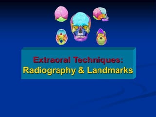
- 1. Extraoral Techniques: Radiography & Landmarks
- 2. Extra-oral Radiography Include all views made for the oro-facial region with film positioned extra-orally. Used to visualize the skull and facial structures, for detection of disease or injury or to evaluate skeletal growth. All extra oral radiographs require screen film and intensifying screen.
- 3. General Indications: Fractures of the skull and maxillofacial skeleton. Cases of Trismus. Detection of gross pathological lesions. Examination of TMJ., Maxillary sinuses, Salivary Glands. Localization of foreign bodies. Multiple impactions and supernumerary teeth. Examination of facial bones and skull bones.
- 4. Mandibular Skull Body Ramus PA AP Lat . PA . Water’s . Reverse Town’s . AP . SMV .Town’s .Trans- orbital . Lat Skull . Lat Ceph . Lat. TMJ PROJETIONS
- 5. Lateral Oblique Jaw Projections
- 6. Mandibular projection (body of the mandible)
- 7. Mandibular projection (body of the mand)
- 11. Indications of lateral oblique: Fractures of the mandible . Detection of gross pathological lesions. Examination of TMJ. Localization of Salivary gland stones. Impacted third molars specially in patients with trismus and gagging sensation.
- 15. 1. Lateral Head Film parallel to sagittal plane of the head The x-rays directed at right angles to the plane of the film
- 18. A) True lateral view Indications: Lateral profile of the skull. Fracture of the outer and inner plates of frontal sinuses. Relationship of the maxilla and mandible. Posterior displacement of maxillary fracture. Foreign bodies in the Oro-pharynx.
- 19. B) lateral Cephalometric view
- 22. Lateral Head holder C1 C3 C2 C4 C5 C6Vertebral body Intervertebral Disk space Frontal sinus (Huge) Sphenoid sinus Nasal bone Sphenoid sinus
- 25. C) Lateral TMJ views Trans-cranial. Trans-pharyngeal.
- 32. Indications: Localizing objects in mediolateral direction. Examination of frontal sinus. Fractures in maxilla and mandible specially in anterior region, angle of mandible, and sub-condylar area. Large pathological lesions and impaction.
- 34. Zyqoma tangential PA skull projection
- 36. Water’s View The Para-nasal Sinuses
- 38. Indications: Examination of para-nasal sinuses (frontal, ethmoidal, sphenoidal, and maxillary). Floors and inferior border of the orbits. Zygomatic bone and arches. Detection of middle third facial fractures. Examination of nasal bones and nasal cavity. Coronoid process fractures.
- 40. Water’s C-spine Nasal septum Frontal sinus Orbital rim Zygomatic arch Lateral wall of the nasal cavity Air-fluid level Ethmoid sinus Hyoid bone Maxillary sinus
- 44. Indications: * Suspection of condylar neck fracture. * Intracapsular fractures of the TMJ. * Condylar hypoplasia or hyperplasia. *Displacement of the condyle.
- 46. 3. Antero-posterior views A. True antero-posterior view. B. Towen’s view. C. Submento vertex view. D. Trans-orbital TMJ view.
- 51. Indications: Zygomatic arches fractures. Lesions affecting the palate or base of the skull. Investigation of sphenoid sinus.
- 52. Submentovertix Foramen Ovale Foramen Spinosum Foramen lacerum Foramen Magnum Occipital condyle Carotid canal Jugular fossa & foramen Mandibular condyle Petrous ridge Nasal septum Lateral wall of orbit Mandible
- 54. Zygomatic arch