PAGE(Polyacrylamide gel electrophoresis).pptx
•Download as PPTX, PDF•
1 like•212 views

Here I have Explained About Polyacrylamide Gel Electrophoresis . What is The Principle of Electrophoresis? What is Gel Electrophoresis? Types of Gel Electrophoresis. What is PAGE? How PAGE Run? Why SDS PAGE?

Recommended
More Related Content
What's hot
What's hot (20)
Similar to PAGE(Polyacrylamide gel electrophoresis).pptx
Similar to PAGE(Polyacrylamide gel electrophoresis).pptx (20)
Recently uploaded
Recently uploaded (20)
PAGE(Polyacrylamide gel electrophoresis).pptx
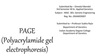
- 1. PAGE (Polyacrylamide gel electrophoresis) Submitted By – Omedul Mondal 3rd Semester M.Sc. Applied Genetics. Subject - MGE- 301: Genetic Engineering Reg. No.-20IAMOS007 Submitted to – Professor Subha Rajiv Department of Genetics Indian Academy Degree College Department of Genetics
- 2. CONTENTS • What is Electrophoresis and Its Principle? • What is Gel Electrophoresis? Types of Gel Electrophoresis • What is PAGE? • How PAGE runs? • Why SDS PAGE? • Difference between PAGE and Agarose Gel? • What is the
- 3. INTRODUCTION OF ELECTROPHORESIS • Migration of a charged particle under the influence of an electric field. • Word Electrophoresis means “to carry with electricity” • Arne Tiselius(father of electrophoresis), with support from the Rockefeller Foundation, developed the "Tiselius apparatus" for moving boundary electrophoresis, which was described in 1937 in the well-known paper "A New Apparatus for Electrophoretic Analysis of Colloidal Mixtures“.
- 5. PRINCIPLE
- 6. According to law of electrostatics, an ion with charge ‘Q’ in an electric field of strength ‘E’ will experience an electric force Resulting migration of the charged molecule through the solution is opposed by a frictional force Frictional coefficient depends on size and shape of the migrating molecule and viscosity of the medium. Felectrical=Q.E Ffrictional=V.f [V =rate of migration of charged molecule; f=frictional coefficient] f=
- 7. In the constant electric field the force on charged molecule balances each other The migration of the charged molecule in the electric field is termed as electrophoretic mobility( ) which is the ratio of the migration rate of a charged molecule to the applied electric field Electrophoretic mobility is directly proportional to the charge and inversely proportional to the viscosity of the medium, size and shape of the molecule. QE=Vf =Qf=VE
- 8. Gel Electrophoresis • Gel electrophoresis is a method for separation and analysis of macromolecules (DNA, RNA and proteins) and their fragments, based on their size and charge. • Used in clinical chemistry to separate proteins by charge or size and in biochemistry and molecular biology to separate a mixed population of DNA and RNA fragments by length, to estimate the size of DNA and RNA fragments or to separate proteins by charge.
- 9. Gel Electrophoresis Agarose gel Electrophoresis Starch Gel Electrophoresis Poly Acrylamide Gel Electrophoresis
- 10. Polyacrylamide gel • Consist of chains of acrylamide monomers crosslinked with Bisacrylamide ( Two units of acrylamide connected by methylene bridge).
- 11. • Polymerization of acrylamide is initiated by the quaternary amine TEMED( N,N,N,N-tetramethylethylenediamine ) which induces free radical formation from ammonium persulphate(APS). • Acrylamide and Bisacrylamide are a neurotoxin in their monomer form. • Resolving power and pore size of a gel depends on the concentration of acrylamide and bisacrylamide. • Lower percentage gel are better for resolving very high molecular weight molecule, higher percentage resolve small molecule. • Protein Mol.Wt range <5k to >200k and polynucleotide from <5 to 3k bp in size. • Gels that have single acrylamide percentage(concentration) throughout the gel :- Uniform Acrylamide concentration gel. • Gels that have different acrylamide percentage through the gel ,a low % of acrylamide at top and high % of acrylamide at bottom:- Gradient Acrylamide Gels.
- 12. Buffer System 1. Gel casting buffer – To cast the gel. 2. Sample Buffer- To prepare sample. 3. Running Buffer – To fill the electrode reserviour.
- 13. PAGE
- 14. SDS PAGE • Relative Movement of protein through PAGE depends on Charge Density(charge per unit mass), mass(or size) and the shape of the molecule. • In SDS PAGE proteins are treated with negatively charged anionic detergent sodium dodecyl sulfate before and during gel electrophoresis. • SDS imparts large net negative charge on proteins. • In SDS-PAGE migration of proteins are not determined by intrinsic electric charge of polypeptide but by molecular weight because SDS treatment eliminates the effect of difference in charge density and shape.
- 15. • Therefore in when current is on all SDS bound proteins in a sample will migrate towards positively charged electrode; proteins of less mass travel more quickly than those with greater mass. • In reducing SDS PAGE, along with SDS protein is treated with reducing Agents such as β-mercaptoethanol, dithiothreitol to break intrachain or interchain disulfide bonds between cysteine residues. • Nonreducing SDS PAGE reducing agents are not used.
- 16. Native PAGE • In native PAGE, proteins are separated according to the net charge, size and shape of their native structure. • Most protein molecules carry a net charge at any pH other than their isoelectric point and hence electrophoretic migration occurs because most proteins carry a net negative charge in alkaline running buffers. • The higher the negative charge density the faster a protein will migrate. At the same time the frictional force of the gel matrix creates a sieving effect, regulating the movement of proteins according to their size and three dimensional shape. • Small proteins face only a small frictional force, while larger proteins face a larger frictional force.
- 17. • Thus, in native PAGE , proteins are prepared in nonreducing, nondenaturing sample buffer, and electrophoresis is perfomed in the absence of denaturing and reducing agents. • Native charge is preserved , separates proteins based upon their charge, mass and shape in their native state and can resolve proteins of the same molecular weight.
- 18. Sample Visualization in the Gel • After electrophoresis run is complete ,proteins are visualized by gel staining. • Coomassie brilliant blue(the lower detectablelimit about 0.1-0.5microgram protein) is the most widely used dye which binds to proteins but not to gel itself. In acidic buffer condition Comassie dye binds to basic and hydrophobic residues of proteins, changing in colour from dull reddish- brown to intense blue. • Silver staining(lower detection limit about 0.5ng of proteins) is the most sensitive method for permanent visible staining of proteins in polyacrylamide gels. Here, silver ions is deposited to give a brown black color. Silver ions interacts and binds with carboxylic acid groups(Asp & Glu), imidazole(His), Sulfhydryls(Cys) and amines(Lys).
- 19. Estimating the molecular weight of a protein
- 20. Immunoblotting
- 21. PROCEDURE
- 22. SET UP GEL Get The purified Sample Load Sample Run Gel Stain and look at UV Light
- 23. Assembling the glass plates: • Assemble the glass plate on clean surface. Lay the longer glass plate (the one with spacer) down first,then place the shorter glass plate on top of it. • Embed them into the casting frame and clamp them properly make sure that the bottom ends of the glass plates are properly aligned • Then place it on the Casting Stand.
- 25. Casting the Gels • Prepare 10% of resolving gel . • Prepare the separating gel solution by combining all reagent. • Add APS and TEMED to the monomer solution(just before pouring) and mix well by swirling gently. Pour the solution till the mark. • Allow the gel to polymerize for 20-30minutes. • Prepare 4.5% stacking gel. Drain the isopropanol with strips of filter paper. • Add APS and TEMED to the monomer solution and mis well by swirling gently. • Place a comb in the stacking gel sandwich. Allow it to polymerize for 10 minutes.
- 26. Preparation of Samples • Mix your protein in the ratio 4:1 with the sample buffer. Heat your sample by either: (a) Boiling for 5-10minutes. (b) 65 C for 10 minutes. (c) 37 C for 30 minutes
- 31. References • Kumar P; Biophysics and Molecular Biology Fundamentals and Techniques; Pathfinder Publication; Third Edition; New Delhi • Wilson.K and Walker.J ;Principles and Techniques of Biochemistry and Molecular Biology; Cambridge University Press; Seventh Edition; UK •