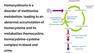
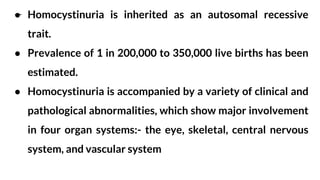
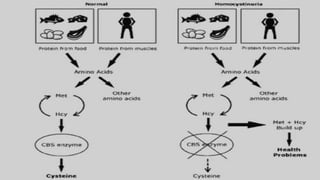
HOMOCYSTINURIA
Dr. V. MAGENDIRA MANI., M.Sc., M.Phil., Ph.D.,TNSET
Assistant Professor of Biochemistry
Islamiah College (Autonomous), Vaniyambadi
magivbio@gmail.com; 9486000227
Inborn errors of metabolism are inherited metabolic
disorders that result from defects in genes coding for
specific enzymes. This defect generates abnormal
chemical reactions that disrupt the normal metabolic
pathways resulting in the toxic accumulation of the
substrate behind the block or a deficiency in the product.
Inborn errors of metabolism
Homocystinuria is a
disorder of methionine
metabolism, leading to an
abnormal accumulation of
homocysteine and its
metabolites (homocystine,
homocysteine-cysteine
complex) in blood and
urine.
● Homocystinuria is inherited as an autosomal recessive
trait.
● Prevalence of 1 in 200,000 to 350,000 live births has been
estimated.
● Homocystinuria is accompanied by a variety of clinical and
pathological abnormalities, which show major involvement
in four organ systems:- the eye, skeletal, central nervous
system, and vascular system
● If both parents carry the faulty gene, for
each child, there is a:
● 25% chance the child will be born with
the disorder
● 50% chance the child will be a carrier
of the faulty gene
● 25% chance the child will neither have
disorder nor would be the carrier.
Inheritance of Homocystinuria
Homocystinuria Caused by Cystathionine
β-Synthase Deficiency (Classic Homocystinuria)
● Methionine is an essential, non-polar α-amino acid.
● Under normal conditions methionine undergoes conversion to
homocysteine.
● This in turn undergoes trans- sulfuration to ultimately yield
cysteine.
● This step is catalyzed by the enzyme Cystathionine beta
synthase (CBS).
● People suffering from this disease are unable to synthesize
CBS, hence leading to an inability to metabolize methionine
● Due to the absence of CBS enzyme, homocysteine
accumulates in the blood serum leading to an increased
excretion of homocystine in the urine
● Normally, most homocysteine, an intermediate compound of
methionine degradation, is remethylated to methionine.
● This methionine sparing reaction is catalyzed by the enzyme
methionine synthase,
● which requires a metabolite of folic acid (5-
methyltetrahydrofolate) as a methyl donor and a metabolite
of vitamin B12 (methylcobalamin), as well as S-
adenosylcobalamin, as cofactors
Other causes
● Homocystinuria Caused by Defects in Methylcobalamin
Formation
● Homocystinuria Caused by Deficiency of Methylene
tetrahydrofolate Reductase
Homocystinuria Caused by Defects
in Methylcobalamin Formation
● Methylcobalamin is the cofactor for the enzyme methionine
synthase,
● which catalyzes remethylation of homocysteine to methionine.
● There are at least 7 distinct defects in the intracellular
metabolism of cobalamin that may interfere with the formation
of methylcobalamin.
Homocystinuria Caused by Deficiency of
Methylene tetrahydrofolate Reductase
● This enzyme reduces 5,10-methylene tetrahydrofolate to form
5- methyltetrahydrofolate, which provides the methyl group
needed for remethylation of homocysteine to methionine
● The severity of the enzyme defect and the clinical
manifestations varies considerably in different families
Symptoms
● Delayed development or failure to thrive,
● Increasing visual problems
● As the child grows, physical deformities become more evident,
such as a peaked or hollowed chest, scoliosis, high arches of
the feet, fine brittle hair, a high palate with crowded teeth,
knock knees, and long thin limbs and fingers.
● Mental retardation,
● Dislocation of the lens of the eye
● Brittle bones that are prone to breaking
● Seizures, movement disorders, and anemia.
● Abnormal Blood clotting, strokes, heart disease and heart
attacks
● osteoporosis, or weakening of the bones
● Learning disabilities
● Long, spindly arms and legs
● Scoliosis - is a sideways curvature of the spine.
● Coronary artery disease - e.g., myocardial infarction
● Fatty infiltration of liver
● Blood clotting which could lead to heart strokes.
DIAGNOSIS
● The test usually looks for high levels of MET, homocystine,
and other sulphur-containing amino acids
● Tests also detect an enzyme deficiency (such as
cystathionine synthetase) can be done as well.
● x-rays to detect osteoporosis, and an eye exam to check for a
dislocated lens.
● Cystine is low or absent in plasma
● Genetic testing may additionally be helpful to identify
carriers in the family and for either prenatal or neonatal
screening for children at high risk of developing the
disease.
TREATMENT
● No specific cure has been discovered for homocystinuria
● Lifelong vitamin B6 supplements (also called pyridoxine)
● Those who do not respond to B6 will need to eat a lifelong low-
methionine diet. No meat, fish, dairy, or eggs are allowed, and
flour, beans, and nuts must be restricted
● A normal dose of folic acid supplement and occasionally adding
cysteine to the diet
● Betaine (N,N,N-trimethylglycine) is used to reduce
concentrations of homocysteine by promoting the conversion
of homocysteine back to methionine
● Special low-methionine flours, breads, and pastas are often
eaten, as well as a highprotein methionine-free formula.
● Administration of large doses of vitamin C (1 g/day) has
improved endothelial function
● The re-formed methionine is then gradually removed by
incorporation into body protein.
● The methionine that is not converted into protein is converted
to S-adenosyl-methionine which goes on to form homocysteine
again.
Prevention
● Genetic counseling is recommended for prospective parents
with a family history of homocystinuria.
● Prenatal diagnosis of homocystinuria is available and is
made by culturing amniotic cells or chorionic villi to test for
the presence or absence of cystathionine synthase
● Screen for homocystinuria in all newborns
Homocystinuria

Homocystinuria

  • 1.
    HOMOCYSTINURIA Dr. V. MAGENDIRAMANI., M.Sc., M.Phil., Ph.D.,TNSET Assistant Professor of Biochemistry Islamiah College (Autonomous), Vaniyambadi magivbio@gmail.com; 9486000227
  • 2.
    Inborn errors ofmetabolism are inherited metabolic disorders that result from defects in genes coding for specific enzymes. This defect generates abnormal chemical reactions that disrupt the normal metabolic pathways resulting in the toxic accumulation of the substrate behind the block or a deficiency in the product. Inborn errors of metabolism
  • 3.
    Homocystinuria is a disorderof methionine metabolism, leading to an abnormal accumulation of homocysteine and its metabolites (homocystine, homocysteine-cysteine complex) in blood and urine.
  • 4.
    ● Homocystinuria isinherited as an autosomal recessive trait. ● Prevalence of 1 in 200,000 to 350,000 live births has been estimated. ● Homocystinuria is accompanied by a variety of clinical and pathological abnormalities, which show major involvement in four organ systems:- the eye, skeletal, central nervous system, and vascular system
  • 6.
    ● If bothparents carry the faulty gene, for each child, there is a: ● 25% chance the child will be born with the disorder ● 50% chance the child will be a carrier of the faulty gene ● 25% chance the child will neither have disorder nor would be the carrier. Inheritance of Homocystinuria
  • 7.
    Homocystinuria Caused byCystathionine β-Synthase Deficiency (Classic Homocystinuria) ● Methionine is an essential, non-polar α-amino acid. ● Under normal conditions methionine undergoes conversion to homocysteine. ● This in turn undergoes trans- sulfuration to ultimately yield cysteine. ● This step is catalyzed by the enzyme Cystathionine beta synthase (CBS).
  • 8.
    ● People sufferingfrom this disease are unable to synthesize CBS, hence leading to an inability to metabolize methionine ● Due to the absence of CBS enzyme, homocysteine accumulates in the blood serum leading to an increased excretion of homocystine in the urine
  • 10.
    ● Normally, mosthomocysteine, an intermediate compound of methionine degradation, is remethylated to methionine. ● This methionine sparing reaction is catalyzed by the enzyme methionine synthase, ● which requires a metabolite of folic acid (5- methyltetrahydrofolate) as a methyl donor and a metabolite of vitamin B12 (methylcobalamin), as well as S- adenosylcobalamin, as cofactors
  • 11.
    Other causes ● HomocystinuriaCaused by Defects in Methylcobalamin Formation ● Homocystinuria Caused by Deficiency of Methylene tetrahydrofolate Reductase
  • 12.
    Homocystinuria Caused byDefects in Methylcobalamin Formation ● Methylcobalamin is the cofactor for the enzyme methionine synthase, ● which catalyzes remethylation of homocysteine to methionine. ● There are at least 7 distinct defects in the intracellular metabolism of cobalamin that may interfere with the formation of methylcobalamin.
  • 13.
    Homocystinuria Caused byDeficiency of Methylene tetrahydrofolate Reductase ● This enzyme reduces 5,10-methylene tetrahydrofolate to form 5- methyltetrahydrofolate, which provides the methyl group needed for remethylation of homocysteine to methionine ● The severity of the enzyme defect and the clinical manifestations varies considerably in different families
  • 14.
    Symptoms ● Delayed developmentor failure to thrive, ● Increasing visual problems ● As the child grows, physical deformities become more evident, such as a peaked or hollowed chest, scoliosis, high arches of the feet, fine brittle hair, a high palate with crowded teeth, knock knees, and long thin limbs and fingers. ● Mental retardation, ● Dislocation of the lens of the eye ● Brittle bones that are prone to breaking ● Seizures, movement disorders, and anemia. ● Abnormal Blood clotting, strokes, heart disease and heart attacks
  • 15.
    ● osteoporosis, orweakening of the bones ● Learning disabilities ● Long, spindly arms and legs ● Scoliosis - is a sideways curvature of the spine. ● Coronary artery disease - e.g., myocardial infarction ● Fatty infiltration of liver ● Blood clotting which could lead to heart strokes.
  • 16.
    DIAGNOSIS ● The testusually looks for high levels of MET, homocystine, and other sulphur-containing amino acids ● Tests also detect an enzyme deficiency (such as cystathionine synthetase) can be done as well. ● x-rays to detect osteoporosis, and an eye exam to check for a dislocated lens.
  • 17.
    ● Cystine islow or absent in plasma ● Genetic testing may additionally be helpful to identify carriers in the family and for either prenatal or neonatal screening for children at high risk of developing the disease.
  • 18.
    TREATMENT ● No specificcure has been discovered for homocystinuria ● Lifelong vitamin B6 supplements (also called pyridoxine) ● Those who do not respond to B6 will need to eat a lifelong low- methionine diet. No meat, fish, dairy, or eggs are allowed, and flour, beans, and nuts must be restricted ● A normal dose of folic acid supplement and occasionally adding cysteine to the diet
  • 19.
    ● Betaine (N,N,N-trimethylglycine)is used to reduce concentrations of homocysteine by promoting the conversion of homocysteine back to methionine ● Special low-methionine flours, breads, and pastas are often eaten, as well as a highprotein methionine-free formula. ● Administration of large doses of vitamin C (1 g/day) has improved endothelial function
  • 20.
    ● The re-formedmethionine is then gradually removed by incorporation into body protein. ● The methionine that is not converted into protein is converted to S-adenosyl-methionine which goes on to form homocysteine again.
  • 21.
    Prevention ● Genetic counselingis recommended for prospective parents with a family history of homocystinuria. ● Prenatal diagnosis of homocystinuria is available and is made by culturing amniotic cells or chorionic villi to test for the presence or absence of cystathionine synthase ● Screen for homocystinuria in all newborns