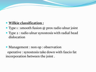
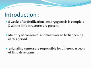
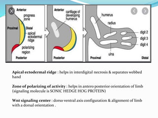
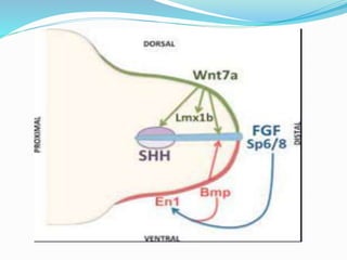
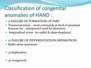
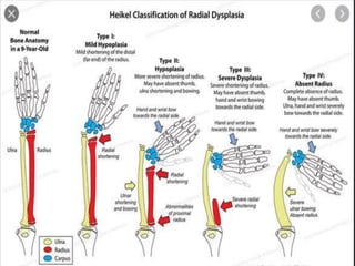
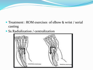
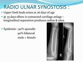
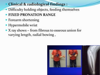
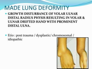
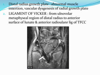
DR VENKATESH V
ASSISTANT PROFESSOR
DEPT OF ORTHOPAEDICS
SSMC
Introduction :
 8 weeks after fertilization , embryogenesis is complete
& all the limb structures are present.
 Majority of congenital anomalies are to be happening
at this period.
 3 signaling centers are responsible for different aspects
of limb development.
Apical ectodermal ridge : helps in interdigital necrosis & separates webbed
hand
Zone of polarizing of activity : helps in antero posterior orientation of limb
(signaling molecule is SONIC HEDGE HOG PROTEIN)
Wnt signaling center : dorso ventral axis configuration & alignment of limb
with a dorsal orientation .
Classification : FRANTZ O RAHILLY
Terminal (T)
LONGITUDINAL
1. Complete paraxial hemimelia (complete absence of
one of the forearm or leg elements, and of the
corresponding portion of the hand or foot)
2. Incomplete paraxial hemimelia (similar to the
above, but part of the defective element is present)—
3. Partial adactylia (absence of one to four digits and
their
metacarpals or metatarsals): 1, 2, 3, 4, or 5
4. Partial aphalangia (absence of one or more
phalanges from one to four digits): 1, 2, 3, 4, or 5
TRANSVERSE
1. Amelia (absence of limb)
2. Hemimelia (absence of forearm and hand or leg and
foot)
3. Partial hemimelia (part of forearm or leg is present)
4. Acheiria or apodia (absence of hand or foot)
5. Complete adactylia (absence of all five digits and
their metacarpals or metatarsals)
6. Complete aphalangia (absence of one or more
phalanges from all five digits)
Intercalary (I)
TRANSVERSE
1. Complete phocomelia (hand or foot attached
directly to trunk)
2. Proximal phocomelia (hand and forearm, or
foot and leg, attached directly to trunk)
3. Distal phocomelia (hand or foot attached
directly to arm or thigh)
LONGITUDINAL
1. Complete paraxial hemimelia (similar to
corresponding terminal defect but hand or
foot is more or less complete
2. Incomplete paraxial hemimelia (similar to
corresponding terminal defect but hand or foot
is more or less complete
3. Partial adactylia (absence of all or part of a
metacarpal or metatarsal
4. Partial aphalangia (absence of proximal or
middle phalanx, or both, from one or more
digits
Classification of congenital
anomalies of HAND :
 1) FAILURE OF FORMATION OF PART
 Transverse arrest – most commonly at level of proximal
forearm (ex : misoprostol used for abortion)
 longitudinal arrest (ex radial & ulnar dysplasia)
 2) FAILURE OF DIFFERENTIATION (SEPARATION)
 Radio ulnar synostosis
 3) duplication
 4) overgrowth
 5) undergrowth
 6) congenital constriction band syndrome
 7) gen abnormalities – MADELUNG DEFORMITY
PHOCOMELIA/SEAL LIMB/FLIPPER:
 Extreme expression of longitudinal reduction of
intercalated segment of limb.
 Hand is suspended from the body (near shoulder)with
only 3 or 4 digits
 M/C due to thalidomide
 FRANTZ O RAHILLY CLASSIFICATION :
1. Complete all 4 limbs affected
2. Extreme hypoplasia of proximal limb bones with
forearm & hand attached to the trunk
3. Hand attached directly to humerus with associated
deficiencies
Treatment : often conservative
Rarely prosthesis
RADIAL DYSPLASIA / RADIAL CLUB HAND :
 Deficiency along the preaxial or radial side of the
extremity.
 Sporadic occurance
 Thalidomide & radiation
 Etiopath : cong absence / 20 to syphillis /abnormal
pressure on embryo / AER defect.
 shortened forearm
 Radially deviated wrist
 Treatment : ROM exercises of elbow & wrist / serial
casting
 Sx Radialization / centralization
RADIO ULNAR SYNOSTOSIS :
 Upper limb buds arises at 26 days of age
 @ 35 days elbow is connected cartilage anlage –
longitudinal separation produces radius & ulna.
 Epidemio : 90% sporadic
90% bilateral
male > female
 Clinical & radiological findings :
 Difficulty holding objects, feeding themselves
 FIXED PRONATION RANGE
 Forearm shortening
 Hypermobile wrist
 X ray shows – from fibrous to osseous union for
varying length, radial bowing ,
 Wilkie classification :
 Type 1 : smooth fusion @ prox radio ulnar joint
 Type 2 : radio ulnar synostosis with radial head
dislocation
 Management : non op : observation
operative : synostosis take down with fascio fat
incorporation between the joint .
MADE LUNG DEFORMITY
 GROWTH DISTURBANCE OF VOLAR ULNAR
DISTAL RADIUS PHYSIS RESULTING IN VOLAR &
ULNAR DRIFTED HAND WITH PROMINENT
DISTAL ULNA.
 Etio : post trauma / dysplastic/ chromosomal /
idiopathic
1. Distal radius growth plate : abnormal muscle
insertion, vascular dysgenesis of radial growth plate
2. LIGAMENT OF VICKER : from ulnovolar
metaphyseal region of distal radius to anterior
surface of lunate & anterior radioulanr lig of TFCC
 Management : distal radio-ulnar arthrodesis with
simultaneous distal ulanr osteotomy (darrachs)
CLEIDO CRANIAL DYSOSTOSIS :
 Aplasia of clavicle
 Exaggerated development of transverse diameter of
cranium
 Delayed closure of fontanelle
 AD
 Etio : membranous bones affected
non fusion of 20 ossification centers
PFFD
 Femur is shorter than normal
 Discontinuity between femoral neck & shaft
 4-6 weeks limb buds appear
 M/C etiology to affect @ this time = anoxia , ischaemia
,infection, toxins, hormonal
 Etiopath : sclerotomal substraction , thalidomide,
maternal diabetes .
 C/F : short limb , FABER, hip & knee dysfunction ,
FFD of hip & knee , bulbous proximal thigh
 Classifications : HIMANISHI
AITKEN
GILLESPIE & TORODE
FIXSEN & BOYD
AITKENS CLASSIFICATION
NINE PAPPAS CLASSIFICATION
 Management :
Equine prosthesis
Ankle disarticulation & prosthesis
Rotationplasty
Knee arthrodesis
Congenital skeletal limb deficiences

Congenital skeletal limb deficiences

  • 1.
    DR VENKATESH V ASSISTANTPROFESSOR DEPT OF ORTHOPAEDICS SSMC
  • 2.
    Introduction :  8weeks after fertilization , embryogenesis is complete & all the limb structures are present.  Majority of congenital anomalies are to be happening at this period.  3 signaling centers are responsible for different aspects of limb development.
  • 3.
    Apical ectodermal ridge: helps in interdigital necrosis & separates webbed hand Zone of polarizing of activity : helps in antero posterior orientation of limb (signaling molecule is SONIC HEDGE HOG PROTEIN) Wnt signaling center : dorso ventral axis configuration & alignment of limb with a dorsal orientation .
  • 5.
  • 6.
    Terminal (T) LONGITUDINAL 1. Completeparaxial hemimelia (complete absence of one of the forearm or leg elements, and of the corresponding portion of the hand or foot) 2. Incomplete paraxial hemimelia (similar to the above, but part of the defective element is present)— 3. Partial adactylia (absence of one to four digits and their metacarpals or metatarsals): 1, 2, 3, 4, or 5 4. Partial aphalangia (absence of one or more phalanges from one to four digits): 1, 2, 3, 4, or 5 TRANSVERSE 1. Amelia (absence of limb) 2. Hemimelia (absence of forearm and hand or leg and foot) 3. Partial hemimelia (part of forearm or leg is present) 4. Acheiria or apodia (absence of hand or foot) 5. Complete adactylia (absence of all five digits and their metacarpals or metatarsals) 6. Complete aphalangia (absence of one or more phalanges from all five digits)
  • 7.
    Intercalary (I) TRANSVERSE 1. Completephocomelia (hand or foot attached directly to trunk) 2. Proximal phocomelia (hand and forearm, or foot and leg, attached directly to trunk) 3. Distal phocomelia (hand or foot attached directly to arm or thigh) LONGITUDINAL 1. Complete paraxial hemimelia (similar to corresponding terminal defect but hand or foot is more or less complete 2. Incomplete paraxial hemimelia (similar to corresponding terminal defect but hand or foot is more or less complete 3. Partial adactylia (absence of all or part of a metacarpal or metatarsal 4. Partial aphalangia (absence of proximal or middle phalanx, or both, from one or more digits
  • 8.
    Classification of congenital anomaliesof HAND :  1) FAILURE OF FORMATION OF PART  Transverse arrest – most commonly at level of proximal forearm (ex : misoprostol used for abortion)  longitudinal arrest (ex radial & ulnar dysplasia)  2) FAILURE OF DIFFERENTIATION (SEPARATION)  Radio ulnar synostosis  3) duplication  4) overgrowth
  • 9.
     5) undergrowth 6) congenital constriction band syndrome  7) gen abnormalities – MADELUNG DEFORMITY
  • 10.
    PHOCOMELIA/SEAL LIMB/FLIPPER:  Extremeexpression of longitudinal reduction of intercalated segment of limb.  Hand is suspended from the body (near shoulder)with only 3 or 4 digits  M/C due to thalidomide
  • 11.
     FRANTZ ORAHILLY CLASSIFICATION : 1. Complete all 4 limbs affected 2. Extreme hypoplasia of proximal limb bones with forearm & hand attached to the trunk 3. Hand attached directly to humerus with associated deficiencies Treatment : often conservative Rarely prosthesis
  • 12.
    RADIAL DYSPLASIA /RADIAL CLUB HAND :  Deficiency along the preaxial or radial side of the extremity.  Sporadic occurance  Thalidomide & radiation  Etiopath : cong absence / 20 to syphillis /abnormal pressure on embryo / AER defect.  shortened forearm  Radially deviated wrist
  • 14.
     Treatment :ROM exercises of elbow & wrist / serial casting  Sx Radialization / centralization
  • 15.
    RADIO ULNAR SYNOSTOSIS:  Upper limb buds arises at 26 days of age  @ 35 days elbow is connected cartilage anlage – longitudinal separation produces radius & ulna.  Epidemio : 90% sporadic 90% bilateral male > female
  • 16.
     Clinical &radiological findings :  Difficulty holding objects, feeding themselves  FIXED PRONATION RANGE  Forearm shortening  Hypermobile wrist  X ray shows – from fibrous to osseous union for varying length, radial bowing ,
  • 17.
     Wilkie classification:  Type 1 : smooth fusion @ prox radio ulnar joint  Type 2 : radio ulnar synostosis with radial head dislocation  Management : non op : observation operative : synostosis take down with fascio fat incorporation between the joint .
  • 18.
    MADE LUNG DEFORMITY GROWTH DISTURBANCE OF VOLAR ULNAR DISTAL RADIUS PHYSIS RESULTING IN VOLAR & ULNAR DRIFTED HAND WITH PROMINENT DISTAL ULNA.  Etio : post trauma / dysplastic/ chromosomal / idiopathic
  • 19.
    1. Distal radiusgrowth plate : abnormal muscle insertion, vascular dysgenesis of radial growth plate 2. LIGAMENT OF VICKER : from ulnovolar metaphyseal region of distal radius to anterior surface of lunate & anterior radioulanr lig of TFCC
  • 20.
     Management :distal radio-ulnar arthrodesis with simultaneous distal ulanr osteotomy (darrachs)
  • 21.
    CLEIDO CRANIAL DYSOSTOSIS:  Aplasia of clavicle  Exaggerated development of transverse diameter of cranium  Delayed closure of fontanelle  AD  Etio : membranous bones affected non fusion of 20 ossification centers
  • 22.
    PFFD  Femur isshorter than normal  Discontinuity between femoral neck & shaft  4-6 weeks limb buds appear  M/C etiology to affect @ this time = anoxia , ischaemia ,infection, toxins, hormonal
  • 23.
     Etiopath :sclerotomal substraction , thalidomide, maternal diabetes .  C/F : short limb , FABER, hip & knee dysfunction , FFD of hip & knee , bulbous proximal thigh  Classifications : HIMANISHI AITKEN GILLESPIE & TORODE FIXSEN & BOYD
  • 24.
  • 25.
     Management : Equineprosthesis Ankle disarticulation & prosthesis Rotationplasty Knee arthrodesis