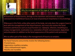
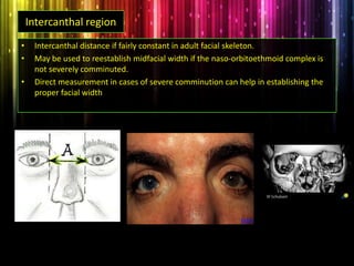
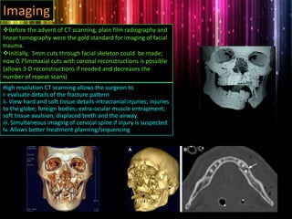
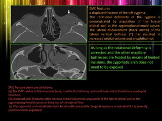
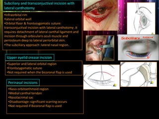
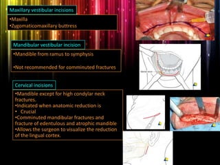
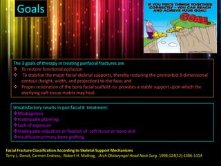
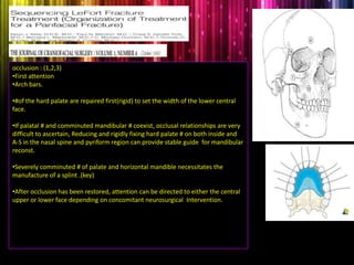
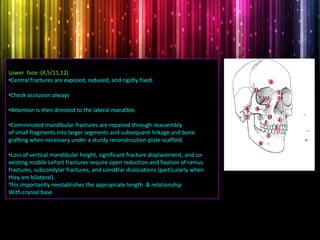
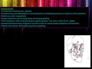
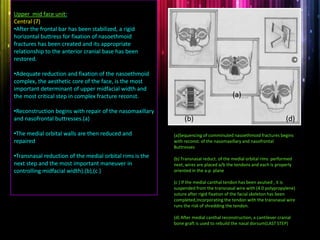
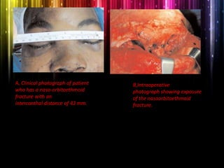
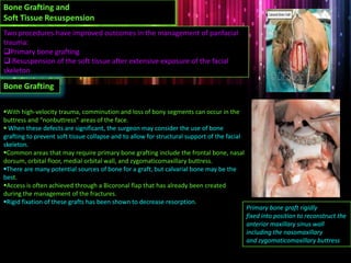
Panfacial fractures involve multiple facial bones, including the frontal bones, zygomaticomaxillary complex, naso-orbitoethmoid region, maxilla and mandible. Due to the complex nature of these injuries, management requires careful planning and sequencing of treatment to restore facial functions, features and symmetry. Key goals are to reestablish occlusion, stabilize major facial supports to restore three-dimensional contour, and provide a stable scaffold for soft tissue healing. Proper imaging, surgical approaches and attention to anatomical landmarks are important to achieve accurate reduction and fixation.
orthognathic surgery is very intresting and well knowing branch in oral surgery ....this presentation is dealing with jaw correction surgery in upper jaw.
Dentist in pune.(BDS. MDS) - Dr. Amit T. Suryawanshi..Zygomaticomaxillary com...All Good Things
Dentist in pune. (BDS. MDS) - Dr. Amit T. Suryawanshi. Seminar-Canine Impaction.
Email ID- amitsuryawanshi999@gmail.com
Contact -Ph no.-9405622455
Subscribe our channel on youtube - https://www.youtube.com/channel/UC_gylEXTrjmEbbOTSXjuZ4Q/videos?view_as=public
Follow us on slideshare
Dr. Ahmed M. Adawy
Professor Emeritus, Dep. Oral & Maxillofacial Surg.
Former Dean, Faculty of Dental Medicine
Al-Azhar UniversityOrthognathic surgery is the art and science of combining orthodontics and maxillofacial surgery to correct dento-facial deformities.The etiology, prevalence,diagnosis and preoperative planning,and Surgical procedures are presented.
Dr. Ahmed M. Adawy, Professor Emeritus, Dep. Oral & Maxillofacial Surgery. Former Dean, Faculty of Dental Medicine, Al-Azhar University. Condylar fractures represent one of the most controversial issues in maxillofacial traumatology regarding classification, diagnoses and therapeutic management. Classification systems of condylar fracture is discussed. Diagnosis is usually based on history clinical examination and radiographic finding. Treatment ranges from observation, jaw exercises to closed or opened interventions. For years closed reduction was thought to be essentially complication-free. Several serious complications however have been reported including temporomandibular joint ankyloses, malocclusion, mandibular deviation and the generative joint pathology. The absolute and relative indications for open reduction is given. The debate between supporters of open or closed reduction is still continuing and the issue has not been resolved. However, the final choice treatment modality should takes into account the location of the fracture, age of the patient, presence or absence of other associated injuries, cosmetic impact of the surgery and presence of other systemic medical conditions.
The naso-orbitoethmoid complex (NOE) fracture represents the most wearisome and challenging of all facial fractures due to the complexity and intricacy of its surgical & anatomic components. A good working knowledge with regards its surgical anatomy, clinical features, sequence of treatment & surgical approaches, potential pitfalls in its treatment & postoperative consideration,. Appropriate diagnosis and timely treatment is crucial to avoid unfavorable & difficult to treat sequelae.
Zygomatic Complex Fractures
Dr. Ahmed M. Adawy
Professor Emeritus, Dept. Oral & Maxillofacial Surg.
Former Dean, Faculty of Dental Medicine
Al-Azhar University
The term “zygomatic complex” refers to zygomatic bone and parts of maxilla, frontal, temporal and sphenoid bone. Fracture of the zygomatic complex, also known as a quadripod fracture, and formerly referred to as a tripod fracture, varies in severity from a simple crack to major disruption. The etiology, clinical presentations, and radiographic findings are presented. Classification systems are mentioned. The management of zygomatic complex fracture depends on the degree of displacement and the resultant esthetical and functional deficit. As a general rule, non- displaced or minimal displaced fracture can usually be treated conservatively. On the other hand, open reduction and internal fixation is applied in all dislocated, instable, and comminuted fractures of the zygomatic bone. Different surgical approaches and fixation methods are discussed.
Dr. Ahmed M. Adawy, Professor Emeritus, Dep. Oral & Maxillofacial Surgery. Former Dean, Faculty of Dental Medicine, Al-Azhar University. Mandibular angle fractures account for 23% to 42% of all facial fractures. Fracture of mandibular angle can be classified as (A) Vertical favorable or unfavorable, (B) Horizontally favorable of unfavorable. Traditionally, mandibular angle fractures have been treated with either closed reduction and inter-maxillary fixation or open reduction and internal fixation with or without inter-maxillary fixation. Patients treated with inter-maxillary fixation have a restricted airway and loose excess weight. Rigid internal fixation and early return to function have eliminated the use of wire osteosenthysis and prolonged use of inter-maxillary fixation. The principal of rigid fixation, however, have inherent set of disadvantages including damage to the inferior alveolar nerve and the marginal mandibular branch of facial nerve. Postoperative malocclusion rates are also high. With the introduction of semi-rigid technique fracture of the mandibular angle could be treated according to Champy’s Ideal lines of osteosenthysis. The technique involves placement of a single monocortial miniplate on the superior border of the mandible. However, some studies suggested using a second miniplate along the inferior border. Wether one or two miniplates should be used is still debatable. The application of 3D plates may provide additional stability in 3 dimension and good resistance against torque forces.
Instructions for Submissions thorugh G- Classroom.pptxJheel Barad
This presentation provides a briefing on how to upload submissions and documents in Google Classroom. It was prepared as part of an orientation for new Sainik School in-service teacher trainees. As a training officer, my goal is to ensure that you are comfortable and proficient with this essential tool for managing assignments and fostering student engagement.
Read| The latest issue of The Challenger is here! We are thrilled to announce that our school paper has qualified for the NATIONAL SCHOOLS PRESS CONFERENCE (NSPC) 2024. Thank you for your unwavering support and trust. Dive into the stories that made us stand out!
How to Create Map Views in the Odoo 17 ERPCeline George
The map views are useful for providing a geographical representation of data. They allow users to visualize and analyze the data in a more intuitive manner.
Welcome to TechSoup New Member Orientation and Q&A (May 2024).pdfTechSoup
In this webinar you will learn how your organization can access TechSoup's wide variety of product discount and donation programs. From hardware to software, we'll give you a tour of the tools available to help your nonprofit with productivity, collaboration, financial management, donor tracking, security, and more.
The Roman Empire A Historical Colossus.pdfkaushalkr1407
The Roman Empire, a vast and enduring power, stands as one of history's most remarkable civilizations, leaving an indelible imprint on the world. It emerged from the Roman Republic, transitioning into an imperial powerhouse under the leadership of Augustus Caesar in 27 BCE. This transformation marked the beginning of an era defined by unprecedented territorial expansion, architectural marvels, and profound cultural influence.
The empire's roots lie in the city of Rome, founded, according to legend, by Romulus in 753 BCE. Over centuries, Rome evolved from a small settlement to a formidable republic, characterized by a complex political system with elected officials and checks on power. However, internal strife, class conflicts, and military ambitions paved the way for the end of the Republic. Julius Caesar’s dictatorship and subsequent assassination in 44 BCE created a power vacuum, leading to a civil war. Octavian, later Augustus, emerged victorious, heralding the Roman Empire’s birth.
Under Augustus, the empire experienced the Pax Romana, a 200-year period of relative peace and stability. Augustus reformed the military, established efficient administrative systems, and initiated grand construction projects. The empire's borders expanded, encompassing territories from Britain to Egypt and from Spain to the Euphrates. Roman legions, renowned for their discipline and engineering prowess, secured and maintained these vast territories, building roads, fortifications, and cities that facilitated control and integration.
The Roman Empire’s society was hierarchical, with a rigid class system. At the top were the patricians, wealthy elites who held significant political power. Below them were the plebeians, free citizens with limited political influence, and the vast numbers of slaves who formed the backbone of the economy. The family unit was central, governed by the paterfamilias, the male head who held absolute authority.
Culturally, the Romans were eclectic, absorbing and adapting elements from the civilizations they encountered, particularly the Greeks. Roman art, literature, and philosophy reflected this synthesis, creating a rich cultural tapestry. Latin, the Roman language, became the lingua franca of the Western world, influencing numerous modern languages.
Roman architecture and engineering achievements were monumental. They perfected the arch, vault, and dome, constructing enduring structures like the Colosseum, Pantheon, and aqueducts. These engineering marvels not only showcased Roman ingenuity but also served practical purposes, from public entertainment to water supply.
Ethnobotany and Ethnopharmacology:
Ethnobotany in herbal drug evaluation,
Impact of Ethnobotany in traditional medicine,
New development in herbals,
Bio-prospecting tools for drug discovery,
Role of Ethnopharmacology in drug evaluation,
Reverse Pharmacology.
How to Split Bills in the Odoo 17 POS ModuleCeline George
Bills have a main role in point of sale procedure. It will help to track sales, handling payments and giving receipts to customers. Bill splitting also has an important role in POS. For example, If some friends come together for dinner and if they want to divide the bill then it is possible by POS bill splitting. This slide will show how to split bills in odoo 17 POS.
Synthetic Fiber Construction in lab .pptxPavel ( NSTU)
Synthetic fiber production is a fascinating and complex field that blends chemistry, engineering, and environmental science. By understanding these aspects, students can gain a comprehensive view of synthetic fiber production, its impact on society and the environment, and the potential for future innovations. Synthetic fibers play a crucial role in modern society, impacting various aspects of daily life, industry, and the environment. ynthetic fibers are integral to modern life, offering a range of benefits from cost-effectiveness and versatility to innovative applications and performance characteristics. While they pose environmental challenges, ongoing research and development aim to create more sustainable and eco-friendly alternatives. Understanding the importance of synthetic fibers helps in appreciating their role in the economy, industry, and daily life, while also emphasizing the need for sustainable practices and innovation.
2. The panfacial injury Conceptually, panfacial fractures are
defined as those involving the upper,middle and lower third of
face(peterson).
In practice, when two out of these three areas are involved,
the term “panfacial fracture” has been applied.These complex
facial injuries are generally result of high velocity trauma and
often produce complex fractures that are extensive and not in
the patterns as cleanly outlined by Le Fort.
What is it if someone says that you have a Pan face??
The face which is flattened due to an extreme blow by a pan
Panfacial fractures
Racist used to describe it as one of a Chinese decent
3. Treatment of facial trauma, damage to the dentition and anatomic structures
subsequent to maxillofacial injury is an issue of paramount importance in traumatology.
Because in this field, unlike other parts of the body, not only does the surgeon have to
deal with the management of the facial fractures, but must also restore the facial
functions and features such as visual function (i.e. diplopia), olfaction, breathing (i.e.
airway management), mastication (i.e. restoration of teeth and occlusion), deglutition
and articulation (in addition to the facial appearance of the patient and symmetry).
In no other part of the body is the management of trauma so complex.
Why the management of this type of trauma is so complex?
Pan facial fractures concurrently involve the following bones :
•Frontal bones,
•Zygomatico-maxillary complex,
• Naso-orbitoethmoid region,
•Maxilla and mandible.
4. Contents
Incidence And Etiology
Anatomical Consideration
Imaging
Timing
Surgical Approaching
Goals & Sequence Of Treatment
Bone Grafting And Soft Tissues Resuspension
Conclusion
5. Motor vehicle collisions
Assault
Sports related accidents
Industrial accidents
Gunshot wounds
INCIDENCE
Kapoor P, Kalra N. A retrospective analysis of maxillofacial injuries
in patients reporting to a tertiary care hospital in East Delhi. Int J
Crit Illn Inj Sci 2012;2:6-10
Raval CB, M. Airway management in patients with
maxillofacial trauma - A retrospective study of 177 cases.
Saudi J Anaesth 2011;5:9-14
ETIOLOGY
INCIDENCE & ETIOLOGY
6. Fossilized cranium and finite element model of
Australopithecus africanus. Bright colors indicate
high strain.
Buttress:A means or device that keeps
something erect, stable, or secure
Anatomical considerations’
Facial buttress
The buttress system of face is formed by strong frontal, maxillary, zygomatic ,sphenoid
and mandible bones and their attachments to one another. The central midface
contains many fragile bones that could easily crumble when subjected to strong
forces. These fragile bones are surrounded by thicker bones of the facial buttress
system lending them some strength and stability. These buttress represent the best
available understanding of the mechanical support of face as they determine how an
impact is distributed over the face
7. For better understanding the components of the facial buttress
system have been divided into:
1. Vertical buttresses
2. Horizontal buttresses
Vertical buttress: These buttresses are very well
developed. Described by manson et al.vertical buttress are
responsible for three dimentional projection of midface .
They include:
1. Nasomaxillary
2. Zygomaticomaxillay
3. Pterygomaxillay
4. Vertical mandible
Majority of the forces absorbed by midface are masticatory in
nature (vertically oriented). Hence the vertical buttresses are
well developed in humans .
Horizontal buttresses:These buttresses interconnect and provide
support for the vertical buttresses. They include:
a. Frontal bar
b. Infraorbital rim & nasal bones
c. Hard palate & maxillary alveolus
8. The buttresses represent areas of relative increased bone thickness that
support the functional units of the face (muscles, eyes, dental
occlusion, airway) in an optimal relation and define the form of the face by
projecting the overlying soft-tissue envelope.
Facial buttress pearls are as follows:
(a) The buttress concept was intended for improved appreciation of facial structure; it
does not replace traditional anatomic terms.
(b) Buttresses have sufficient bone thickness to accommodate metal screw fixation.
(c) Buttresses are all linked either directly or through another buttress to the cranium or
cranial base as a stable reference point.
(d) Transverse buttress reduction restores facial profile and width; vertical buttress
reduction restores facial height.
(e) Buttress reduction establishes a functional support for the teeth and globes.
So restoration of 3-D shape of face after panfacial fracture requires precise reduction of
these buttress against stable cranial base or mandible
Source : Diagnosis of Midface Fractures with CT: What the Surgeon Needs to Know.Richard A. Hopper. RadioGraphics 2006; 26:783–793
9. When there is panfacial fractures
,reconstruction should be approached as a
puzzle. Known landmarks can be used to
reconstruct more precisely those areas that
have been damaged. These landmarks may help
in establishing the proper positioning of facial
skeleton:
1. Dental arches
2. The Mandible
3. Sphenozygomatic suture
4. Intercanthal region
Anatomical considerations’
Key Landmarks
10. Dental arches
• When one or both dental arches are intact they can be used to a guide to establish proper
dental width.
• Clinical scenario of Midpalatal split + fracture of the tooth bearing region of the mandible +
condylar fracture. 3 options:
1. Establish maxillary width by exposing the palatal fracture and doing reduction and rigid
fixation.
2. Take impressions for fabrication of dental models . Perform simulated surgery on upper and
lower arches and fabricate a surgical splint.
If the patient has dental models from preinjury orthodontic or prosthodontic
rehabilitation, these can provide good clues to establishing proper arch form.
3. Reconstruct the mandible first as it is a very robust bone that can be anatomically reduced if
attention is paid to detail.
11. The mandible
• Aim to achieve anatomical reduction of both lingual and buccal cortical surfaces
prior to fixation.
• Bilateral subcondylar fractures must be treated to establish posterior facial height
and facial width.
• Bilateral subcondylar fracture + fracture of the symphysis and or body- the
mandible may undergo splaying (widening).The condyle can be reconstituted to
ramus to help establish facial height and width.
12. Sphenozygomatic suture
• Situated along the internal surface of the lateral orbital wall.
• Is a key landmark for reduction and fixation of the zygomaticomaxillary complex
provided the orbital roof and lateral orbit are intact .
• Likewise the zygomatic buttress is important in establishing the proper position of
the zygoma and or maxilla.
• If there is gross bone loss in this are, primary bone grafting may be indicated to
reestablish the buttress.
The surgeon should pay particular attention to the alignment of the zygoma
and sphenoid at the lateral orbital wall, since angulation here after fixation of the remaining
buttresses reflects a residual rotational deformity and an associated increased orbital volume.
13. Intercanthal region
• Intercanthal distance if fairly constant in adult facial skeleton.
• May be used to reestablish midfacial width if the naso-orbitoethmoid complex is
not severely comminuted.
• Direct measurement in cases of severe comminution can help in establishing the
proper facial width
14. Before the advent of CT scanning, plain film radiography and
linear tomography were the gold standard for imaging of facial
trauma.
Initially, 5mm cuts through facial skeleton could be made;
now 0.75mmaxial cuts with coronal reconstructions is possible
(allows 3-D reconstructions if needed and decreases the
number of repeat scans)
Imaging
High resolution CT scanning allows the surgeon to
i. evaluate details of the fracture pattern
ii. View hard and soft tissue details-intracranial injuries; injuries
to the globe; foreign bodies; extra-ocular muscle entrapment;
soft tissue avulsion; displaced teeth and the airway.
iii. Simultaneous imaging of cervical spine if injury is suspected.
Iv. Allows better treatment planning/sequencing
15. If the rectus remains flattened in cross-
section and in the correct position, the
fascial sling is likely intact and the surgeon
will encounter minimal entrapped
periorbital tissue (Fig 10a). However, if
the inferior rectus is round and inferiorly
displaced, the fascial sling is disrupted and
the periorbita and muscle have prolapsed
into the orbital floor defect
ORBITS
16. Orbital apex.
(a) normal anatomy of the orbital apices
(b) impingement of the orbital apex secondary to
a sphenoid–skull base fracture.
An isolated blow-in fracture of the left orbital roof
.The associated exophthalmos and dural tears were
treated with an intracranial approach.
Orbital fracture pearls are as follows:
(a) Orbital fractures can occur in isolation or with other fracture patterns.
(b) The position and shape of the medial and inferior rectus muscles can indicate whether entrapment and
clinical diplopia are likely.
(c) Pediatric trapdoor orbital fractures are a surgical emergency.
(d) The size of the orbital floor defect can be underestimated in severely Impacted ZMC fractures.
(e) Medial orbital wall blow-out fractures cause enophthalmos if the posterior- medial orbital bulge is lost.
(f) Orbital apex compression with clinical decreasing vision is a surgical emergency.
17. NOE fractures
Radiologic description of NOE fractures should comment on the
degree of comminution of the medial vertical maxillary buttress,
specifically in the region of the lacrimal fossa, where the medial
canthus attaches.
Nasofrontal ducts.
NOE fracture pearls are as follows:
(a) NOE fractures are distinguished from simple nasal fractures by posterior disruption of the medial canthal
region, the ethmoids, and the medial orbital walls.
(b) Clinically, the most obvious deformity is loss of nasal projection in profile and apparent increased distance between
the inner corners of the eyes.
(c) NOE fractures can be classified by the degree of injury to the region where the medial canthus attaches around the
lacrimal fossa.
(d) Although the frontal sinus may not be directly injured, if the nasofrontal ducts are disrupted, then frontal sinus
surgery is needed to prevent a mucocele in the future.
18. ZMC fractures
a displaced fracture of the left zygoma.
The rotational deformity of the zygoma is
demonstrated by angulation of the lateral
orbital wall at the zygomaticosphenoid suture.
The lateral displacement (black arrow) of the
lateral vertical buttress (*) has resulted in
increased orbital volume and enophthalmos
As long as the rotational deformity is
corrected and the other maxillary
buttresses are fixated by means of limited
incisions, the zygomatic arch does not
need to be exposed
ZMC fracture pearls are as follows:
(a) The ZMC relates to the temporal bone, maxilla, frontal bone, and skull base and is therefore a quadripod
structure.
(b) Displaced ZMC fractures often increase orbital volume by angulation of the lateral orbital wall at the
zygomaticosphenoid suture or blow-out of the orbital floor.
(c) The zygomatic arch establishes both facial width and profile. Surgical exposure is indicated if it is severely
comminuted or angulated.
19. Bilateral Le Fort I, II, and III fractures. The lateral and
medial maxillary buttresses (white lines) are fractured
inferiorly and superiorly (junctions of white lines and
black lines).
To confirm the diagnosis, pterygomaxillary
disjunction and fractures of the zygomatic arches
would need to be observed on axial images.
A right-sided unilateral pterygomaxillary disjunction, which
has resulted in separation of the posterior vertical maxillary
buttress (*) from the rest of the maxilla; this appearance is
indicative of a Le Fort fracture.
The contralateral pterygomaxillary junction is intact because
the fracture exited in the form of a parasagittal palate
fracture
Le Fort fracture pearls are as follows:
(a) All Le Fort fractures require disruption of the pterygoids from the posterior maxilla, as seen at
axial imaging.
(b) Any combination of Le Fort I, II, and III patterns can occur.
(c) A sagittal or parasagittal hard palate fracture with a Le Fort pattern will result in a widened
maxillary arch.
(d) Displaced unilateral Le Fort fractures are possible only with a sagittal or parasagittal palate
fracture.
20. Fracture repair should be initiated as soon as the patient's other injuries permit.
Particularly in midfacial fracture repair Paul Manson’s quote: “you never get a second
chance” has to be kept in mind .Early management of fractures facilitates reduction and
avoids the insult of a second injury to soft tissues in a vulnerable period of early wound
healing. Reduction and fixation of complex injuries within 48 hours is ideal; management
within 10 days is critical because soft-tissue stiffening and interfragmentary healing make
later corrections very difficult.
It is not so much the fracture morphology in the midfacial area that limits the intended
treatment but mainly the preexisting general health status and the severity of associated
accompanying injuries or in the vicinity of the midface (optic nerve trauma, CSF leakage,
bleeding, etc) or in independent locations.
TIMING
21. Surgical Approaches
Designed to achieve wide exposure of the
fracture lines which is essential for accurate
anatomic reduction.
The location and extent of exposure are
dependent upon fracture severity and
combination.
22. Bicoronal flap
•Frontal sinus
•Superior part of naso-orbito ethmoid
•Medial canthal tendon
•Supraorbital rim
•Orbital roof
•Superior aspect of lateral orbital wall
•Zygomatic arch
•Mandibular condyle (with
preauricular extension)
23. Subciliary and transconjuctival incision with
lateral canthotomy
•Infraorbital rim
•lateral orbital wall
•Orbital floor & frontozygomatic suture:
transconjuctival incision with lateral canthotomy .It
requires detachment of lateral canthal ligament and
incision through orbicularis oculi muscle and
periosteum deep to lateral periorbital skin.
•The subciliary approach :lateral nasal region.
Upper eyelid crease incision
•Superior and lateral orbital region
•Frontozygomatic suture
•Not required when the bicoronal flap is used
Perinasal incisions
•Naso-orbitoethmoid region
•Medial canthal tendon
•Nasolacrimal sac
•Disadvantage: significant scarring occurs
•Not required if Bicoronal flap is used
24. Maxillary vestibular incisions
•Maxilla
•Zygomaticomaxillary buttress
Mandibular vestibular incision
•Mandible from ramus to symphysis
•Not recommended for comminuted fractures
Cervical incisions
•Mandible except for high condylar neck
fractures.
•Indicated when anatomic reduction is
• Crucial
•Comminuted mandibular fractures and
fracture of edentulous and atrophic mandible
•Allows the surgeon to visualize the reduction
of the lingual cortex.
25. The 3 goals of therapy in treating panfacial fractures are
To restore functional occlusion
To stabilize the major facial skeletal supports, thereby restoring the premorbid 3-dimensional
contour (height, width, and projection) to the face; and
Proper restoration of the bony facial scaffold to provides a stable support upon which the
overlying soft tissue matrix may heal.
Facial Fracture Classification According to Skeletal Support Mechanisms
Terry L. Donat, Carmen Endress, Robert H. Mathog, .Arch Otolaryngol Head Neck Surg. 1998;124(12):1306-1314
Goals
Unsatisfactory results in pan facial # treatment:
Misdiagnosis
Inadequate planning
Lack of exposure
Inadequate reduction or fixation of soft tissue or bone and
Insufficient primary bone grafting
26. Crucial decision
Dictated by fracture pattern, extent of other injuries
Extensive head injuries and prolonged intubation anticipated-
tracheostomy(it also facilitates management of multiple facial #.
Extensive injuries in NOE Region make nasal intubation difficult
If IMF is not possible or not indicated- oral intubation /submental
/retromolar (in c/o symphysis/body #submental intubation hinders
access
Airway management
27. Sequence Of Repair
Manson says that when multiple areas of face are fractured ,an order of treatment
needs to be developed.
The exact order of treatment is not as important as the development of the plan that
permits both flexibily and reproducibly accurate positioning of the various fracture
segments.
Different orders of treatment have been proposed ,any of which are satisfactory if
one understands the anatomy ,goals, and procedures.
This issue however relate more to the experience and habits of surgeon and
prevention of common treatment errors.
Much has been written about proper sequencing of treatment for Panfacial
fractures. “Bottom up & inside out” or “Top down & outside in” have been used to
describe 2 of classic approaches for management of Panfacial fractures
28. Traditionally, complex reconstruction began with the reestablishment of
occlusion and repair of mandibular fractures. From this foundation, the
upper face was reconstructed.
Another strategy supported by craniofacial surgeons began
reconstruction with the external frame of the face, including the frontal
bar, zygomatic arches, and orbital rims .This approach emphasized the
importance of the zygomatic arch in the control of facial width and its
reciprocal, facial projection.
One strategy focuses on repair of the central upper midface after
occlusion has been reestablished . This technique, although
emphasizing the importance of controlling facial width, recognizes that
, the NOE , is the most difficult region to narrow acutely. Minor
deformities in this aesthetic core, which is one of the primary focuses of
visual attention in human interaction, are easily noticeable and
extremely difficult to repair secondarily.
Addressing lateral midface first risks compounding small unavoidable
imperfections in reduction, thus compromising the central core. With
this in mind, the lateral zones including the zygomatic arches and orbital
rims are repaired after frontal and naso-orbital-ethmoid repair has
been optimized.
How these approaches came in use with time?
SEQUENCING AND ORGANIZATION OF THE REPAIR OF PANFACIAL FRACTURES.MICHAEL A. FRITZ, MD, PETER J. KOLTAI, MD
.OPERATIVETECHNIQUES IN OTOLARYNGOLOGY-HEAD AND NECK SURGERY, VOL 13, NO 4 (DEC), 2002: PP 261-264
29. It is important to recognize the
contributions of each facial component to
critical dimensions of facial width,
projection and height.
Key contributors to central facial width
are the naso-orbital-ethmoid complex,
the palate, and the mandibular arch.
Lateral facial width :The frontal bar,
zygomatic arches, malar eminences, and
mandibular angles
Projection, the reciprocal of width,
:frontal bar, frontonasomaxillary
buttresses, zygomatic arches, and
mandible from angle to symphysis.
Facial height: The frontal bone, midface
buttresses, and mandibular angles and
condyles.
SEQUENCING AND ORGANIZATION OF THE REPAIR OF PANFACIAL FRACTURES.MICHAEL A. FRITZ, MD, PETER J. KOLTAI, MD
.OPERATIVETECHNIQUES IN OTOLARYNGOLOGY-HEAD AND NECK SURGERY, VOL 13, NO 4 (DEC), 2002: PP 261-264
An exploded view of a child's
facial skeleton highlighting
component units. For severe
injuries, comminuted
component units are first
reconstructed individually
Units are then connected to each
other and to the cranium via their
associated
buttresses.
30. Review of facial subunits
The face is divided into upper and lower half at
lefort –I level.
Each facial half is divided into two facial units:
1.Lower face-
occlusal unit-teeth,palate,dentition ,alveoler
process of maxilla and mandible
mandibular units-1.horizontal(basal mandible)-
distal angle,body,symphysis,parasymphysis
2.vertical section-condyle
2.Upper face
Cranial unit-frontal ,ant. Temporal bones ,
supraorbital rims, orbital roofs, frontal sinus.
Upper midface-zygoma laterally, nasoethmoid
area centrally ans medially, lat. & inferior portion of
the orbits bilaterally.
31. occlusion : (1,2,3)
•First attention
•Arch bars.
•#of the hard palate are repaired first(rigid) to set the width of the lower central
face.
•If palatal # and comminuted mandibular # coexist, occlusal relationships are very
difficult to ascertain, Reducing and rigidly fixing hard palate # on both inside and
A-S in the nasal spine and pyriform region can provide stable guide for mandibular
reconst.
•Severely comminuted # of palate and horizontal mandible necessitates the
manufacture of a splint .(key)
•After occlusion has been restored, attention can be directed to either the central
upper or lower face depending on concomitant neurosurgical Intervention.
32. Lower face: (4,5/11,12)
•Central fractures are exposed, reduced, and rigidly fixed.
•Check occlusion always
•Attention is then directed to the lateral mandible.
•Comminuted mandibular fractures are repaired through reassembly
of small fragments into larger segments and subsequent linkage and bone
grafting when necessary under a sturdy reconstruction plate scaffold.
•Loss of vertical mandibular height, significant fracture displacement, and co-
existing mobile LeFort fractures require open reduction and fixation of ramus
fractures, subcondylar fractures, and cond¥lar dislocations (particularly when
they are bilateral).
This importantly reestablishes the appropriate length & relationship
With cranial base
1
2
3
4
5
6
7
8 9
10
11
12
13
14
3
33. Cranial unit (6)
• Frontal bone fractures are reduced
•Frontal sinuses are obliterated or cranialized when mandated by presense or absense of the posterior
frontal sinus wall, respectively.
•Isolate nose from cranial cavity by by cranial base grafting
•The frontal bar is then reconstructed by stabilizing lower ant. Sinus with S-O rim.-stable
landmark.(temporal bone alingment must be correct to assure proper projection of frontal bar.
•Orbital roof reconst. With grafts.(avoid over grafting)
1
2
3
4
5
6
7
8 9
10
11
12
13
14
3
35. A, Clinical photograph of patient
who has a naso-orbitoethmoid
fracture with an
intercanthal distance of 43 mm.
B,Intraoperative
photograph showing exposure
of the nasoorbitoethmoid
fracture.
36. •Upper mid face unit:
•Lateral(8,9,10,11)
•Accurate repositioning of the zygomatic complex ensures
•the restoration of lateral facial width and projection.
•The zygomatic arches are reconstructed and reunited to the
temporal bone posteriorly.
•Key to proper reduction is alignment of orbital portion of
the zygoma and the greater wing of the sphenoid at the
lateral orbital wall.“
• The inferior orbital rim is then stabilized.
•The last area of the lateral midface to be addressed is the Z-
F suture because this relationship contains the strongest
bone and is the poorest guide to proper reduction.
1
2
3
4
5
6
7
8 9
10
11
12
13
14
3
37. Linking upper and lower face:
•The upper and lower midface are linked at the
LeFort I level through fixation of the four anterior maxillary
buttresses. (13)
•Midfacial height is determined using an intact or reconstructed
maxillary buttress as a guide.
• Liptooth position may provide information about facial
height if extensive comminution or bone loss is present.“
•Buttress gaps exceeding 5 mm should be bone-grafted.
•After this has been accomplished, the orbital floors are
addressed with reduction, fixation, and bone grafting as
indicated. (12)
•The nasal dorsum is then reconstructed with a
cantilever cranial bone or rib graft with columellar strut
grafting and reattachment of the septum to the nasal spine
as necessary.(14)
1
2
3
4
5
6
7
8 9
10
11
12
13
14
3
38. Facial width is the most important component of facial dimentions .In treated #,facial height ↓,projection ↓,but
width ↑ ,as a result face looses its elongated, sophisticated look and becomes more spherical.
Control o f Width allows projection to be reciprocally established.
Malar eminence projection is assessed by inspecting Sphenozygomatic suture.
Pterygoid buttress are not addressed in any current facial repair scheme. Its stab. Is achieved indirectly by
relating u/l alveoli by IMF
In severe hypertelorism ,it may not be possible to reduce palate until upper face is reduced. Muscular origins
must be reduced before their insertions can be narrowed.
In edentulous max. #, proper projection is only confirmed by relating U/L ridges by splints/dentures as buttress
are guide for max . height not projection.
The fracture pattern occurring in symphysis/parasymphysis region associated with fracture of condyle(s) result in
retrodisplacement of mandible with widening at angles. Under such conditions all fractures should be exposed
prior to reduction and fixation of anyone of them. Pressure should be applied at gonial angles to close any lingual
gap to establish lower facial width and achieve correct anterior projection.
Some important points
39. BOTTOM UP ,INSIDE OUT
•Repair of palatal fracture
•Maxillomandibular fixation
•Repair of mandibular #
•Repair of condyle #
•Repair of frontal sinus #
•Repair of NOE complex
•Repair of ZMC # including arches
•Repair of maxilla
TOP-DOWN,OUTSIDE IN
•Repair of frontal sinus fracture
•Repair of ZMC(bileteral) # including arches
•Repair of NOE complex
•Repair of le fort including mid palatal split
•Maxillomandibular fixation
•Repair of bicondyle #
•Repair of mandibular #
1
2
3
4
5
6
7
8 9
10
11
12
13
14
3
1
2
3 4
5
6
7
8
9
10
11
12
13
14
Re-establish the maxillo-mandibular unit as
the first major step of the sequencing
Starting with the reduction and fixation at the level
of the calvarium and working in a caudal direction
40. Top- down ,outside in
Advantage:
Open treatment of condyle may not be necessary. The patient is treated with varying period of
IMF ,which may be valid approach in c/o comminuted intracapsular #
Potential complication:
1.unrecognized rotation of body or ramus of mandible ,resulting in widening.
2.TMJ ankylosis caused by inability to begin early physical therapy-compromised result.
41. With high-velocity trauma, comminution and loss of bony segments can occur in the
buttress and “nonbuttress” areas of the face.
When these defects are significant, the surgeon may consider the use of bone
grafting to prevent soft tissue collapse and to allow for structural support of the facial
skeleton.
Common areas that may require primary bone grafting include the frontal bone, nasal
dorsum, orbital floor, medial orbital wall, and zygomaticomaxillary buttress.
There are many potential sources of bone for a graft, but calvarial bone may be the
best.
Access is often achieved through a Bicoronal flap that has already been created
during the management of the fractures.
Rigid fixation of these grafts has been shown to decrease resorption.
Bone Grafting and
Soft Tissue Resuspension
Two procedures have improved outcomes in the management of panfacial
trauma:
Primary bone grafting
Resuspension of the soft tissue after extensive exposure of the facial
skeleton
Bone Grafting
Primary bone graft rigidly
fixed into position to reconstruct the
anterior maxillary sinus wall
including the nasomaxillary
and zygomaticomaxillary buttress
42. Soft tissue resuspension after surgical access to facial fractures is
important for long-term facial esthetics.
For repair of PANFACIAL face fractures, usually large exposure of
# sites is required.
The soft tissue attachment over the midface is almost
completely stripped.
This frequently results in sagging of the soft tissue, with
reattachment at a more inferior position.
Manson stated two steps to placing the soft tissue back into
proper position after exposure :
Refixation of the periosteum or fascia to the skeleton,
Closure of the periosteum, muscle fascia, and skin where
incisions have been made.
The periosteum is inflexible and limits soft tissue lengthening
and migration. Its reattachment is usually accomplished by drilling
holes in key locations to fix the periosteum to the bone.
Areas where periosteal reattachment should be obtained include
- malar eminence and infraorbital rim, temporal fascia over the
zygomatic arch, medial and lateral canthi, and mentalis musc
Areas where periosteal closure should be obtained include - f-z
suture, infraorbital rim, deep temporal fascia, and muscular layers
of maxillary and mandibular incisions.
Soft tissue resuspension
43. Reconstruction of pan facial injuries is simplified by a highly organized treatment sequence
that conceptualizes the face in two groups of two subunits.
Each unit is divided into sections and each section is assembled in three dimentions.
Sections are integrated into units and units into a single reconstruction.
Conceptually ,in each unit.facial width must first be controlled by orientation from cranial
base landmarks .Projection is then established.
Finally ,facial length is set both in individual units and in the upper and lower face.
Soft tissue is considered as the “fourth dimention” of facial reconstruction.
Bone reconstruction shd be completed as early as possible to minimize soft tissues in non
anatomic positions . S/T that heals from a single insult over anatomically constructed bone
support provides the most natural facial appearance.
Conclusion
44. Neither one of these techniques will achieve optimal results in
every situation.
Instead, an approach that goes from known to unknown is
certainly more accurate.
For e.g. if there is significant calvarial injury , it may be
difficult to start from cranium and proceed caudally. In this
case ,a sequence that starts caudally and proceeds cranially
may achieve more optimal results, allowing the surgeon to
reconstruct the damaged cranial portion last.
Conversely, if there communication of mandible ,it maybe
more appropriate to start cranially and proceed caudally.
Thus a maxillofacial trauma surgeon must be comfortable
with both approaches and use known landmarks to achieve
optimal results.
45.
46. Incisions frequently used for orbital surgery. Deep approaches:
A, Stallard-Wright lateral orbitotomy incision; B, lid crease with
lateral extension; C, modified Berke lateral canthotomy
incision; D, transcaruncular incision; E, frontoethmoidal “Lynch”
incision. Anterior approaches: F, upper lid crease incision, G,
vertical lid split incision; H, transconjunctival medial
orbitotomy; I, lateral canthotomy incision; J, lower lid
percutaneous incision; K, transconjunctival lower lid incision.