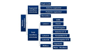
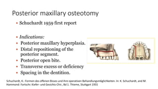
Maxillary Orthognathic
Surgery
Moderator : Dr (Col) Suresh Menon
Presented by : Dr Rayan Mallick
Contents
• Introduction
• Orthognathic surgery – History
• Anatomical considerations
• Biology of wound healing
• Maxillary procedures
1. Segmental procedures
2. Lefort I osteotomy
3. Lefort II osteotomy
4. Surgically assisted maxillary expansion
• Complications
• Conclusion
• References
Introduction
Orthognathic surgery :
The word ‘ orthognathic’ comes from the Greek word ‘ ortho’ meaning
to straighten and ‘ gnathia’ meaning jaws. Orthognathic surgery is
defined as ‘ the art and science of diagnosis, treatment planning and
execution of treatment to correct musculoskeletal , dento-osseous and
soft tissue deformities of the jaws and associated structures’
• Dentofacial deformities affect 20% of the population
• Patients with dentofacial deformities may demonstrate varying
degrees of functional and aesthetic compromise.
• Orthognathic surgery gives an opportunity to obtain better occlusal ,
skeletal and cosmetic results.
• Many psychosocial studies have shown that cosmetic motives for
seeking treatment seem to be quite common.
Goals of orthognathic surgery
• Function – mastication, speech, respiration, ocular etc
• Aesthetics – facial harmony and balance
• Stability – prevention of short and long term relapse
• Minimising of treatment time
Indications
• Dysphagia
• Speech abnormalities
• Malocclusion
• Masticatory dysfunction or malocclusion
• Anteroposterior discrepancies
• Vertical discrepancies
• Transverse discrepancies
• Asymmetries.
Orthognathic surgery - History
• In 1859 by Von Langenbeck -- for the removal of nasopharyngeal polyps.
• The first American report -- Cheever in 1867 -- for the treatment of
complete nasal obstruction secondary to recurrent epistaxis for which a
right hemimaxillary downfracture was used.
• In 1927 Wassmund -- LeFort I osteotomy for the correction of the midfacial
deformities and used orthopedic force postsurgically
• In 1934 Axhausen described total mobilization of the maxilla with
immediate repositioning for an open bite case
• In 1942 Schuchardt first advocated the pterygomaxillary dysjunction.
• In 1949 Moore and Ward -- horizontal transaction of the pterygoid plates
for advancement
• In 1965 Obwegeser -- complete mobilization of the maxilla so that
repositioning could be accomplished without tension.
• Bone grafting to enhance stabilization for LeFort and anterior osteotomies --
by Cupar, Gilles and Rowe and Obwegeser.
• Early description of the rigid fixation of maxillary osteotomies were
published by Michelet and colleagues in 1973, Horster in 1980, Drommer
and Luhr in 1981, and Luyk and Ward- Booth in 1985.
Wassmund’s procedure for the
correction of maxillary protrusion
Outline of the posterior
maxillary osteotomy which was described
by Schuchhardt.
E. W. Steinhauser : Historical development of orthognathic surgery ; Journal of Cranio-Maxillofacial Surgery (1996) 24, 195-204
Le Fort I osteotomy with additional vertical
bone cut for the correction of a malunited
fracture this technique was first
side. described by Axhausen
Total maxillary advancement by a Le Fort I
osteotomy in a cleft palate patient as published by
Obwegeser.
E. W. Steinhauser : Historical development of orthognathic surgery ; Journal of Cranio-Maxillofacial Surgery (1996) 24, 195-204
SEQUENCE OF TREATMENT PLANNING
• Dental and periodontal treatment
• Extractions
• Presurgical orthodontics
Position teeth over their respective basal bone
Align and level the teeth
Adjust for tooth size discrepancies
Divergence of roots adjacent to surgical sites.
• Orthognathic surgery
• Post-surgical orthodontics
Final tooth alignment and root parallelism
Maximal interdigitation
Ideal over bite and overjet
Centric occlusion - centric relation
Anatomical considerations
• BLOOD SUPPLY ( vascularization)
Bell and Levey 1969 and 1970 have shown in
a study that periosteum is necessary for
maintaining the blood supply to the teeth of a
mobile jaw segment. Even when the labial
periosteum is raised, care should be taken not
to cause any tension or tears.
Restoration of blood supply after 1 week
postoperatively – Dodson 1994. Anterior maxillary osteotomy performed
after reflection of the labial and buccal
mucoperiosteum.
• It is advised to handle the soft tissues with care so that adequate collateral blood
supply to the osteotomized segment is maintained and injury to other vital structures
is avoided.
• Prominent vessels to consider when planning orthognathic surgery are the posterior
superior alveolar (PSA) artery, greater palatine artery, maxillary artery, pterygoid
venous plexus.
Nerves
Facial nerve branches are rarely damaged in orthognathic surgery, but great care should be
taken not to inadvertently damage nerves.
Bradycardia and asystole may occur during down fracture or mobilization of the maxilla due to
the trigeminal‐cardiac reflex.
This can happen as a result of manipulation of the central or peripheral portions of the
trigeminal nerve during mobilization of the maxilla.
Patients who undergo Le Fort II and III osteotomies may experience infraorbital nerve sensory
dysfunction .
Lang, S., Lanigan, D. T., & van der Wal, M. (1991). Trigeminocardiac reflexes: maxillary and mandibular variants of the
oculocardiac reflex. Canadian Journal of Anaesthesia, 38(6), 757–760.
Biology of wound healing
Anterior maxillary osteotomy
• Attempts to move the anterior maxilla
were first made by Cohn-Stock,
Wassmund, and Spanier, who were
unaware of the biologic basis for the
healing of such surgically created
wounds.
• In 1962, animal and clinical
investigations were initiated to
delineate the biology of maxillary
osteotomy wound healing.
From 1962 to 1965, revascularization and bone healing were studied on animal
models after clinical simulations of three variations of anterior maxillary osteotomy
techniques
• Intraosseous and intrapulpal circulation to the anterior maxillary segment was
maintained when soft tissue was kept intact. Osteonecrosis was minimal and
vascular ischemia was only transient when the anterior maxillary bone segment
was pedicled to the labiobuccal mucoperiosteum, palatal mucoperiosteum or a
combination of both.
• Circulation to the dental pulp was maintained when the bone cuts were made away
from the apices of the teeth
• Six weeks after the osteotomies, there was no detectable intraosseous or
intrapulpal ischemia.
Posterior maxillary osteotomy
• In 1971, Bell and Levy reported on the biology of wound healing in posterior
maxillary osteotomies.
• Their microangiographic and histologic study of single-stage posterior
maxillary osteotomies in adult rhesus monkeys revealed minimal osteonecrosis
• When the bone cuts were made away from the apices of the teeth, pulpal
circulation was preserved.
• Within 4 weeks after the palatal surgery, the palatal mucoperiosteum was
reattached to the underlying bone, as evidenced by the revascularization of
the raised buccal and palatal soft tissue flaps to the underlying bone
• The results of these clinically analogous animal studies indicated that
single-stage posterior maxillary osteotomies are biologically sound.
• Thus, wound healing in both anterior and posterior maxillary osteotomies
are a biologically sound clinical procedures when the circulation to the
mobilized bone segment is maintained by attached mucoperiosteum
Maxillary Orthognathic procedures
Segmental maxillary surgeries
• Single tooth dento-osseous osteotomies.
• Corticotomy
• Anterior segmental maxillary osteotomy
• Posterior segmental maxillary osteotomy
• Horse shoe osteotomy.
Total maxillary osteotomies:
• Le Fort I osteotomies
• Le Fort II osteotomies
Segmental maxillary osteotomies
• Single tooth osteotomy
• Indicated in tooth malposition
• Dental ankylosis
• Closure of diastema
Anterior segmental osteotomy
• Cohn-Stock (1920)
• Wassmund (1935)
• Wunderer (1963)
• Cupar (1955)
• Epker and wolford (1980)
INDICATIONS
1. Anterior vertical maxillary excess in cases with acceptable posterior
occlusion
2. Sagittal maxillary excess with acceptable posterior occlusion
3. Maxillary anterior protrusion of anterior teeth with normal incisor axial
inclination to bone and acceptable posterior occlusion
4. Excessive proclination of anterior teeth
5. Dentoalveolar bimaxillary protrusion when an acceptable posterior
occlusion
6. Anterior open bite without vertical maxillary excess and normal posterior
occlusion
7. When retraction of anterior teeth is indicated but cannot be accomplished
with conventional orthodontic treatment (e.g., because of root resorption as
a result of previous orthodontic treatment, tooth ankylosis, malpositioned
dental implants)
8. Reduction of upper lip prominence relative to the nose and lower face
9. Maxillary excess combined with wide interdental spaces (malformed teeth,
oligodontia)
10. Preprosthetic procedure: augmentation and repositioning of anterior
edentulous atrophic maxillary ridge for dental implants
Cohn-Stock (1920)
Günther Cohn-Stock tried to surgically “correct a marked overjet and
overbite of the central maxillary teeth.”
In his pioneering article in 1921, he described the evolution of his idea to
perform an osteotomy of the anterior segment of the maxilla while
preserving the vestibular pedicle and, in a later design, also the palatal
artery.
Cohn-Stock, G.: Die chirurgische Immediatregulierung der Kiefer, speziell die chirurgische Behandlung der Prognathie.
Viertelj. schr. Zahnheilk. 3 (1921) 320
• Cohn-Stock presented two surgical cases
performed under local anesthesia in his Berlin
practice in May and June 1920. In his definitive
version, “Cohn III,” he described a transverse
palatal wedge ostectomy palatal to the anterior
teeth, performed through a subperiosteal
tunnel, and then a manual manipulation to
create a greenstick fracture at the ostectomy
site to retract the anterior maxilla.
• Contemporary authors suggest that Cohn-
Stock’s greenstick fracture method resulted in
significant relapse after removal of the fixation
splint because the anterior maxilla was not
adequately mobilized.
Wassmund technique(1935)
1927 Wassmund improved Cohn-Stock’s design by
creating a direct approach to the labial premaxillary
cortex using three vertical incisions and subperiosteal
tunneling for completion of the labial osteotomy
without reflection of labial or palatal flaps.
Both the labial and palatal blood supply is maintained;
however, the osteotomy is made in a relatively blind
fashion.
This method may be indicated for closure of multiple
interdental spaces and for anteroposterior
repositioning of the premaxilla. It was found to
maintain the best vascularity of the repositioned
segment in comparison to all other ASMO methods.
Wassmund, M.: Lehrbuch der praktischen Chirurgie des Mundes und der Kiefer. (Vol. 1) Meusser, Leipzig (1935) 282
 Preserves both buccal and palatal pedicle.
 Buccal as well as anterior vertical incision
 Tunneling between anterior and buccal
incisions
 Trans palatal osteotomy through buccal
vertical osteotomy.
 Occasional mid palatal sagittal incision.
Wandurer (1963)
 Relies on intact buccal pedicle.
 Transpalatal incision combined with
buccal vertical incision.
 Modification: Midline vertical incision
combined with buccal vertical incision.
Wunderer, S.: Die Prognathieoperation mittels frontal gestieltem Maxillafragment. Osterreich Z. Stomatol. 59 (1962) 98
Cupar method
 Buccal vestibular incision
 Nasal septum is first released
 Horizontal osteotomy
followed by vertical buccal
osteotomy.
 Trans palatal osteotomy
under direct vision.
 Advantages:
 Direct access to the nasal structures and superior maxilla
 Preservation of the palatal pedicle
 Ease of placement of rigid fixation
 Ability to remove bone from palate under direct visualization
 Good access to the superior maxilla for reduction of vertical
maxillary access
 Complications of the AMO:
 Loss of teeth vitality.
 Persistent periodontal defects
 Communication with nasal cavity or antrum
 Occlusal steps formation.
Corticotomy/Ostetomy
• In 1892 , Cunningham, first defined it as a linear cutting technique in the
cortical plates surrounding the teeth to produce mobilization of the teeth for
immediate movement.
• Köle (1959) thoroughly described the clinical application of orthodontically
moving teeth after interproximal bone segmentation as a means to expedite
tooth movement. He suggested that teeth can be segmented and moved as
“small boxes” through bone remodeling without involving the periodontal
ligament.
• Technique was described as an adjunct in the correction of numerous
types of malocclusions, with different treatment protocols such as non-
extraction and space closure approaches.
• Using this method, he claimed orthodontic treatment could be
accomplished in six to twelve weeks.
• Wilcko and colleagues (2001) reported, Patients with moderate to
severe crowding to accelerate tooth movement. The surgical procedure
consisted of interproximal vertical grooves on the labial and lingual
cortices of all teeth. A subapical horizontal scalloped corticotomy
connected the vertical grooves.
Posterior maxillary osteotomy
 Schuchardt 1959 first report
 Indications:
 Posterior maxillary hyperplasia.
 Distal repositioning of the
posterior segment.
 Posterior open bite.
 Transverse excess or deficiency
 Spacing in the dentition.
Schuchardt, K.: Formen des offenen Bisses und ihre operativen Behandlungsm6glichkeiten. In: K. Schuchardt, and M.
Hammond: Fortschr. Kiefer- und Gesichts-Chir., Bd 1. Thieme, Stuttgart 1955
Pterygomaxillary dysjunction
 Surgical technique:
 Buccal vestibular incision below the buttress.
 Palatal osteotomy through the buccal osteotomy site.
 Occasional palatal incision.
 Principles are same as for the total maxillary osteotomies.
 Segmental osteotomies are indicated for isolated dento facial
deformities when there is good dento skeletal relationships in
the non affected areas.
 Decreased morbidity when compared to total maxillary
osteotomies.
 For isolated dentofacial deformities and prosthetic problems,
the segmental osteotomies should be in the armamentarium of
the surgeon
Combination of anterior and posterior maxillary
osteotomy (Horseshoe osteotomy)
• Paul Tessier in 1969 reported this procedure for midface hypoplasia. It has also
been described and further developed by West and Epker 1972, Hall and Roddy
1975, Wolford and Epker 1975, West and McNeil 1975, Hall and West 1976,
and Maloney 1982.
• Palatal parasagittal osteotomies are performed with a piezoelectric device. The
hard palate is untouched staying in position.
• The method creates a three‐piece maxilla with the central nasal portion left
undisturbed. This is a complicated technique since multiple areas of bone
contacts exist. The indication is maxillary alveolar hyperplasia or transverse
hypoplasia without a vertical component. The method has more or less been
replaced by the traditional Le Fort I osteotomy.
Le Fort I
• Wassmund 1927 described this
osteotomy. In 1969, Obwegeser
reviewed and modified the
technique.
• Indications
• Vertical maxillary
excess/deficiency
• Maxillary hypoplasia
• Facial asymmetry
• An intraoral incision, 3–4 mm above the attached gingiva at maxillary
vestibular fornix, is made from the second premolar of one side to the
opposite side. A mucoperiosteal flap is raised exposing maxillary
walls.
• The infraorbital nerve must be identified, and the dissection will then
extend to the level of infraorbital nerve to simplify the following
osteotomies and to achieve direct control of periorbital tissues.
• During the exposure, great care should be taken not to expose the buccal
fat pad. Before the bone osteotomy, a nasal mucosa mobilization is
performed from the wall of the nasal cavity.
• The osteotomy is carried out with a burr, saw, or piezo machine, starting
from the lateral aspect of the piriform aperture and extended to the
posterior aspect of the maxilla towards the zygomatic buttress as
backwards as possible and inferiorly.
Frontal view of a Le Fort I osteotomy performed by a
piezo saw with a nice cut
• The same is done on the opposite side. These osteotomies are completed with
a chisel, to detach the nasal septum and lateral wall of the nasal cavity. The
osteotomy of the nasal septum and separation of septal cartilage from the
anterior nasal spine is usually made by chisel.
• Finally, it is important to separate the pterygomaxillary junction by using a
curved chisel lateromedially to simplify the mobilization of the maxilla.
• After the last detachment of the maxilla is done, downfracture can be
performed. The advantages of this osteotomy are full mobilization, speed,
simplicity, direct vision, the safety of segmentation, and reduced risk for
relapse.
• An alternative to the separation of the pterygomaxillary junction is a vertical
osteotomy in the tuber region of the alveolar crest. This method will, however,
not enable the same degree of mobility. It is fixated using miniplates.
Segmented Le Fort I osteotomy
In cases of open bite or when a transversal expansion is
required in the maxilla, a Le Fort I osteotomy approach can be
combined with a multiple‐piece osteotomy to correct an
unfavorable curve of Spee or a transverse discrepancy.
The maxilla can be sectioned into 2, 3, 4, 5, or 6 segments
depending on the indications.
A median osteotomy is made by a Lindeman burr or piezo at the
lateral aspect of the median palatal suture. When a
two‐segment osteotomy is planned, one median cut is
performed only on the side of the palatal suture.
This cut will be connected consequently to a vertical interdental
osteotomy that will be placed between the canine and first
bicuspid teeth on the same side
A vertical interdental osteotomy placed between the
canine and first bicuspid teeth on the same side
Clinical view of segmented maxilla
In some cases, a high Le Fort I osteotomy is required for advancement of
the entire midface to improve the extraoral profile
• Indications:
• Vertical maxillary excess in bimax protrusion.
• Superior re positioning of the maxilla to correct vertical excess.
• To advance maxilla in cleft palate and post traumatic patients.
• To correct open bites when combined with mandibular
procedures.
• Correction of cants.
• Advancement of the maxilla in class III patients.
Causes of failure
•Mobilized maxilla in maxillomandibular fixation
with manual manipulation demonstrating
posterior bony interferences preventing desired
positioning
Inadequate seating with maxilla placed too forward
despite proper bone trimming because of improper
condylar positioning during fixation, with
immediate relapse following appropriate condylar
seating (Condylar sag)
Incomplete posterior bone trimming
with unsatisfactory maxillary
positoning and resultant posterior
occlusal premature contacts
Appropriately positioned maxilla through proper bone
trimming and bimanual mandibular positioning with
downward and posterior pressure on chin and upward
forward pressure on the ramus
Lefort II
• The indication for this osteotomy is when a forward, downward movement of the nasal and
maxillary complex is necessary for correction of the midface. This osteotomy is performed in the
upper midface, between the frontal facial unit and above Le Fort I
• The approach has its place where there is a need for the correction of nasomaxillary hypoplasia.
• This osteotomy is also called a pyramidal naso‐orbital maxillary osteotomy.
• The Le Fort II osteotomy includes the naso‐orbital ethmoidal (NOE) fracture line, the zygoma
laterally, and internal part of the orbit.
• This osteotomy was first presented by Henderson and Jackson in 1973.
• Surgically, an incision is performed obliquely to the paranasal region extending to the
infraorbital rim to the medial canthus and over the nasal bone
• The Le Fort II osteotomy is relatively rare because it is not required as often
(only in 2% of dentofacial anomalies cases, such as in Apert, Crouzon
Treacher Collins syndromes).
• Other indications are a skeletal class III malocclusion in combination with
maxillary‐zygomatic deficiency, maxillary‐alveolar‐palatal cleft deformity,
and nasomaxillary deficiency.
• The osteotomy allows lengthening of the nose along with the movement of
the upper jaw in selected cases where this effect is desired.
• Steinhauser, 1980, described three different surgical approaches namely
anterio, pyramidal, and quadrangular osteotomies
• To get surgical access for the Le Fort II osteotomy, a V‐shaped incision with the apex at the glabella is made to
extend bilaterally along both sides of the nose to reach just above the alar base.
• The columella of the nose is pulled down, and the cartilaginous and bony part is separated.
• Osteotomy starts at the bottom of the nasal bone towards the medial wall of orbit towards the floor of orbit
posterior to the nasolacrimal apparatus.
• Then it continues to the infraorbital margin medial to the infraorbital nerve and extends to the alveolar bone
posterior to the second premolar. A flap in the posterior buccal area is raised. The osteotomy is completed
through the intra oral incision towards the pterygoid plates.
• In cases with deficiency in the infraorbital area, the cut can be continued to the zygomatic buttress before
going down towards the pterygoid plates.
• Downfracture of the midface can be done. The segment can be advanced after mobilization. Fixation can be
done either by an acrylic splint or by fixation plates. In this approach, bone grafts should be used to restore
the bone deficiencies. It is crucial to have skin coverage, and nasal lining must be provided.
Le Fort II osteotomy is performed in the upper midface,
between the frontal facial unit and above Le Fort I
Indications
• Patient with:
• Short nose
• Nasomaxillary retrusion
• Skeletal class III occlusion
• Posttraumatic defects
• Maxillonasal dysplasia in which a class I occlusion exists
• Secondary correction of cleft deformity
Exposure is gained through
 Bicoronal incision: more extensive
procedure, possibly entailing medial
• canthal disruption
 Bilateral Paranasal (inner canthus)
skin incision: results in potentially
visible external scar.
 Intraoral access was also described for
Le Fort II osteotomy.
 Exposure of the nasofrontal region
 Exposure of :
 Medial orbital floor
 Infraorbital rim
 Medial orbital wall
 Lacrimal sac
 Elevation of the nasal periosteum with a fine Obwegeser periosteal elevator
 Horizontal glabella osteotomy : below the level of the frontonasal suture
 Osteotomy extended:
• Orbital rim osteotomy continued downwards and posteriorly
• Medial wall osteotomy sparing the lacrimal sac
 Anteriorly: detachment of the anterior and superior arms of the medial canthal tendon,
 Posteriorly into the ethmoid bone and inferiorly behind the lacrimal sac by detachment of the
complete medial canthal area
Fixation
 In the nasofrontal region
 H- shaped 1.5-mm titanium miniplate
 Inverted T-shaped 1.5-mm titanium miniplate
 two short, straight 1.5-mm plates extending from
the glabella onto the lateral aspect of the nasal
complex bilaterally
 6mm screws
• In maxillary buttress region:
• Long L-shaped 2-0 titanium plate and 6-
mm screws
• Calvarial graft can be secured with lag
screws
Surgically Assisted Rapid Maxillary
Expansion (SARME)
• Assists to correct deformities in transverse dimension.
• First described by Angel in 1860
• This procedure is in essence combination of distraction osteogenisis and
controlled soft tissue expansion.
• Diagnosis and clinical evaluation:
• paranasal hallowing
• narrowed alar base
• deepening of nasolabial folds
• zygomatic difficiancy
• Treatment options: based on skeletal maturity
• Slow dentoalveolar expansion
• Orthopedic rapid maxillary expansion
• SAME
• Segmental maxillary osteotomy Advantages of SAME
• improved stability
• non extraction alignment of dentition
• elimination of negative space
• improved periodontal health and nasal respiration
Indications of SAME:
• Skeletal maxillomandibular transverse discrepancy greater than 5mm
• Significant TMD asstd with a narrow maxilla and wide mandible
• Failed or orthodontic expansion
• Necessity for a large amount more than 7mm of expansion
• Extremely thin and delicate gingival tissues with buccal gingival
recession
• Significant nasal stenosis
Technique
• Subtotal Le Fort 1 osteotomy
• Mandibular dentition should be decompensated
• Maxillary expansion appliance – preoperatively
Surgical technique
• B/L maxillary osteotomy
• Release of nasal septum
• Midline palatal osteotomy
• Lateral nasal wall osteotomy
• B/L release of the pterygoid plates
• Activation of the appliance : 1-1.5 mm
• Soft tissue closure
• Maxilla should remain stationary for 5 days postoperatively.
• Pt should feel discomfort while activation.
• Expansion at a rate of 0.5 mm/day
• Over correction is not recommended.
Retention:
• 6 to 12 months after expansion
APPLIANCES FOR RME
These are banded and bonded appliances. The banded appliance are
attached to teeth with bands on the maxillary first molar and first
premolars. The banded appliances are hygienic as there is no palatal
coverage. The banded RME are of two types:
1. Tooth and tissue borne
2. Tooth borne.
Tooth and tissue borne
Tooth borne
TOOTH BORNE RME
They consist of only bands and wires without any acrylic covering.
• HYRAX expander: It is a tooth borne appliance, which was introduced by
William Biederman in 1968. This type of appliance makes use of a special screw
called HYRAX (Hygenic Rapid Expander).
• The Hyrax Expander is essentially a non-spring loaded jackscrew
• The main advantage of this expander is that it does not irritate the palatal
mucosa and is easy to keep clean.
• It is capable of providing sutural separation of 11 mm within a very short period
of wear and a maximum of 13 mm can also be achieved.
• Each activation of the screw produces approximately 0.2 mm of lateral
expansion and it is activated from front to back.
Hyrax expander
Issacson expander:
• It is a tooth borne appliance without any palatal covering. This
expander makes use of a spring loaded screw called Minne expander,
which is soldered directly to the bands on first premolar and molars.
• The Minne expander is a heavily calibrated coil spring expanded by
turning a nut to compress the coil. Two metal flanges perpendicular
to the coil are soldered to the bands on abutment teeth.
TOOTH AND TISSUE BORNE RME
They consist of an expansion screw with acrylic abutting on alveolar ridges.
Haas, in 1970, gave the following advantages of tooth and tissue RME:
1. Produces more parallel expansion
2. Less relapse
3. Greater nasal cavity and apical base gain
4. More favorable relationship of the denture bases in width and
frequently in the anteroposterior plane as well
5. Creates more mobility of the maxilla instead of teeth.
Types of Tooth and Tissue Borne RME
Haas: The basis for the rapid expansion procedure is
to produce immediate mid-palatal suture separation by
disruption of the sutural connective tissue.
The rapid palatal expander as described by Haas is a rigid
appliance designed for maximum dental anchorage that
uses a jackscrew to produce expansion in 10 to 14 days.
He believed that this will maximize the orthopedic effects
and forces produced by this appliance have been reported
in the range of 3 to 10 pounds.
2. Derichsweiler: The first premolar and molars are
banded. Wire tags are soldered to these bands and then
inserted to the split palatal acrylic, which contains the
screw.
Haas expander
BONDED RAPID PALATAL EXPANDER
The Bonded RPE were first described by Cohen and Silverman in 1973. The
bonded appliance has become increasingly popular because of its
advantages:
1. It can be easily cemented during the mixed dentition stage, when
retention from other appliances can be poor.
2. Number of appointments are reduced.
3. There is reduced posterior teeth tipping and extrusion.
The buccal capping limits molar extrusion during treatment
4. It provides Bite block effect to facilitate the correction of anterior
crossbite.
Bonded palatal expander
IPC RAPID PALATAL EXPANDER
IPC is designed for orthopedic expansion along with labial alignment of
incisors. As expansion occurs, the IPC controls the NiTi open coil spring
force applied to the lingual surface of the anterior teeth. Wire around
the distal end of the lateral incisors limits the midline diastema that
often occurs during RPE treatment
IPC palatal expander
• Complications:
• Those due to inadequate surgery:
• pain
• dental tipping
• periodontal breakdown
• post orthodontic relapse Those due to expansion
• lack of appliance expansion
• deformation of the appliance due to processing errors
• stripping or loosening of midpalatal screw
Complications of Maxillary Osteotomies :
• Malpositioning
• Bleeding
• Perfusion deficiencies
• Periodontal defects
• Devitalized tooth
• Nerve injury
• Unfavorable fracture
• Nasolacrimal injuries
• Oronasal and oroantral fistulas
• Nasal septal deviation
• Maxillary sinusitis
• Unaesthetic soft tissue changes
• Non union
Intra cranial and ophthalmic complications
following maxillary osteotomies
• Complications can be grouped into four categories:
• (a) loss of function of the lacrimal gland,
• (b) cranial nerve palsies,
• (c)damage to the internal carotid artery,
• (d) loss of vision.
• Adie’s pupil
• Dry eye (Tomasetti et al 1976)
• Internal carotid thrombosis (Brady et al. 1981)
• Carotid cavernous sinus fistula (Habal 1986)
• Orbital complications previously described involve fractures that extended to the
orbital apex, and injury to the III and VI cranial nerves
• Mydriasis and accommodation paresis (Sirikumara and Sugar 1990)
• Blindness (Lanigan et al. 1993)
• Left corneal anesthesia (Lanigan et al. 1993)
• Haemolacria, “bloody tears” (Bruno Ramos Chrcanovica et al 2013)
• CSF leak (Bhaskaran et al 2010)
• Fracture of the clivus (Seung-Won Chung 2014)
Bibliography
• Bell WH. Revascularization and bone healing after anterior maxillary
osteotomy: a study using adult rhesus monkeys. J. Oral Surg.Editor
Reyneke J.P 1969; 27:249.
• Lang, S., Lanigan, D. T., & van der Wal, M.
(1991). Trigeminocardiac reflexes: maxillary and mandibular
variants of the oculocardiac reflex. Canadian Journal of
Anaesthesia, 38(6), 757–760.
• Orthognathic surgery - Fonseca
• Textbook of Maxillofacial surgery – Peterward booth
• Essentials of Orthognathic surgery – Johan .P . Reyneke
Carr RJ, Gilbert P. Isolated partial third nerve palsy following Le FortI
maxillary osteotomy in a patient with cleft lip and palate. Br J
OralMaxillofac Surg 1986;24:206–11.
Watts PG. Unilateral abducent nerve palsy: a rare complication follow-
ing a Le Fort I maxillary osteotomy. Br J Oral Maxillofac Surg
1984;22:212–5

Maxillary Orthognathic Surgery

  • 1.
    Maxillary Orthognathic Surgery Moderator :Dr (Col) Suresh Menon Presented by : Dr Rayan Mallick
  • 2.
    Contents • Introduction • Orthognathicsurgery – History • Anatomical considerations • Biology of wound healing • Maxillary procedures 1. Segmental procedures 2. Lefort I osteotomy 3. Lefort II osteotomy 4. Surgically assisted maxillary expansion • Complications • Conclusion • References
  • 3.
    Introduction Orthognathic surgery : Theword ‘ orthognathic’ comes from the Greek word ‘ ortho’ meaning to straighten and ‘ gnathia’ meaning jaws. Orthognathic surgery is defined as ‘ the art and science of diagnosis, treatment planning and execution of treatment to correct musculoskeletal , dento-osseous and soft tissue deformities of the jaws and associated structures’
  • 4.
    • Dentofacial deformitiesaffect 20% of the population • Patients with dentofacial deformities may demonstrate varying degrees of functional and aesthetic compromise. • Orthognathic surgery gives an opportunity to obtain better occlusal , skeletal and cosmetic results. • Many psychosocial studies have shown that cosmetic motives for seeking treatment seem to be quite common.
  • 5.
    Goals of orthognathicsurgery • Function – mastication, speech, respiration, ocular etc • Aesthetics – facial harmony and balance • Stability – prevention of short and long term relapse • Minimising of treatment time
  • 6.
    Indications • Dysphagia • Speechabnormalities • Malocclusion • Masticatory dysfunction or malocclusion • Anteroposterior discrepancies • Vertical discrepancies • Transverse discrepancies • Asymmetries.
  • 7.
    Orthognathic surgery -History • In 1859 by Von Langenbeck -- for the removal of nasopharyngeal polyps. • The first American report -- Cheever in 1867 -- for the treatment of complete nasal obstruction secondary to recurrent epistaxis for which a right hemimaxillary downfracture was used. • In 1927 Wassmund -- LeFort I osteotomy for the correction of the midfacial deformities and used orthopedic force postsurgically • In 1934 Axhausen described total mobilization of the maxilla with immediate repositioning for an open bite case
  • 8.
    • In 1942Schuchardt first advocated the pterygomaxillary dysjunction. • In 1949 Moore and Ward -- horizontal transaction of the pterygoid plates for advancement • In 1965 Obwegeser -- complete mobilization of the maxilla so that repositioning could be accomplished without tension. • Bone grafting to enhance stabilization for LeFort and anterior osteotomies -- by Cupar, Gilles and Rowe and Obwegeser. • Early description of the rigid fixation of maxillary osteotomies were published by Michelet and colleagues in 1973, Horster in 1980, Drommer and Luhr in 1981, and Luyk and Ward- Booth in 1985.
  • 9.
    Wassmund’s procedure forthe correction of maxillary protrusion Outline of the posterior maxillary osteotomy which was described by Schuchhardt. E. W. Steinhauser : Historical development of orthognathic surgery ; Journal of Cranio-Maxillofacial Surgery (1996) 24, 195-204
  • 10.
    Le Fort Iosteotomy with additional vertical bone cut for the correction of a malunited fracture this technique was first side. described by Axhausen Total maxillary advancement by a Le Fort I osteotomy in a cleft palate patient as published by Obwegeser. E. W. Steinhauser : Historical development of orthognathic surgery ; Journal of Cranio-Maxillofacial Surgery (1996) 24, 195-204
  • 11.
    SEQUENCE OF TREATMENTPLANNING • Dental and periodontal treatment • Extractions • Presurgical orthodontics Position teeth over their respective basal bone Align and level the teeth Adjust for tooth size discrepancies Divergence of roots adjacent to surgical sites. • Orthognathic surgery • Post-surgical orthodontics Final tooth alignment and root parallelism Maximal interdigitation Ideal over bite and overjet Centric occlusion - centric relation
  • 12.
    Anatomical considerations • BLOODSUPPLY ( vascularization) Bell and Levey 1969 and 1970 have shown in a study that periosteum is necessary for maintaining the blood supply to the teeth of a mobile jaw segment. Even when the labial periosteum is raised, care should be taken not to cause any tension or tears. Restoration of blood supply after 1 week postoperatively – Dodson 1994. Anterior maxillary osteotomy performed after reflection of the labial and buccal mucoperiosteum.
  • 13.
    • It isadvised to handle the soft tissues with care so that adequate collateral blood supply to the osteotomized segment is maintained and injury to other vital structures is avoided. • Prominent vessels to consider when planning orthognathic surgery are the posterior superior alveolar (PSA) artery, greater palatine artery, maxillary artery, pterygoid venous plexus.
  • 14.
    Nerves Facial nerve branchesare rarely damaged in orthognathic surgery, but great care should be taken not to inadvertently damage nerves. Bradycardia and asystole may occur during down fracture or mobilization of the maxilla due to the trigeminal‐cardiac reflex. This can happen as a result of manipulation of the central or peripheral portions of the trigeminal nerve during mobilization of the maxilla. Patients who undergo Le Fort II and III osteotomies may experience infraorbital nerve sensory dysfunction . Lang, S., Lanigan, D. T., & van der Wal, M. (1991). Trigeminocardiac reflexes: maxillary and mandibular variants of the oculocardiac reflex. Canadian Journal of Anaesthesia, 38(6), 757–760.
  • 15.
  • 16.
    Anterior maxillary osteotomy •Attempts to move the anterior maxilla were first made by Cohn-Stock, Wassmund, and Spanier, who were unaware of the biologic basis for the healing of such surgically created wounds. • In 1962, animal and clinical investigations were initiated to delineate the biology of maxillary osteotomy wound healing.
  • 17.
    From 1962 to1965, revascularization and bone healing were studied on animal models after clinical simulations of three variations of anterior maxillary osteotomy techniques • Intraosseous and intrapulpal circulation to the anterior maxillary segment was maintained when soft tissue was kept intact. Osteonecrosis was minimal and vascular ischemia was only transient when the anterior maxillary bone segment was pedicled to the labiobuccal mucoperiosteum, palatal mucoperiosteum or a combination of both. • Circulation to the dental pulp was maintained when the bone cuts were made away from the apices of the teeth • Six weeks after the osteotomies, there was no detectable intraosseous or intrapulpal ischemia.
  • 18.
  • 19.
    • In 1971,Bell and Levy reported on the biology of wound healing in posterior maxillary osteotomies. • Their microangiographic and histologic study of single-stage posterior maxillary osteotomies in adult rhesus monkeys revealed minimal osteonecrosis • When the bone cuts were made away from the apices of the teeth, pulpal circulation was preserved. • Within 4 weeks after the palatal surgery, the palatal mucoperiosteum was reattached to the underlying bone, as evidenced by the revascularization of the raised buccal and palatal soft tissue flaps to the underlying bone
  • 20.
    • The resultsof these clinically analogous animal studies indicated that single-stage posterior maxillary osteotomies are biologically sound. • Thus, wound healing in both anterior and posterior maxillary osteotomies are a biologically sound clinical procedures when the circulation to the mobilized bone segment is maintained by attached mucoperiosteum
  • 21.
    Maxillary Orthognathic procedures Segmentalmaxillary surgeries • Single tooth dento-osseous osteotomies. • Corticotomy • Anterior segmental maxillary osteotomy • Posterior segmental maxillary osteotomy • Horse shoe osteotomy. Total maxillary osteotomies: • Le Fort I osteotomies • Le Fort II osteotomies
  • 23.
    Segmental maxillary osteotomies •Single tooth osteotomy • Indicated in tooth malposition • Dental ankylosis • Closure of diastema
  • 25.
    Anterior segmental osteotomy •Cohn-Stock (1920) • Wassmund (1935) • Wunderer (1963) • Cupar (1955) • Epker and wolford (1980)
  • 26.
    INDICATIONS 1. Anterior verticalmaxillary excess in cases with acceptable posterior occlusion 2. Sagittal maxillary excess with acceptable posterior occlusion 3. Maxillary anterior protrusion of anterior teeth with normal incisor axial inclination to bone and acceptable posterior occlusion 4. Excessive proclination of anterior teeth 5. Dentoalveolar bimaxillary protrusion when an acceptable posterior occlusion 6. Anterior open bite without vertical maxillary excess and normal posterior occlusion
  • 27.
    7. When retractionof anterior teeth is indicated but cannot be accomplished with conventional orthodontic treatment (e.g., because of root resorption as a result of previous orthodontic treatment, tooth ankylosis, malpositioned dental implants) 8. Reduction of upper lip prominence relative to the nose and lower face 9. Maxillary excess combined with wide interdental spaces (malformed teeth, oligodontia) 10. Preprosthetic procedure: augmentation and repositioning of anterior edentulous atrophic maxillary ridge for dental implants
  • 28.
    Cohn-Stock (1920) Günther Cohn-Stocktried to surgically “correct a marked overjet and overbite of the central maxillary teeth.” In his pioneering article in 1921, he described the evolution of his idea to perform an osteotomy of the anterior segment of the maxilla while preserving the vestibular pedicle and, in a later design, also the palatal artery. Cohn-Stock, G.: Die chirurgische Immediatregulierung der Kiefer, speziell die chirurgische Behandlung der Prognathie. Viertelj. schr. Zahnheilk. 3 (1921) 320
  • 29.
    • Cohn-Stock presentedtwo surgical cases performed under local anesthesia in his Berlin practice in May and June 1920. In his definitive version, “Cohn III,” he described a transverse palatal wedge ostectomy palatal to the anterior teeth, performed through a subperiosteal tunnel, and then a manual manipulation to create a greenstick fracture at the ostectomy site to retract the anterior maxilla. • Contemporary authors suggest that Cohn- Stock’s greenstick fracture method resulted in significant relapse after removal of the fixation splint because the anterior maxilla was not adequately mobilized.
  • 32.
    Wassmund technique(1935) 1927 Wassmundimproved Cohn-Stock’s design by creating a direct approach to the labial premaxillary cortex using three vertical incisions and subperiosteal tunneling for completion of the labial osteotomy without reflection of labial or palatal flaps. Both the labial and palatal blood supply is maintained; however, the osteotomy is made in a relatively blind fashion. This method may be indicated for closure of multiple interdental spaces and for anteroposterior repositioning of the premaxilla. It was found to maintain the best vascularity of the repositioned segment in comparison to all other ASMO methods. Wassmund, M.: Lehrbuch der praktischen Chirurgie des Mundes und der Kiefer. (Vol. 1) Meusser, Leipzig (1935) 282
  • 33.
     Preserves bothbuccal and palatal pedicle.  Buccal as well as anterior vertical incision  Tunneling between anterior and buccal incisions  Trans palatal osteotomy through buccal vertical osteotomy.  Occasional mid palatal sagittal incision.
  • 34.
    Wandurer (1963)  Relieson intact buccal pedicle.  Transpalatal incision combined with buccal vertical incision.  Modification: Midline vertical incision combined with buccal vertical incision. Wunderer, S.: Die Prognathieoperation mittels frontal gestieltem Maxillafragment. Osterreich Z. Stomatol. 59 (1962) 98
  • 37.
    Cupar method  Buccalvestibular incision  Nasal septum is first released  Horizontal osteotomy followed by vertical buccal osteotomy.  Trans palatal osteotomy under direct vision.
  • 38.
     Advantages:  Directaccess to the nasal structures and superior maxilla  Preservation of the palatal pedicle  Ease of placement of rigid fixation  Ability to remove bone from palate under direct visualization  Good access to the superior maxilla for reduction of vertical maxillary access
  • 39.
     Complications ofthe AMO:  Loss of teeth vitality.  Persistent periodontal defects  Communication with nasal cavity or antrum  Occlusal steps formation.
  • 40.
    Corticotomy/Ostetomy • In 1892, Cunningham, first defined it as a linear cutting technique in the cortical plates surrounding the teeth to produce mobilization of the teeth for immediate movement. • Köle (1959) thoroughly described the clinical application of orthodontically moving teeth after interproximal bone segmentation as a means to expedite tooth movement. He suggested that teeth can be segmented and moved as “small boxes” through bone remodeling without involving the periodontal ligament.
  • 41.
    • Technique wasdescribed as an adjunct in the correction of numerous types of malocclusions, with different treatment protocols such as non- extraction and space closure approaches. • Using this method, he claimed orthodontic treatment could be accomplished in six to twelve weeks. • Wilcko and colleagues (2001) reported, Patients with moderate to severe crowding to accelerate tooth movement. The surgical procedure consisted of interproximal vertical grooves on the labial and lingual cortices of all teeth. A subapical horizontal scalloped corticotomy connected the vertical grooves.
  • 43.
    Posterior maxillary osteotomy Schuchardt 1959 first report  Indications:  Posterior maxillary hyperplasia.  Distal repositioning of the posterior segment.  Posterior open bite.  Transverse excess or deficiency  Spacing in the dentition. Schuchardt, K.: Formen des offenen Bisses und ihre operativen Behandlungsm6glichkeiten. In: K. Schuchardt, and M. Hammond: Fortschr. Kiefer- und Gesichts-Chir., Bd 1. Thieme, Stuttgart 1955
  • 46.
  • 47.
     Surgical technique: Buccal vestibular incision below the buttress.  Palatal osteotomy through the buccal osteotomy site.  Occasional palatal incision.  Principles are same as for the total maxillary osteotomies.
  • 48.
     Segmental osteotomiesare indicated for isolated dento facial deformities when there is good dento skeletal relationships in the non affected areas.  Decreased morbidity when compared to total maxillary osteotomies.  For isolated dentofacial deformities and prosthetic problems, the segmental osteotomies should be in the armamentarium of the surgeon
  • 49.
    Combination of anteriorand posterior maxillary osteotomy (Horseshoe osteotomy) • Paul Tessier in 1969 reported this procedure for midface hypoplasia. It has also been described and further developed by West and Epker 1972, Hall and Roddy 1975, Wolford and Epker 1975, West and McNeil 1975, Hall and West 1976, and Maloney 1982. • Palatal parasagittal osteotomies are performed with a piezoelectric device. The hard palate is untouched staying in position. • The method creates a three‐piece maxilla with the central nasal portion left undisturbed. This is a complicated technique since multiple areas of bone contacts exist. The indication is maxillary alveolar hyperplasia or transverse hypoplasia without a vertical component. The method has more or less been replaced by the traditional Le Fort I osteotomy.
  • 51.
    Le Fort I •Wassmund 1927 described this osteotomy. In 1969, Obwegeser reviewed and modified the technique. • Indications • Vertical maxillary excess/deficiency • Maxillary hypoplasia • Facial asymmetry
  • 52.
    • An intraoralincision, 3–4 mm above the attached gingiva at maxillary vestibular fornix, is made from the second premolar of one side to the opposite side. A mucoperiosteal flap is raised exposing maxillary walls.
  • 53.
    • The infraorbitalnerve must be identified, and the dissection will then extend to the level of infraorbital nerve to simplify the following osteotomies and to achieve direct control of periorbital tissues. • During the exposure, great care should be taken not to expose the buccal fat pad. Before the bone osteotomy, a nasal mucosa mobilization is performed from the wall of the nasal cavity. • The osteotomy is carried out with a burr, saw, or piezo machine, starting from the lateral aspect of the piriform aperture and extended to the posterior aspect of the maxilla towards the zygomatic buttress as backwards as possible and inferiorly.
  • 54.
    Frontal view ofa Le Fort I osteotomy performed by a piezo saw with a nice cut
  • 55.
    • The sameis done on the opposite side. These osteotomies are completed with a chisel, to detach the nasal septum and lateral wall of the nasal cavity. The osteotomy of the nasal septum and separation of septal cartilage from the anterior nasal spine is usually made by chisel. • Finally, it is important to separate the pterygomaxillary junction by using a curved chisel lateromedially to simplify the mobilization of the maxilla. • After the last detachment of the maxilla is done, downfracture can be performed. The advantages of this osteotomy are full mobilization, speed, simplicity, direct vision, the safety of segmentation, and reduced risk for relapse. • An alternative to the separation of the pterygomaxillary junction is a vertical osteotomy in the tuber region of the alveolar crest. This method will, however, not enable the same degree of mobility. It is fixated using miniplates.
  • 56.
    Segmented Le FortI osteotomy In cases of open bite or when a transversal expansion is required in the maxilla, a Le Fort I osteotomy approach can be combined with a multiple‐piece osteotomy to correct an unfavorable curve of Spee or a transverse discrepancy. The maxilla can be sectioned into 2, 3, 4, 5, or 6 segments depending on the indications. A median osteotomy is made by a Lindeman burr or piezo at the lateral aspect of the median palatal suture. When a two‐segment osteotomy is planned, one median cut is performed only on the side of the palatal suture. This cut will be connected consequently to a vertical interdental osteotomy that will be placed between the canine and first bicuspid teeth on the same side
  • 57.
    A vertical interdentalosteotomy placed between the canine and first bicuspid teeth on the same side Clinical view of segmented maxilla
  • 58.
    In some cases,a high Le Fort I osteotomy is required for advancement of the entire midface to improve the extraoral profile
  • 59.
    • Indications: • Verticalmaxillary excess in bimax protrusion. • Superior re positioning of the maxilla to correct vertical excess. • To advance maxilla in cleft palate and post traumatic patients. • To correct open bites when combined with mandibular procedures. • Correction of cants. • Advancement of the maxilla in class III patients.
  • 60.
    Causes of failure •Mobilizedmaxilla in maxillomandibular fixation with manual manipulation demonstrating posterior bony interferences preventing desired positioning Inadequate seating with maxilla placed too forward despite proper bone trimming because of improper condylar positioning during fixation, with immediate relapse following appropriate condylar seating (Condylar sag)
  • 61.
    Incomplete posterior bonetrimming with unsatisfactory maxillary positoning and resultant posterior occlusal premature contacts Appropriately positioned maxilla through proper bone trimming and bimanual mandibular positioning with downward and posterior pressure on chin and upward forward pressure on the ramus
  • 62.
    Lefort II • Theindication for this osteotomy is when a forward, downward movement of the nasal and maxillary complex is necessary for correction of the midface. This osteotomy is performed in the upper midface, between the frontal facial unit and above Le Fort I • The approach has its place where there is a need for the correction of nasomaxillary hypoplasia. • This osteotomy is also called a pyramidal naso‐orbital maxillary osteotomy. • The Le Fort II osteotomy includes the naso‐orbital ethmoidal (NOE) fracture line, the zygoma laterally, and internal part of the orbit. • This osteotomy was first presented by Henderson and Jackson in 1973. • Surgically, an incision is performed obliquely to the paranasal region extending to the infraorbital rim to the medial canthus and over the nasal bone
  • 63.
    • The LeFort II osteotomy is relatively rare because it is not required as often (only in 2% of dentofacial anomalies cases, such as in Apert, Crouzon Treacher Collins syndromes). • Other indications are a skeletal class III malocclusion in combination with maxillary‐zygomatic deficiency, maxillary‐alveolar‐palatal cleft deformity, and nasomaxillary deficiency. • The osteotomy allows lengthening of the nose along with the movement of the upper jaw in selected cases where this effect is desired. • Steinhauser, 1980, described three different surgical approaches namely anterio, pyramidal, and quadrangular osteotomies
  • 64.
    • To getsurgical access for the Le Fort II osteotomy, a V‐shaped incision with the apex at the glabella is made to extend bilaterally along both sides of the nose to reach just above the alar base. • The columella of the nose is pulled down, and the cartilaginous and bony part is separated. • Osteotomy starts at the bottom of the nasal bone towards the medial wall of orbit towards the floor of orbit posterior to the nasolacrimal apparatus. • Then it continues to the infraorbital margin medial to the infraorbital nerve and extends to the alveolar bone posterior to the second premolar. A flap in the posterior buccal area is raised. The osteotomy is completed through the intra oral incision towards the pterygoid plates. • In cases with deficiency in the infraorbital area, the cut can be continued to the zygomatic buttress before going down towards the pterygoid plates. • Downfracture of the midface can be done. The segment can be advanced after mobilization. Fixation can be done either by an acrylic splint or by fixation plates. In this approach, bone grafts should be used to restore the bone deficiencies. It is crucial to have skin coverage, and nasal lining must be provided.
  • 65.
    Le Fort IIosteotomy is performed in the upper midface, between the frontal facial unit and above Le Fort I
  • 66.
    Indications • Patient with: •Short nose • Nasomaxillary retrusion • Skeletal class III occlusion • Posttraumatic defects • Maxillonasal dysplasia in which a class I occlusion exists • Secondary correction of cleft deformity
  • 67.
    Exposure is gainedthrough  Bicoronal incision: more extensive procedure, possibly entailing medial • canthal disruption  Bilateral Paranasal (inner canthus) skin incision: results in potentially visible external scar.  Intraoral access was also described for Le Fort II osteotomy.
  • 68.
     Exposure ofthe nasofrontal region  Exposure of :  Medial orbital floor  Infraorbital rim  Medial orbital wall  Lacrimal sac  Elevation of the nasal periosteum with a fine Obwegeser periosteal elevator  Horizontal glabella osteotomy : below the level of the frontonasal suture  Osteotomy extended: • Orbital rim osteotomy continued downwards and posteriorly • Medial wall osteotomy sparing the lacrimal sac  Anteriorly: detachment of the anterior and superior arms of the medial canthal tendon,  Posteriorly into the ethmoid bone and inferiorly behind the lacrimal sac by detachment of the complete medial canthal area
  • 69.
    Fixation  In thenasofrontal region  H- shaped 1.5-mm titanium miniplate  Inverted T-shaped 1.5-mm titanium miniplate  two short, straight 1.5-mm plates extending from the glabella onto the lateral aspect of the nasal complex bilaterally  6mm screws
  • 70.
    • In maxillarybuttress region: • Long L-shaped 2-0 titanium plate and 6- mm screws • Calvarial graft can be secured with lag screws
  • 71.
    Surgically Assisted RapidMaxillary Expansion (SARME) • Assists to correct deformities in transverse dimension. • First described by Angel in 1860 • This procedure is in essence combination of distraction osteogenisis and controlled soft tissue expansion. • Diagnosis and clinical evaluation: • paranasal hallowing • narrowed alar base • deepening of nasolabial folds • zygomatic difficiancy
  • 72.
    • Treatment options:based on skeletal maturity • Slow dentoalveolar expansion • Orthopedic rapid maxillary expansion • SAME • Segmental maxillary osteotomy Advantages of SAME • improved stability • non extraction alignment of dentition • elimination of negative space • improved periodontal health and nasal respiration
  • 73.
    Indications of SAME: •Skeletal maxillomandibular transverse discrepancy greater than 5mm • Significant TMD asstd with a narrow maxilla and wide mandible • Failed or orthodontic expansion • Necessity for a large amount more than 7mm of expansion • Extremely thin and delicate gingival tissues with buccal gingival recession • Significant nasal stenosis
  • 74.
    Technique • Subtotal LeFort 1 osteotomy • Mandibular dentition should be decompensated • Maxillary expansion appliance – preoperatively
  • 75.
    Surgical technique • B/Lmaxillary osteotomy • Release of nasal septum • Midline palatal osteotomy • Lateral nasal wall osteotomy • B/L release of the pterygoid plates • Activation of the appliance : 1-1.5 mm • Soft tissue closure
  • 79.
    • Maxilla shouldremain stationary for 5 days postoperatively. • Pt should feel discomfort while activation. • Expansion at a rate of 0.5 mm/day • Over correction is not recommended. Retention: • 6 to 12 months after expansion
  • 80.
    APPLIANCES FOR RME Theseare banded and bonded appliances. The banded appliance are attached to teeth with bands on the maxillary first molar and first premolars. The banded appliances are hygienic as there is no palatal coverage. The banded RME are of two types: 1. Tooth and tissue borne 2. Tooth borne.
  • 81.
    Tooth and tissueborne Tooth borne
  • 82.
    TOOTH BORNE RME Theyconsist of only bands and wires without any acrylic covering. • HYRAX expander: It is a tooth borne appliance, which was introduced by William Biederman in 1968. This type of appliance makes use of a special screw called HYRAX (Hygenic Rapid Expander). • The Hyrax Expander is essentially a non-spring loaded jackscrew • The main advantage of this expander is that it does not irritate the palatal mucosa and is easy to keep clean. • It is capable of providing sutural separation of 11 mm within a very short period of wear and a maximum of 13 mm can also be achieved. • Each activation of the screw produces approximately 0.2 mm of lateral expansion and it is activated from front to back.
  • 83.
  • 84.
    Issacson expander: • Itis a tooth borne appliance without any palatal covering. This expander makes use of a spring loaded screw called Minne expander, which is soldered directly to the bands on first premolar and molars. • The Minne expander is a heavily calibrated coil spring expanded by turning a nut to compress the coil. Two metal flanges perpendicular to the coil are soldered to the bands on abutment teeth.
  • 85.
    TOOTH AND TISSUEBORNE RME They consist of an expansion screw with acrylic abutting on alveolar ridges. Haas, in 1970, gave the following advantages of tooth and tissue RME: 1. Produces more parallel expansion 2. Less relapse 3. Greater nasal cavity and apical base gain 4. More favorable relationship of the denture bases in width and frequently in the anteroposterior plane as well 5. Creates more mobility of the maxilla instead of teeth.
  • 86.
    Types of Toothand Tissue Borne RME Haas: The basis for the rapid expansion procedure is to produce immediate mid-palatal suture separation by disruption of the sutural connective tissue. The rapid palatal expander as described by Haas is a rigid appliance designed for maximum dental anchorage that uses a jackscrew to produce expansion in 10 to 14 days. He believed that this will maximize the orthopedic effects and forces produced by this appliance have been reported in the range of 3 to 10 pounds. 2. Derichsweiler: The first premolar and molars are banded. Wire tags are soldered to these bands and then inserted to the split palatal acrylic, which contains the screw. Haas expander
  • 87.
    BONDED RAPID PALATALEXPANDER The Bonded RPE were first described by Cohen and Silverman in 1973. The bonded appliance has become increasingly popular because of its advantages: 1. It can be easily cemented during the mixed dentition stage, when retention from other appliances can be poor. 2. Number of appointments are reduced. 3. There is reduced posterior teeth tipping and extrusion. The buccal capping limits molar extrusion during treatment 4. It provides Bite block effect to facilitate the correction of anterior crossbite.
  • 88.
  • 89.
    IPC RAPID PALATALEXPANDER IPC is designed for orthopedic expansion along with labial alignment of incisors. As expansion occurs, the IPC controls the NiTi open coil spring force applied to the lingual surface of the anterior teeth. Wire around the distal end of the lateral incisors limits the midline diastema that often occurs during RPE treatment
  • 90.
  • 91.
    • Complications: • Thosedue to inadequate surgery: • pain • dental tipping • periodontal breakdown • post orthodontic relapse Those due to expansion • lack of appliance expansion • deformation of the appliance due to processing errors • stripping or loosening of midpalatal screw
  • 92.
    Complications of MaxillaryOsteotomies : • Malpositioning • Bleeding • Perfusion deficiencies • Periodontal defects • Devitalized tooth • Nerve injury • Unfavorable fracture • Nasolacrimal injuries • Oronasal and oroantral fistulas • Nasal septal deviation • Maxillary sinusitis • Unaesthetic soft tissue changes • Non union
  • 93.
    Intra cranial andophthalmic complications following maxillary osteotomies • Complications can be grouped into four categories: • (a) loss of function of the lacrimal gland, • (b) cranial nerve palsies, • (c)damage to the internal carotid artery, • (d) loss of vision.
  • 94.
    • Adie’s pupil •Dry eye (Tomasetti et al 1976) • Internal carotid thrombosis (Brady et al. 1981) • Carotid cavernous sinus fistula (Habal 1986) • Orbital complications previously described involve fractures that extended to the orbital apex, and injury to the III and VI cranial nerves • Mydriasis and accommodation paresis (Sirikumara and Sugar 1990) • Blindness (Lanigan et al. 1993) • Left corneal anesthesia (Lanigan et al. 1993) • Haemolacria, “bloody tears” (Bruno Ramos Chrcanovica et al 2013) • CSF leak (Bhaskaran et al 2010) • Fracture of the clivus (Seung-Won Chung 2014)
  • 95.
    Bibliography • Bell WH.Revascularization and bone healing after anterior maxillary osteotomy: a study using adult rhesus monkeys. J. Oral Surg.Editor Reyneke J.P 1969; 27:249. • Lang, S., Lanigan, D. T., & van der Wal, M. (1991). Trigeminocardiac reflexes: maxillary and mandibular variants of the oculocardiac reflex. Canadian Journal of Anaesthesia, 38(6), 757–760. • Orthognathic surgery - Fonseca • Textbook of Maxillofacial surgery – Peterward booth • Essentials of Orthognathic surgery – Johan .P . Reyneke
  • 96.
    Carr RJ, GilbertP. Isolated partial third nerve palsy following Le FortI maxillary osteotomy in a patient with cleft lip and palate. Br J OralMaxillofac Surg 1986;24:206–11. Watts PG. Unilateral abducent nerve palsy: a rare complication follow- ing a Le Fort I maxillary osteotomy. Br J Oral Maxillofac Surg 1984;22:212–5

Editor's Notes

  • #18 Osseous union between most of the sectioned segments occurred within 6 weeks without immobilization of the mandible. (5 mm when feasible, which was thought intuitively to be “safe”).