Downloaded 415 times
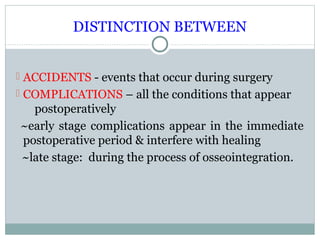
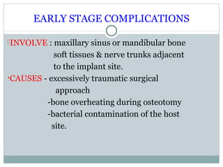


This document discusses early and late stage complications that can occur after dental implant surgery. Early stage complications within the immediate postoperative period include edema, exudate, pain, and infection caused by bacterial contamination during surgery. Late stage complications occur during osseointegration and include bone defects, periapical lesions, failed osseointegration, and mandibular fractures. Prevention of complications focuses on strict asepsis during surgery, atraumatic surgical techniques, appropriate treatment planning, and proper management of healing.


